Karimi Ali, Shahrooz Rasoul, Hobbenagh Rahim, Delirezh Nowruz, Amani Saeede, Garssen Johan, Mortaz Esmaeil, M Adcock Ian
Department of Basic Science, Faculty of Veterinary Medicine, Urmia University, Urmia, Iran.
Department of Basic Science, Faculty of Veterinary Medicine, Urmia University, Urmia, Iran.Electronic Address:
Cell J. 2020 Jan;21(4):391-400. doi: 10.22074/cellj.2020.6287. Epub 2019 Jul 29.
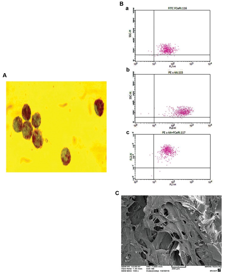
Peripheral arterial disease results from obstructed blood flow in arteries and increases the risk of amputation in acute cases. Therapeutic angiogenesis using bioengineered tissues composed of a chitosan scaffold that was enriched with mast cells (MCs) and/or platelet-rich plasma (PRP) was used to assess the formation of vascular networks and subsequently improved the functional recovery following hindlimb ischemia. This study aimed to find an optimal approach for restoring local vascularization.
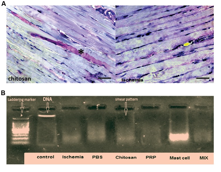
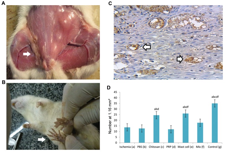
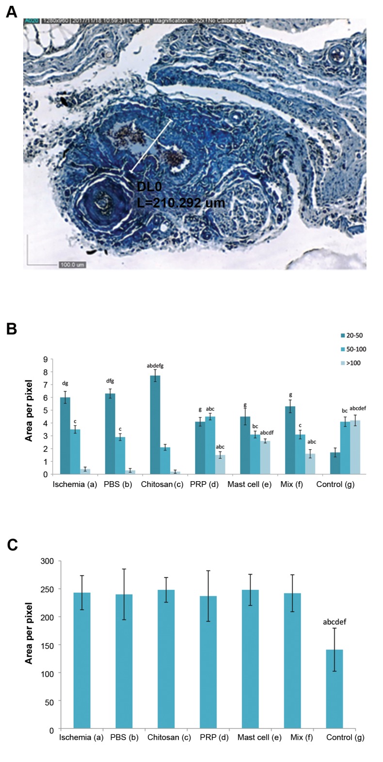
In this experimental study, thirty rats were randomly divided into six experimental groups: a. Ischemic control group with right femoral artery transection, b. Ischemia with phosphate-buffered saline (PBS) control group, c. Ischemia with chitosan scaffold, d. Ischemia with chitosan and MCs, e. Ischemia with chitosan and PRP, and f. Ischemia with chitosan, PRP, and MCs. The left hind limbs served as non-ischemic controls. The analysis of capillary density, arterial diameter, histomorphometric analysis and immunohistochemistry at the transected locations and in gastrocnemius muscles was performed.
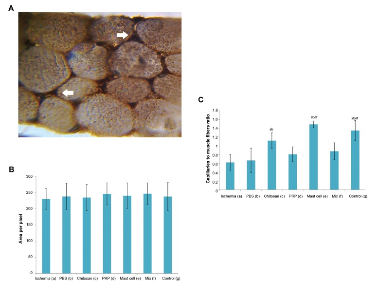
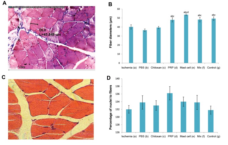
The group treated with chitosan/MC significantly increased capillary density and the mean number of large blood vessels at the site of femoral artery transection compared with other experimental groups (P<0.05). The treatment with chitosan/MC also significantly increased the muscle fiber diameter and the capillary-to-muscle fiber ratio in gastrocnemius muscles compared with all other ischemic groups (P<0.05).
These findings suggested that chitosan and MCs together could offer a new approach for the therapeutic induction of angiogenesis in cases of peripheral arterial diseases.
外周动脉疾病是由动脉血流受阻引起的,在急性病例中会增加截肢风险。使用由富含肥大细胞(MCs)和/或富血小板血浆(PRP)的壳聚糖支架组成的生物工程组织进行治疗性血管生成,以评估血管网络的形成,并随后改善后肢缺血后的功能恢复。本研究旨在找到一种恢复局部血管化的最佳方法。
在本实验研究中,30只大鼠被随机分为六个实验组:a. 右股动脉横断的缺血对照组;b. 缺血加磷酸盐缓冲盐水(PBS)对照组;c. 缺血加壳聚糖支架组;d. 缺血加壳聚糖和MCs组;e. 缺血加壳聚糖和PRP组;f. 缺血加壳聚糖、PRP和MCs组。左后肢作为非缺血对照。对横断部位和腓肠肌进行毛细血管密度分析、动脉直径测量、组织形态计量分析和免疫组织化学分析。
与其他实验组相比,壳聚糖/MC治疗组在股动脉横断部位的毛细血管密度和大血管平均数量显著增加(P<0.05)。与所有其他缺血组相比,壳聚糖/MC治疗组在腓肠肌中的肌纤维直径和毛细血管与肌纤维的比例也显著增加(P<0.05)。
这些发现表明,壳聚糖和MCs共同可为外周动脉疾病病例的治疗性诱导血管生成提供一种新方法。