• 文献检索
  • 文档翻译
  • 深度研究
  • 学术资讯
  • Suppr Zotero 插件Zotero 插件
  • 邀请有礼
  • 套餐&价格
  • 历史记录
应用&插件
Suppr Zotero 插件Zotero 插件浏览器插件Mac 客户端Windows 客户端微信小程序
定价
高级版会员购买积分包购买API积分包
服务
文献检索文档翻译深度研究API 文档MCP 服务
关于我们
关于 Suppr公司介绍联系我们用户协议隐私条款
关注我们

Suppr 超能文献

核心技术专利:CN118964589B侵权必究
粤ICP备2023148730 号-1Suppr @ 2026

文献检索

告别复杂PubMed语法,用中文像聊天一样搜索,搜遍4000万医学文献。AI智能推荐,让科研检索更轻松。

立即免费搜索

文件翻译

保留排版,准确专业,支持PDF/Word/PPT等文件格式,支持 12+语言互译。

免费翻译文档

深度研究

AI帮你快速写综述,25分钟生成高质量综述,智能提取关键信息,辅助科研写作。

立即免费体验

接受新辅助化疗的乳腺癌患者中用于肿瘤定位的乳腺组织标志物比较。

Comparison of breast tissue markers for tumor localization in breast cancer patients undergoing neoadjuvant chemotherapy.

作者信息

Koo Ja Ho, Kim Eun-Kyung, Moon Hee Jung, Yoon Jung Hyun, Park Vivian Youngjean, Kim Min Jung

机构信息

Department of Radiology, Severance Hospital, Yonsei University College of Medicine, Seoul, Korea.

出版信息

Ultrasonography. 2019 Oct;38(4):336-344. doi: 10.14366/usg.19004. Epub 2019 Apr 7.

DOI:10.14366/usg.19004
PMID:31378014
原文链接:https://pmc.ncbi.nlm.nih.gov/articles/PMC6769188/
Abstract

PURPOSE

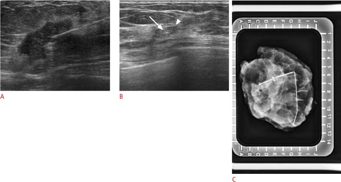
The purpose of this study was to compare the visibility of breast tissue markers in cases of breast cancer on ultrasonography (US) after neoadjuvant chemotherapy (NAC) and to analyze whether the type of marker affected the choice of localization method after NAC.

METHODS

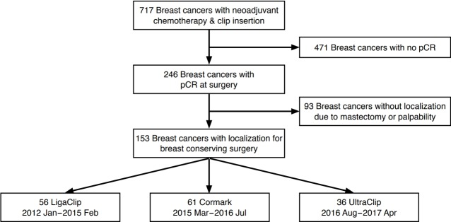
We included 153 tissue markers inserted within breast cancers that showed pathologically complete response (pCR) after NAC from January 2012 to April 2017. One of three types of markers (a surgical clip, Cormark, or UltraClip) was inserted. Medical records and imaging findings were retrospectively reviewed. We compared the visibility of the different types of tissue markers on US after NAC, and also compared the imaging modalities used in the preoperative localization. The chi-square test, Fisher exact test, and multiple logistic regression were used for analysis.

RESULTS

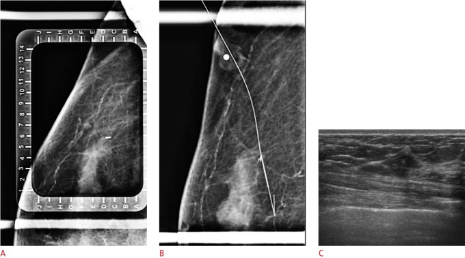
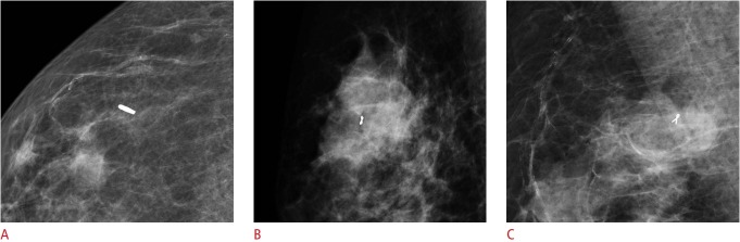
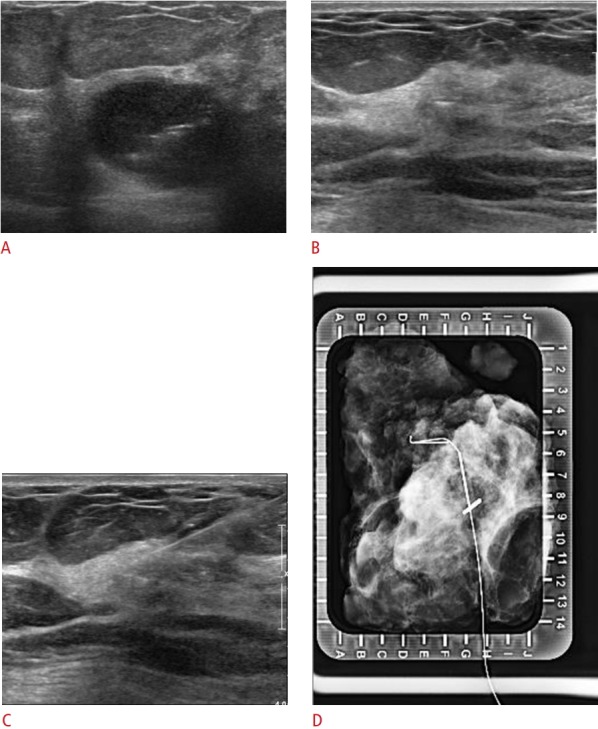
Of the 153 tissue markers, 56 were surgical clips, 61 Cormark, and 36 UltraClip. After NAC, residual lesions were not seen on US in 42 cases (27.5%). In multivariate analysis, the visibility of the surgical clips and Cormark markers was better than that of the UltraClip markers (odds ratio [OR], 5.467; 95% confidence interal [CI], 1.717 to 17.410; P=0.004 and OR, 3.045; 95% CI, 1.074 to 8.628; P=0.036, respectively). Among the 131 cases where localization targeting the marker was required, the proportion of US-guided localizations was significantly higher when a surgical clip was used than when an UltraClip marker was used (OR, 5.566; 95% CI, 1.610 to 19.246; P=0.007) in the multivariate analysis.

CONCLUSION

The type of breast tissue marker affected its visibility on US in cases with pCR after NAC, which in turn affected the localization methodology.

摘要

目的

本研究旨在比较新辅助化疗(NAC)后乳腺癌病例中乳腺组织标记物在超声检查(US)中的可见性,并分析标记物类型是否会影响NAC后定位方法的选择。

方法

我们纳入了2012年1月至2017年4月期间在NAC后病理显示完全缓解(pCR)的乳腺癌中插入的153个组织标记物。插入的标记物为三种类型之一(手术夹、Cormark或UltraClip)。对病历和影像学检查结果进行回顾性分析。我们比较了NAC后不同类型组织标记物在超声检查中的可见性,并比较了术前定位所使用的成像方式。采用卡方检验、Fisher精确检验和多元逻辑回归进行分析。

结果

153个组织标记物中,56个为手术夹,61个为Cormark,36个为UltraClip。NAC后,42例(27.5%)超声检查未见残留病变。在多变量分析中,手术夹和Cormark标记物的可见性优于UltraClip标记物(优势比[OR]分别为5.467;95%置信区间[CI]为1.717至17.410;P = 0.004和OR为3.045;95%CI为1.074至8.628;P = 0.036)。在需要针对标记物进行定位的131例病例中,多变量分析显示,使用手术夹时超声引导定位的比例显著高于使用UltraClip标记物时(OR为5.566;95%CI为1.610至19.246;P = 0.007)。

结论

乳腺组织标记物的类型会影响NAC后pCR病例中其在超声检查中的可见性,进而影响定位方法。

https://cdn.ncbi.nlm.nih.gov/pmc/blobs/4d1e/6769188/196cfdf06993/usg-19004f5.jpg
https://cdn.ncbi.nlm.nih.gov/pmc/blobs/4d1e/6769188/a3ef35dc4bd1/usg-19004f1.jpg
https://cdn.ncbi.nlm.nih.gov/pmc/blobs/4d1e/6769188/92d8227c85aa/usg-19004f2.jpg
https://cdn.ncbi.nlm.nih.gov/pmc/blobs/4d1e/6769188/297147edc895/usg-19004f3.jpg
https://cdn.ncbi.nlm.nih.gov/pmc/blobs/4d1e/6769188/e1311e7c0fb5/usg-19004f4.jpg
https://cdn.ncbi.nlm.nih.gov/pmc/blobs/4d1e/6769188/196cfdf06993/usg-19004f5.jpg

相似文献

1
Comparison of breast tissue markers for tumor localization in breast cancer patients undergoing neoadjuvant chemotherapy.接受新辅助化疗的乳腺癌患者中用于肿瘤定位的乳腺组织标志物比较。
Ultrasonography. 2019 Oct;38(4):336-344. doi: 10.14366/usg.19004. Epub 2019 Apr 7.
2
Ultrasonography-guided surgical clip placement for tumor localization in patients undergoing neoadjuvant chemotherapy for breast cancer.超声引导下手术夹定位在接受新辅助化疗乳腺癌患者中的应用。
J Breast Cancer. 2015 Mar;18(1):44-9. doi: 10.4048/jbc.2015.18.1.44. Epub 2015 Mar 27.
3
Comparative Evaluation of USG-Guided Single Tissue Marker Versus Multiple Tissue Marker Placements in Breast Malignancy Patients Undergoing Neoadjuvant Chemotherapy for Tumor Localization.超声引导下单组织标志物与多组织标志物放置在接受新辅助化疗的乳腺癌患者肿瘤定位中的比较评估
Cureus. 2024 Jul 25;16(7):e65355. doi: 10.7759/cureus.65355. eCollection 2024 Jul.
4
Evaluation of the sonographic visibility and sonographic appearance of the breast biopsy marker (UltraClip) placed in phantoms and patients.评估置于体模和患者体内的乳腺活检标记物(UltraClip)的超声可见性及超声表现。
Breast Cancer. 2017 Jul;24(4):585-592. doi: 10.1007/s12282-016-0741-0. Epub 2016 Nov 12.
5
Comparison of the Ultrasound Visibility of Tissue Markers in Metastatic Lymph Nodes after Neoadjuvant Chemotherapy in Patients with Breast Cancer.乳腺癌患者新辅助化疗后转移性淋巴结中组织标志物的超声可见性比较
Diagnostics (Basel). 2022 Oct 7;12(10):2424. doi: 10.3390/diagnostics12102424.
6
Feasibility of Preoperative Axillary Lymph Node Marking with a Clip in Breast Cancer Patients Before Neoadjuvant Chemotherapy: A Preliminary Study.新辅助化疗前乳腺癌患者术前用夹子标记腋窝淋巴结的可行性:一项初步研究
World J Surg. 2018 Feb;42(2):582-589. doi: 10.1007/s00268-017-4171-8.
7
Evaluation of a Marker Clip System in Sonographically Guided Core Needle Biopsy for Breast Cancer Localization Before and After Neoadjuvant Chemotherapy.在新辅助化疗前后,对用于超声引导下乳腺癌定位的核心针活检中的标记夹系统进行评估。
Geburtshilfe Frauenheilkd. 2017 Feb;77(2):169-175. doi: 10.1055/s-0042-124191.
8
CT-Guided Wire Localization for Involved Axillary Lymph Nodes After Neo-adjuvant Chemotherapy in Patients With Initially Node-Positive Breast Cancer.新辅助化疗后初始淋巴结阳性乳腺癌患者受累腋窝淋巴结的CT引导下金属丝定位
Breast J. 2016 Jul;22(4):390-6. doi: 10.1111/tbj.12597. Epub 2016 Apr 9.
9
Surgery after Neoadjuvant Chemotherapy: A Clip-Based Technique to Improve Surgical Outcomes, a Single-Center Experience.新辅助化疗后的手术:一种基于夹子的技术以改善手术效果,单中心经验
Cancers (Basel). 2022 Apr 29;14(9):2229. doi: 10.3390/cancers14092229.
10
Usefulness of tissue marker clips in patients undergoing neoadjuvant chemotherapy for breast cancer.组织标记夹在接受乳腺癌新辅助化疗患者中的应用价值
AJR Am J Roentgenol. 1999 Oct;173(4):911-7. doi: 10.2214/ajr.173.4.10511147.

引用本文的文献

1
Can the organic coating of breast tissue markers be sampled by an ultrasound-guided core cut biopsy without removing the clip marker? A proof-of-concept evaluation of two coated clip marker types in turkey breast tissue to improve representativity of ultrasound-guided core cut biopsies.能否在不移除夹式标记物的情况下,通过超声引导下的芯针切割活检对乳腺组织标记物的有机涂层进行采样?对火鸡乳腺组织中两种涂层夹式标记物进行概念验证评估,以提高超声引导下芯针切割活检的代表性。
Ultrasound. 2025 May 9:1742271X251337701. doi: 10.1177/1742271X251337701.
2
Underlying factors that affect ultrasound conspicuity of breast biopsy markers: an exploratory study.影响乳腺活检标记物超声显影的潜在因素:一项探索性研究。
Transl Breast Cancer Res. 2025 Jan 21;6:2. doi: 10.21037/tbcr-24-48. eCollection 2025.
3

本文引用的文献

1
Evaluation of the sonographic visibility and sonographic appearance of the breast biopsy marker (UltraClip) placed in phantoms and patients.评估置于体模和患者体内的乳腺活检标记物(UltraClip)的超声可见性及超声表现。
Breast Cancer. 2017 Jul;24(4):585-592. doi: 10.1007/s12282-016-0741-0. Epub 2016 Nov 12.
2
Ultrasonography-guided surgical clip placement for tumor localization in patients undergoing neoadjuvant chemotherapy for breast cancer.超声引导下手术夹定位在接受新辅助化疗乳腺癌患者中的应用。
J Breast Cancer. 2015 Mar;18(1):44-9. doi: 10.4048/jbc.2015.18.1.44. Epub 2015 Mar 27.
3
Presurgical localization of breast abnormalities: an overview and analysis of 202 cases.
The Evaluation of a Cost-Effective Method for Tumour Marking Prior to Neo-Adjuvant Chemotherapy Using Silver Rods.使用银棒对新辅助化疗前肿瘤标记的一种经济有效方法的评估。
Eur J Breast Health. 2023 Jan 1;19(1):99-105. doi: 10.4274/ejbh.galenos.2022.2022-10-3. eCollection 2023 Jan.
4
Comparison of the Ultrasound Visibility of Tissue Markers in Metastatic Lymph Nodes after Neoadjuvant Chemotherapy in Patients with Breast Cancer.乳腺癌患者新辅助化疗后转移性淋巴结中组织标志物的超声可见性比较
Diagnostics (Basel). 2022 Oct 7;12(10):2424. doi: 10.3390/diagnostics12102424.
5
Surgery after Neoadjuvant Chemotherapy: A Clip-Based Technique to Improve Surgical Outcomes, a Single-Center Experience.新辅助化疗后的手术:一种基于夹子的技术以改善手术效果,单中心经验
Cancers (Basel). 2022 Apr 29;14(9):2229. doi: 10.3390/cancers14092229.
6
Surgical outcomes after radioactive 125I seed versus hookwire localization of non-palpable breast cancer: a multicentre randomized clinical trial.放射性 125I 种子与钩丝定位非扪及性乳腺癌的手术结果:一项多中心随机临床试验。
Br J Surg. 2021 Jan 27;108(1):40-48. doi: 10.1093/bjs/znaa008.
乳腺异常的术前定位:202例病例概述与分析
Indian J Surg Oncol. 2010 Dec;1(4):278-83. doi: 10.1007/s13193-010-0016-8. Epub 2010 Dec 30.
4
Recommendations from an international consensus conference on the current status and future of neoadjuvant systemic therapy in primary breast cancer.国际原发性乳腺癌新辅助全身治疗现状与未来共识会议推荐意见。
Ann Surg Oncol. 2012 May;19(5):1508-16. doi: 10.1245/s10434-011-2108-2. Epub 2011 Dec 23.
5
A plea for the biopsy marker: how, why and why not clipping after breast biopsy?呼吁使用活检标志物:乳腺活检后为何及为何不进行夹闭?
Breast Cancer Res Treat. 2012 Apr;132(3):881-93. doi: 10.1007/s10549-011-1847-x. Epub 2011 Nov 1.
6
Current neoadjuvant treatment options for HER2-positive breast cancer.HER2阳性乳腺癌的当前新辅助治疗方案。
Biologics. 2011;5:87-94. doi: 10.2147/BTT.S22917. Epub 2011 Aug 9.
7
Placement of radiopaque clips for tumor localization in patients undergoing neoadjuvant chemotherapy and breast conservation therapy.在接受新辅助化疗和保乳治疗的患者中放置不透射线夹用于肿瘤定位。
Cancer. 2007 Dec 1;110(11):2420-7. doi: 10.1002/cncr.23068.
8
Recommendations from an international expert panel on the use of neoadjuvant (primary) systemic treatment of operable breast cancer: an update.国际专家小组关于可手术乳腺癌新辅助(原发性)全身治疗应用的建议:更新版
J Clin Oncol. 2006 Apr 20;24(12):1940-9. doi: 10.1200/JCO.2005.02.6187.
9
Sonographically guided metallic clip placement after core needle biopsy of the breast.超声引导下乳腺粗针活检后金属夹置入术
AJR Am J Roentgenol. 2000 Nov;175(5):1353-5. doi: 10.2214/ajr.175.5.1751353.
10
Sonographically guided clip placement facilitates localization of breast cancer after neoadjuvant chemotherapy.超声引导下夹子放置有助于新辅助化疗后乳腺癌的定位。
AJR Am J Roentgenol. 2000 Feb;174(2):539-40. doi: 10.2214/ajr.174.2.1740539.