• 文献检索
  • 文档翻译
  • 深度研究
  • 学术资讯
  • Suppr Zotero 插件Zotero 插件
  • 邀请有礼
  • 套餐&价格
  • 历史记录
应用&插件
Suppr Zotero 插件Zotero 插件浏览器插件Mac 客户端Windows 客户端微信小程序
定价
高级版会员购买积分包购买API积分包
服务
文献检索文档翻译深度研究API 文档MCP 服务
关于我们
关于 Suppr公司介绍联系我们用户协议隐私条款
关注我们

Suppr 超能文献

核心技术专利:CN118964589B侵权必究
粤ICP备2023148730 号-1Suppr @ 2026

文献检索

告别复杂PubMed语法,用中文像聊天一样搜索,搜遍4000万医学文献。AI智能推荐,让科研检索更轻松。

立即免费搜索

文件翻译

保留排版,准确专业,支持PDF/Word/PPT等文件格式,支持 12+语言互译。

免费翻译文档

深度研究

AI帮你快速写综述,25分钟生成高质量综述,智能提取关键信息,辅助科研写作。

立即免费体验

急性后极部多灶性鳞状色素上皮病变具有Vogt-小柳-原田病的特征性光学相干断层扫描表现

Acute Posterior Multifocal Placoid Pigment Epitheliopathy Sharing Characteristic OCT Findings of Vogt-Koyanagi-Harada Disease.

作者信息

Kitamura Yuta, Oshitari Toshiyuki, Kitahashi Masayasu, Baba Takayuki, Yamamoto Shuichi

机构信息

Department of Ophthalmology and Visual Science, Chiba University Graduate School of Medicine, Japan.

Department of Ophthalmology, International University of Health and Welfare, School of Medicine, Japan.

出版信息

Case Rep Ophthalmol Med. 2019 Jul 9;2019:9217656. doi: 10.1155/2019/9217656. eCollection 2019.

DOI:10.1155/2019/9217656
PMID:31380133
原文链接:https://pmc.ncbi.nlm.nih.gov/articles/PMC6652076/
Abstract

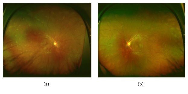
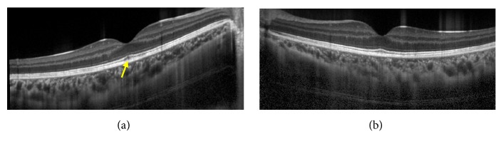
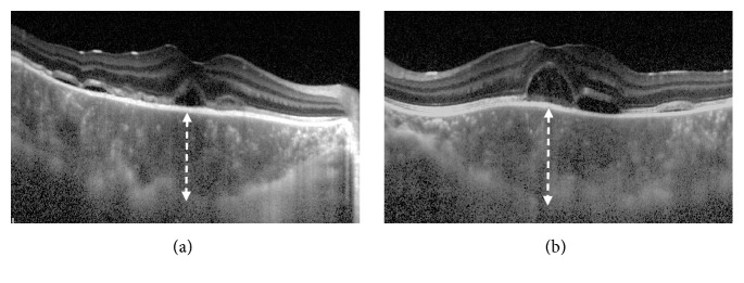
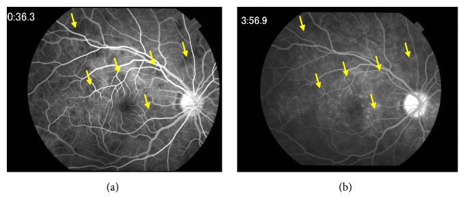
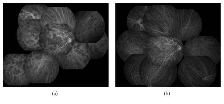
A 17-year-old male presented with acute bilateral paracentral scotomata and blurred vision. Funduscopic examination showed bilateral macular serous retinal detachment and yellow-white placoid lesions at the level of retinal pigment epithelium. OCT study showed typical VKH disease findings with marked choroidal thickening and macular serous retinal detachment partly with subretinal septa in both eyes. FA demonstrated hypofluorescence at the placoid lesions in the early phase and hyperfluorescence in the late phase. Laboratory investigation showed negative result for HLA-DR4 serotype and the patient's cerebrospinal fluid test values were within normal range. We made the diagnosis of APMPPE from these results. At 2-month follow-up without the use of corticosteroids, OCT reexamination showed complete amelioration of subretinal fluid in both eyes. Patchy pigmentary lesions also resolved clinically with partial chorioretinal scars. The results in this case suggested OCT findings in APMPPE patients could be similar to characteristic features usually found in acute VKH disease. We recommend comprehensive assessments such as FA, cerebral spinal fluid analysis, and HLA typing which help in leading proper diagnosis.

摘要

一名17岁男性患者出现急性双侧旁中心暗点和视力模糊。眼底检查显示双侧黄斑区浆液性视网膜脱离以及视网膜色素上皮层的黄白色类脂质性病变。光学相干断层扫描(OCT)研究显示典型的Vogt-小柳-原田(VKH)病表现,双眼脉络膜显著增厚,黄斑区浆液性视网膜脱离,部分伴有视网膜下间隔。荧光素眼底血管造影(FA)显示类脂质性病变在早期呈低荧光,晚期呈高荧光。实验室检查显示HLA-DR4血清型结果为阴性,患者脑脊液检测值在正常范围内。根据这些结果我们诊断为急性后极部多发性鳞状色素上皮病变(APMPPE)。在未使用皮质类固醇的2个月随访中,OCT复查显示双眼视网膜下液完全吸收。斑片状色素沉着性病变在临床上也消退,伴有部分脉络膜视网膜瘢痕。该病例结果提示APMPPE患者的OCT表现可能与急性VKH病通常所见的特征相似。我们建议进行全面评估,如FA、脑脊液分析和HLA分型,这有助于做出正确诊断。

https://cdn.ncbi.nlm.nih.gov/pmc/blobs/12f4/6652076/8b2462fc0416/CRIOPM2019-9217656.005.jpg
https://cdn.ncbi.nlm.nih.gov/pmc/blobs/12f4/6652076/02ad22814663/CRIOPM2019-9217656.001.jpg
https://cdn.ncbi.nlm.nih.gov/pmc/blobs/12f4/6652076/e653042ebc1f/CRIOPM2019-9217656.002.jpg
https://cdn.ncbi.nlm.nih.gov/pmc/blobs/12f4/6652076/4259af6b3c48/CRIOPM2019-9217656.003.jpg
https://cdn.ncbi.nlm.nih.gov/pmc/blobs/12f4/6652076/1eab8b81ed03/CRIOPM2019-9217656.004.jpg
https://cdn.ncbi.nlm.nih.gov/pmc/blobs/12f4/6652076/8b2462fc0416/CRIOPM2019-9217656.005.jpg

相似文献

1
Acute Posterior Multifocal Placoid Pigment Epitheliopathy Sharing Characteristic OCT Findings of Vogt-Koyanagi-Harada Disease.急性后极部多灶性鳞状色素上皮病变具有Vogt-小柳-原田病的特征性光学相干断层扫描表现
Case Rep Ophthalmol Med. 2019 Jul 9;2019:9217656. doi: 10.1155/2019/9217656. eCollection 2019.
2
A case of acute posterior multifocal placoid pigment epitheliopathy demonstrating vogt-koyanagi-harada disease-like optical coherence tomography findings in the acute stage.一例急性后极部多灶性鳞状色素上皮病变,在急性期表现出伏格特-小柳-原田病样光学相干断层扫描结果。
Case Rep Ophthalmol. 2013 Oct 11;4(3):172-9. doi: 10.1159/000356051. eCollection 2013.
3
Role of multimodal imaging in differentiating unilateral APMPPE from unilateral Harada disease - a case report.多模态成像在鉴别单侧急性后极部多发性鳞状色素上皮病变与单侧原田病中的作用——病例报告
Ann Med Surg (Lond). 2023 Apr 7;85(4):1286-1290. doi: 10.1097/MS9.0000000000000548. eCollection 2023 Apr.
4
Acute posterior multifocal placoid pigment epitheliopathy associated with serous retinal detachment: A case report.急性后极部多灶性鳞状色素上皮病变伴浆液性视网膜脱离:一例报告。
Ann Med Surg (Lond). 2022 Apr 8;77:103600. doi: 10.1016/j.amsu.2022.103600. eCollection 2022 May.
5
A case of unilateral and spontaneously resolving posterior uveitis with overlapping features of Vogt-Koyanagi-Harada disease and Acute Posterior Multifocal Placoid Pigment Epitheliopathy.一例具有Vogt-小柳原田病和急性后极部多发性鳞状色素上皮病变重叠特征的单侧自发性消退性后葡萄膜炎病例。
Springerplus. 2016 Sep 1;5(1):1471. doi: 10.1186/s40064-016-3132-2. eCollection 2016.
6
Acute posterior multifocal placoid pigment epitheliopathy associated with infectious mononucleosis: A rare presentation.与传染性单核细胞增多症相关的急性后部多灶性扁平色素上皮病变:一种罕见的表现。
Saudi J Ophthalmol. 2021 Nov 17;36(4):387-389. doi: 10.4103/sjopt.sjopt_95_21. eCollection 2022 Oct-Dec.
7
Choroidal perfusion abnormalities associated with Acute Posterior Multifocal Placoid Pigment Epitheliopathy: a case report.急性后极部多灶性鳞状色素上皮病变相关的脉络膜灌注异常:一例报告
BMC Ophthalmol. 2018 Apr 10;18(1):87. doi: 10.1186/s12886-018-0756-8.
8
Serous retinal detachment as a complication of acute posterior multifocal placoid pigment epitheliopathy.浆液性视网膜脱离作为急性后极部多灶性扁平色素上皮病变的一种并发症。
Retin Cases Brief Rep. 2010 Spring;4(2):129-31. doi: 10.1097/ICB.0b013e318196b313.
9
[Pathophysiology of macular diseases--morphology and function].黄斑疾病的病理生理学——形态与功能
Nippon Ganka Gakkai Zasshi. 2011 Mar;115(3):238-74; discussion 275.
10
A case of APMPPE-like panuveitis presenting with extensive outer retinal layer impairment following COVID-19 vaccination.一例 COVID-19 疫苗接种后出现广泛的外层视网膜损伤的 APMPPE 样全葡萄膜炎。
BMC Ophthalmol. 2023 May 24;23(1):233. doi: 10.1186/s12886-023-02978-2.

引用本文的文献

1
Proposing a Neurotropic Etiology for Acute Posterior Multifocal Placoid Pigment Epitheliopathy and Relentless Placoid Chorioretinitis.提出急性后极部多灶性扁平色素上皮病变及顽固性扁平脉络膜视网膜病变的嗜神经病因学。
Front Ophthalmol (Lausanne). 2022 Jan 10;1:802962. doi: 10.3389/fopht.2021.802962. eCollection 2021.
2
Challenges in posterior uveitis-tips and tricks for the retina specialist.后葡萄膜炎的挑战——视网膜专科医生的小贴士和技巧
J Ophthalmic Inflamm Infect. 2023 Aug 17;13(1):35. doi: 10.1186/s12348-023-00342-5.
3
Association of Retinal Pigment Epithelium Reflectivity on Optical Coherence Tomography with Recurrence of Vogt-Koyanagi-Harada Disease: A Retrospective Observational Study.

本文引用的文献

1
Choroidal perfusion abnormalities associated with Acute Posterior Multifocal Placoid Pigment Epitheliopathy: a case report.急性后极部多灶性鳞状色素上皮病变相关的脉络膜灌注异常:一例报告
BMC Ophthalmol. 2018 Apr 10;18(1):87. doi: 10.1186/s12886-018-0756-8.
2
Application of OCT-angiography to characterise the evolution of chorioretinal lesions in acute posterior multifocal placoid pigment epitheliopathy.光学相干断层扫描血管造影术在急性后极部多灶性扁平状色素上皮病变脉络膜视网膜病变演变特征分析中的应用
Eye (Lond). 2017 Oct;31(10):1399-1408. doi: 10.1038/eye.2017.180. Epub 2017 Oct 6.
3
ACUTE POSTERIOR MULTIFOCAL PLACOID PIGMENT EPITHELIOPATHY ON OPTICAL COHERENCE TOMOGRAPHY ANGIOGRAPHY.
光学相干断层扫描中视网膜色素上皮反射率与Vogt-小柳-原田病复发的相关性:一项回顾性观察研究
Clin Ophthalmol. 2023 Jul 21;17:2071-2079. doi: 10.2147/OPTH.S419546. eCollection 2023.
4
Role of multimodal imaging in differentiating unilateral APMPPE from unilateral Harada disease - a case report.多模态成像在鉴别单侧急性后极部多发性鳞状色素上皮病变与单侧原田病中的作用——病例报告
Ann Med Surg (Lond). 2023 Apr 7;85(4):1286-1290. doi: 10.1097/MS9.0000000000000548. eCollection 2023 Apr.
5
Relentless placoid chorioretinitis: A review of four cases in pediatric and young adult patients with a discussion of therapeutic strategies.持续性扁平苔藓样脉络膜视网膜炎:4例儿科及青年患者病例回顾并探讨治疗策略
Front Pediatr. 2023 Mar 27;11:885230. doi: 10.3389/fped.2023.885230. eCollection 2023.
6
Acute posterior multifocal placoid pigment epitheliopathy associated with serous retinal detachment: A case report.急性后极部多灶性鳞状色素上皮病变伴浆液性视网膜脱离:一例报告。
Ann Med Surg (Lond). 2022 Apr 8;77:103600. doi: 10.1016/j.amsu.2022.103600. eCollection 2022 May.
7
Acute posterior multifocal placoid pigment epitheliopathy (APMPPE).急性后极部多灶性鳞状色素上皮病变
J Ophthalmic Inflamm Infect. 2021 Nov 1;11(1):31. doi: 10.1186/s12348-021-00263-1.
光学相干断层扫描血管造影术显示的急性后极部多灶性扁平状色素上皮病变
Retina. 2017 Nov;37(11):2084-2094. doi: 10.1097/IAE.0000000000001487.
4
A case of unilateral and spontaneously resolving posterior uveitis with overlapping features of Vogt-Koyanagi-Harada disease and Acute Posterior Multifocal Placoid Pigment Epitheliopathy.一例具有Vogt-小柳原田病和急性后极部多发性鳞状色素上皮病变重叠特征的单侧自发性消退性后葡萄膜炎病例。
Springerplus. 2016 Sep 1;5(1):1471. doi: 10.1186/s40064-016-3132-2. eCollection 2016.
5
Phase-Variance Optical Coherence Tomographic Angiography Imaging of Choroidal Perfusion Changes Associated With Acute Posterior Multifocal Placoid Pigment Epitheliopathy.急性后极部多灶性鳞状色素上皮病变相关脉络膜灌注变化的相位方差光学相干断层扫描血管造影成像
JAMA Ophthalmol. 2016 Aug 1;134(8):943-5. doi: 10.1001/jamaophthalmol.2016.1645.
6
Vogt-Koyanagi-Harada syndrome: Perspectives for immunogenetics, multimodal imaging, and therapeutic options.Vogt-Koyanagi-Harada 综合征:免疫遗传学、多模态成像和治疗选择的视角。
Autoimmun Rev. 2016 Aug;15(8):809-19. doi: 10.1016/j.autrev.2016.04.001. Epub 2016 Apr 7.
7
Increased choroidal blood flow velocity with regression of acute posterior multifocal placoid pigment epitheliopathy.急性后极部多灶性扁平色素上皮病变消退时脉络膜血流速度增加。
Jpn J Ophthalmol. 2016 May;60(3):172-8. doi: 10.1007/s10384-016-0440-6. Epub 2016 Mar 28.
8
Choroidal Involvement in Acute Posterior Multifocal Placoid Pigment Epitheliopathy.急性后极部多灶性鳞状色素上皮病变中的脉络膜受累情况
Ophthalmic Surg Lasers Imaging Retina. 2016 Jan;47(1):20-6. doi: 10.3928/23258160-20151214-03.
9
Analysis of choroidal folds in acute Vogt-Koyanagi-Harada disease using high-penetration optical coherence tomography.利用高穿透光学相干断层扫描分析急性小柳原田病的脉络膜皱褶
Graefes Arch Clin Exp Ophthalmol. 2015 Jun;253(6):959-64. doi: 10.1007/s00417-015-2945-y. Epub 2015 Feb 10.
10
Comparison of the optical coherence tomographic characters between acute Vogt-Koyanagi-Harada disease and acute central serous chorioretinopathy.急性 Vogt-小柳原田病与急性中心性浆液性脉络膜视网膜病变的光学相干断层扫描特征比较。
BMC Ophthalmol. 2014 Jun 28;14:87. doi: 10.1186/1471-2415-14-87.