• 文献检索
  • 文档翻译
  • 深度研究
  • 学术资讯
  • Suppr Zotero 插件Zotero 插件
  • 邀请有礼
  • 套餐&价格
  • 历史记录
应用&插件
Suppr Zotero 插件Zotero 插件浏览器插件Mac 客户端Windows 客户端微信小程序
定价
高级版会员购买积分包购买API积分包
服务
文献检索文档翻译深度研究API 文档MCP 服务
关于我们
关于 Suppr公司介绍联系我们用户协议隐私条款
关注我们

Suppr 超能文献

核心技术专利:CN118964589B侵权必究
粤ICP备2023148730 号-1Suppr @ 2026

文献检索

告别复杂PubMed语法,用中文像聊天一样搜索,搜遍4000万医学文献。AI智能推荐,让科研检索更轻松。

立即免费搜索

文件翻译

保留排版,准确专业,支持PDF/Word/PPT等文件格式,支持 12+语言互译。

免费翻译文档

深度研究

AI帮你快速写综述,25分钟生成高质量综述,智能提取关键信息,辅助科研写作。

立即免费体验

用于脑肿瘤手术的共聚焦辅助多光谱荧光显微镜

Confocal-Assisted Multispectral Fluorescent Microscopy for Brain Tumor Surgery.

作者信息

Charalampaki Patra, Nakamura Makoto, Athanasopoulos Dimitrios, Heimann Axel

机构信息

Department of Neurosurgery, Cologne Medical Center, University Witten-Herdecke, Witten, Germany.

Institute of Neurosurgical Pathophysiology, Medical University Mainz, Mainz, Germany.

出版信息

Front Oncol. 2019 Jul 18;9:583. doi: 10.3389/fonc.2019.00583. eCollection 2019.

DOI:10.3389/fonc.2019.00583
PMID:31380264
原文链接:https://pmc.ncbi.nlm.nih.gov/articles/PMC6657348/
Abstract

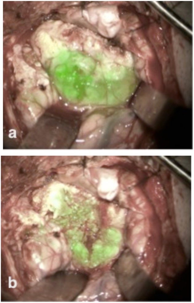
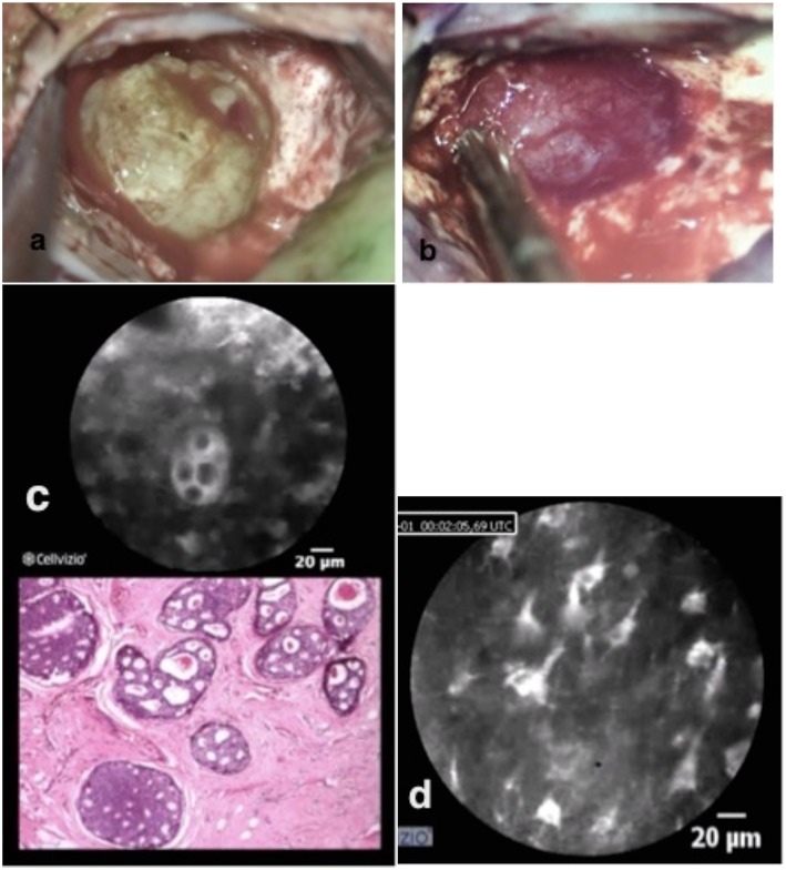
Optimal surgical therapy for brain tumors is the combination of complete resection with minimal invasion and damage to the adjacent normal tissue. To achieve this goal, we need advanced imaging techniques on a scale from macro- to microscopic resolution. In the last decade, the development of fluorescence-guided surgery has been the most influential breakthrough, marginally improving outcomes in brain tumor surgery. Multispectral fluorescence microscopy (MFL) is a novel imaging technique that allows the overlapping of a fluorescent image and a white light image in real-time, with delivery of the merged image to the surgeon through the eyepieces of a surgical microscope. MFL permits the detection and characterization of brain tumors using fluorescent molecular markers such as 5-aminolevulinic acid (5-ALA) or indocyanine green (ICG), while simultaneously obtaining high definition white light images to create a pseudo-colored composite image in real-time. Limitations associated with the use of MFL include decreased light imaging intensity and decreased levels of magnification that may compromise maximal tumor resection on a cellular scale. Confocal laser endomicroscopy (CLE) is another novel advanced imaging technique that is based on miniaturization of the microscope imaging head in order to provide the possibility of microscopy at the cellular level. Clear visualization of the cellular cytoarchitecture can be achieved with 400-fold-1,000-fold magnification. CLE allows on the one hand the intra-operative detection and differentiation of single tumor cells (without the need for intra-operative histologic analysis of biopsy specimens) as well as the definition of borders between tumor and normal tissue at a cellular level, dramatically improving the accuracy of surgical resection. The application and implementation of CLE-assisted surgery in surgical oncology increases not only the number of options for real-time diagnostic imaging, but also the therapeutic options by extending the resection borders of cancer at a cellular level and, more importantly, by protecting the functionality of normal tissue in the adjacent areas of the human brain. In this article, we describe our experience using these new techniques of confocal-assisted fluorescent surgery including analysis on the technology, usability, indications, limitations, and further developments.

摘要

脑肿瘤的最佳手术治疗方法是在对相邻正常组织侵袭和损伤最小的情况下进行完全切除。为实现这一目标,我们需要从宏观到微观分辨率的先进成像技术。在过去十年中,荧光引导手术的发展是最具影响力的突破,在一定程度上改善了脑肿瘤手术的结果。多光谱荧光显微镜(MFL)是一种新型成像技术,它允许荧光图像和白光图像实时重叠,并通过手术显微镜的目镜将合并后的图像传送给外科医生。MFL允许使用荧光分子标记物(如5-氨基乙酰丙酸(5-ALA)或吲哚菁绿(ICG))来检测和表征脑肿瘤,同时获得高清晰度白光图像以实时创建伪彩色合成图像。与使用MFL相关的局限性包括光成像强度降低和放大倍数降低,这可能会影响细胞水平上肿瘤的最大切除。共聚焦激光内镜显微镜(CLE)是另一种新型先进成像技术,它基于显微镜成像头的小型化,以便在细胞水平上提供显微镜检查的可能性。通过400倍至1000倍的放大倍数,可以清晰地观察细胞的细胞结构。CLE一方面允许术中检测和区分单个肿瘤细胞(无需对活检标本进行术中组织学分析),以及在细胞水平上定义肿瘤与正常组织之间的边界,从而显著提高手术切除的准确性。CLE辅助手术在外科肿瘤学中的应用和实施不仅增加了实时诊断成像的选择数量,还通过在细胞水平上扩展癌症切除边界,更重要的是通过保护人脑相邻区域正常组织的功能,增加了治疗选择。在本文中,我们描述了我们使用这些共聚焦辅助荧光手术新技术的经验,包括对技术、可用性、适应症、局限性和进一步发展的分析。

https://cdn.ncbi.nlm.nih.gov/pmc/blobs/033d/6657348/063747eebca1/fonc-09-00583-g0004.jpg
https://cdn.ncbi.nlm.nih.gov/pmc/blobs/033d/6657348/8214d4c773e3/fonc-09-00583-g0001.jpg
https://cdn.ncbi.nlm.nih.gov/pmc/blobs/033d/6657348/39575877c154/fonc-09-00583-g0002.jpg
https://cdn.ncbi.nlm.nih.gov/pmc/blobs/033d/6657348/92519bdd3a04/fonc-09-00583-g0003.jpg
https://cdn.ncbi.nlm.nih.gov/pmc/blobs/033d/6657348/063747eebca1/fonc-09-00583-g0004.jpg

相似文献

1
Confocal-Assisted Multispectral Fluorescent Microscopy for Brain Tumor Surgery.用于脑肿瘤手术的共聚焦辅助多光谱荧光显微镜
Front Oncol. 2019 Jul 18;9:583. doi: 10.3389/fonc.2019.00583. eCollection 2019.
2
Progress in Confocal Laser Endomicroscopy for Neurosurgery and Technical Nuances for Brain Tumor Imaging With Fluorescein.用于神经外科的共聚焦激光内镜检查进展及使用荧光素进行脑肿瘤成像的技术细节
Front Oncol. 2019 Jul 3;9:554. doi: 10.3389/fonc.2019.00554. eCollection 2019.
3
Intraoperative confocal laser endomicroscopy during 5-aminolevulinic acid-guided glioma surgery: significant considerations for resection at the tumor margin.5-氨基酮戊酸引导的胶质瘤手术中的术中共聚焦激光内镜检查:肿瘤边缘切除的重要考量因素
J Neurosurg. 2024 Sep 27;142(2):429-442. doi: 10.3171/2024.5.JNS24140. Print 2025 Feb 1.
4
Clinical feasibility of miniaturized Lissajous scanning confocal laser endomicroscopy for indocyanine green-enhanced brain tumor diagnosis.小型化李萨如扫描共聚焦激光内镜显微镜用于吲哚菁绿增强脑肿瘤诊断的临床可行性
Front Oncol. 2023 Jan 13;12:994054. doi: 10.3389/fonc.2022.994054. eCollection 2022.
5
Real-Time Overlapping of Indocyanine Green-Video Angiography With White Light Imaging for Vascular Neurosurgery: Technique, Implementation, and Clinical Experience.吲哚菁绿视频血管造影与白光成像在血管神经外科手术中的实时重叠:技术、实施及临床经验
Oper Neurosurg (Hagerstown). 2020 Sep 15;19(4):453-460. doi: 10.1093/ons/opaa050.
6
Use of in vivo near-infrared laser confocal endomicroscopy with indocyanine green to detect the boundary of infiltrative tumor.应用近红外激光共聚焦体内显微镜联合吲哚菁绿检测浸润性肿瘤边界。
J Neurosurg. 2011 Dec;115(6):1131-8. doi: 10.3171/2011.8.JNS11559. Epub 2011 Sep 16.
7
Probe-based three-dimensional confocal laser endomicroscopy of brain tumors: technical note.基于探头的脑肿瘤三维共聚焦激光内镜检查:技术说明。
Cancer Manag Res. 2018 Aug 30;10:3109-3123. doi: 10.2147/CMAR.S165980. eCollection 2018.
8
Confocal laser endomicroscopy for diagnosis and histomorphologic imaging of brain tumors in vivo.共聚焦激光显微内镜在体内诊断和脑肿瘤组织形态成像中的应用。
PLoS One. 2012;7(7):e41760. doi: 10.1371/journal.pone.0041760. Epub 2012 Jul 24.
9
5-aminolevulinic acid, fluorescein sodium, and indocyanine green for glioma margin detection: analysis of operating wide-field and confocal microscopy in glioma models of various grades.用于神经胶质瘤边缘检测的5-氨基乙酰丙酸、荧光素钠和吲哚菁绿:不同级别神经胶质瘤模型中的手术广角显微镜和共聚焦显微镜分析
Front Oncol. 2023 May 23;13:1156812. doi: 10.3389/fonc.2023.1156812. eCollection 2023.
10
Role of In Vivo Reflectance Confocal Microscopy in the Analysis of Melanocytic Lesions.体内反射共聚焦显微镜在黑素细胞性病变分析中的作用
Acta Dermatovenerol Croat. 2018 Apr;26(1):64-67.

引用本文的文献

1
Stable distance regression via spatial-frequency state space model for robot-assisted endomicroscopy.基于空间频率状态空间模型的机器人辅助内镜检查稳定距离回归
Int J Comput Assist Radiol Surg. 2025 Jun;20(6):1167-1174. doi: 10.1007/s11548-025-03353-w. Epub 2025 Apr 12.
2
Multimodal imaging platform for enhanced tumor resection in neurosurgery: integrating hyperspectral and pCLE technologies.用于神经外科手术中增强肿瘤切除的多模态成像平台:整合高光谱和pCLE技术
Int J Comput Assist Radiol Surg. 2025 Apr 3. doi: 10.1007/s11548-025-03340-1.
3
Confocal Laser Endomicroscopy: Enhancing Intraoperative Decision Making in Neurosurgery.

本文引用的文献

1
A Pilot Comparison of Multispectral Fluorescence to Indocyanine Green Videoangiography and Other Modalities for Intraoperative Assessment in Vascular Neurosurgery.多光谱荧光与吲哚菁绿视频血管造影术及其他模式在血管神经外科术中评估中的初步比较。
Oper Neurosurg (Hagerstown). 2019 Jul 1;17(1):103-109. doi: 10.1093/ons/opy237.
2
Utilization of intraoperative confocal laser endomicroscopy in brain tumor surgery.术中共聚焦激光内镜检查在脑肿瘤手术中的应用。
J Neurosurg Sci. 2018 Dec;62(6):704-717. doi: 10.23736/S0390-5616.18.04553-8. Epub 2018 Aug 28.
3
Confocal scanning microscopy provides rapid, detailed intraoperative histological assessment of brain neoplasms: Experience with 106 cases.
共聚焦激光内镜显微镜:增强神经外科手术中的术中决策
Diagnostics (Basel). 2025 Feb 19;15(4):499. doi: 10.3390/diagnostics15040499.
4
Envisioning the Future of the Neurosurgical Operating Room with the Concept of the Medical Metaverse.用医学元宇宙概念展望神经外科手术室的未来。
J Korean Neurosurg Soc. 2025 Mar;68(2):137-149. doi: 10.3340/jkns.2024.0160. Epub 2024 Nov 4.
5
Fluorescence saturation imaging microscopy: molecular fingerprinting with a standard confocal microscope.荧光饱和成像显微镜:用标准共聚焦显微镜进行分子指纹识别。
Biomed Opt Express. 2024 May 16;15(6):3755-3769. doi: 10.1364/BOE.512188. eCollection 2024 Jun 1.
6
FF-ViT: probe orientation regression for robot-assisted endomicroscopy tissue scanning.FF-ViT:用于机器人辅助内窥组织扫描的探头方向回归。
Int J Comput Assist Radiol Surg. 2024 Jun;19(6):1137-1145. doi: 10.1007/s11548-024-03113-2. Epub 2024 Apr 10.
7
A prospective multicenter assessor blinded pilot study using confocal laser endomicroscopy for intraoperative brain tumor diagnosis.一项使用共聚焦激光内镜检查术进行术中脑肿瘤诊断的前瞻性多中心评估员盲法试点研究。
Sci Rep. 2024 Mar 21;14(1):6784. doi: 10.1038/s41598-024-52494-6.
8
Fluorescein-stained confocal laser endomicroscopy versus conventional frozen section for intraoperative histopathological assessment of intracranial tumors.用于颅内肿瘤术中组织病理学评估的荧光素染色共聚焦激光内镜检查与传统冰冻切片的比较
Neuro Oncol. 2024 May 3;26(5):922-932. doi: 10.1093/neuonc/noae006.
9
Meningiomas with CNS invasion.伴有中枢神经系统侵犯的脑膜瘤。
Front Neurosci. 2023 Jun 29;17:1189606. doi: 10.3389/fnins.2023.1189606. eCollection 2023.
10
Clinical feasibility of miniaturized Lissajous scanning confocal laser endomicroscopy for indocyanine green-enhanced brain tumor diagnosis.小型化李萨如扫描共聚焦激光内镜显微镜用于吲哚菁绿增强脑肿瘤诊断的临床可行性
Front Oncol. 2023 Jan 13;12:994054. doi: 10.3389/fonc.2022.994054. eCollection 2022.
共聚焦扫描显微镜术可提供快速、详细的脑肿瘤术中组织学评估:106例经验。
Clin Neurol Neurosurg. 2018 Jun;169:21-28. doi: 10.1016/j.clineuro.2018.03.015. Epub 2018 Mar 21.
4
The second window ICG technique demonstrates a broad plateau period for near infrared fluorescence tumor contrast in glioblastoma.第二代吲哚菁绿(ICG)技术显示,胶质母细胞瘤中近红外荧光肿瘤对比度存在一个广泛的平台期。
PLoS One. 2017 Jul 24;12(7):e0182034. doi: 10.1371/journal.pone.0182034. eCollection 2017.
5
Intraoperative Near-Infrared Optical Contrast Can Localize Brain Metastases.术中近红外光学造影可定位脑转移瘤。
World Neurosurg. 2017 Oct;106:120-130. doi: 10.1016/j.wneu.2017.06.128. Epub 2017 Jun 30.
6
Near-infrared fluorescent image-guided surgery for intracranial meningioma.近红外荧光引导下的颅内脑膜瘤手术。
J Neurosurg. 2018 Feb;128(2):380-390. doi: 10.3171/2016.10.JNS161636. Epub 2017 Apr 7.
7
Intraoperative Fluorescence Imaging for Personalized Brain Tumor Resection: Current State and Future Directions.术中荧光成像用于个性化脑肿瘤切除术:现状与未来方向
Front Surg. 2016 Oct 17;3:55. doi: 10.3389/fsurg.2016.00055. eCollection 2016.
8
Presurgical mapping of basal cell carcinoma or squamous cell carcinoma by confocal laser endomicroscopy compared to traditional micrographic surgery: a single-centre prospective feasibility study.与传统显微手术相比,共聚焦激光内镜显微镜对基底细胞癌或鳞状细胞癌的术前定位:一项单中心前瞻性可行性研究。
Eur J Dermatol. 2016 Dec 1;26(6):572-579. doi: 10.1684/ejd.2016.2874.
9
Noninvasive histological imaging of head and neck squamous cell carcinomas using confocal laser endomicroscopy.使用共聚焦激光内镜对头颈部鳞状细胞癌进行无创组织学成像。
Eur Arch Otorhinolaryngol. 2016 Dec;273(12):4473-4483. doi: 10.1007/s00405-016-4145-8. Epub 2016 Jun 15.
10
A Pilot Study of In Vivo Confocal Laser Endomicroscopy of Upper Tract Urothelial Carcinoma.上尿路尿路上皮癌的体内共聚焦激光内镜检查的初步研究。
J Endourol. 2015 Dec;29(12):1418-23. doi: 10.1089/end.2015.0523. Epub 2015 Oct 6.