School of Medicine, Stony Brook University, Stony Brook, New York, United States of America.
Global Health Institute, Stony Brook University, Stony Brook, New York, United States of America.
PLoS Negl Trop Dis. 2019 Aug 5;13(8):e0007577. doi: 10.1371/journal.pntd.0007577. eCollection 2019 Aug.
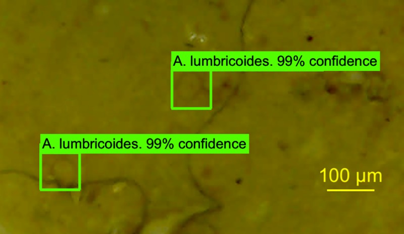
Endemic areas for soil-transmitted helminthiases often lack the tools and trained personnel necessary for point-of-care diagnosis. This study pilots the use of smartphone microscopy and an artificial neural network-based (ANN) object detection application named Kankanet to address those two needs.
METHODOLOGY/PRINCIPAL FINDINGS: A smartphone was equipped with a USB Video Class (UVC) microscope attachment and Kankanet, which was trained to recognize eggs of Ascaris lumbricoides, Trichuris trichiura, and hookworm using a dataset of 2,078 images. It was evaluated for interpretive accuracy based on 185 new images. Fecal samples were processed using Kato-Katz (KK), spontaneous sedimentation technique in tube (SSTT), and Merthiolate-Iodine-Formaldehyde (MIF) techniques. UVC imaging and ANN interpretation of these slides was compared to parasitologist interpretation of standard microscopy.Relative to a gold standard defined as any positive result from parasitologist reading of KK, SSTT, and MIF preparations through standard microscopy, parasitologists reading UVC imaging of SSTT achieved a comparable sensitivity (82.9%) and specificity (97.1%) in A. lumbricoides to standard KK interpretation (97.0% sensitivity, 96.0% specificity). The UVC could not accurately image T. trichiura or hookworm. Though Kankanet interpretation was not quite as sensitive as parasitologist interpretation, it still achieved high sensitivity for A. lumbricoides and hookworm (69.6% and 71.4%, respectively). Kankanet showed high sensitivity for T. trichiura in microscope images (100.0%), but low in UVC images (50.0%).
CONCLUSIONS/SIGNIFICANCE: The UVC achieved comparable sensitivity to standard microscopy with only A. lumbricoides. With further improvement of image resolution and magnification, UVC shows promise as a point-of-care imaging tool. In addition to smartphone microscopy, ANN-based object detection can be developed as a diagnostic aid. Though trained with a limited dataset, Kankanet accurately interprets both standard microscope and low-quality UVC images. Kankanet may achieve sensitivity comparable to parasitologists with continued expansion of the image database and improvement of machine learning technology.
土壤传播性蠕虫病的流行地区通常缺乏用于即时诊断的工具和经过培训的人员。本研究试用了智能手机显微镜和基于人工神经网络(ANN)的目标检测应用程序 Kankanet,以满足这两个需求。
方法/主要发现:将智能手机配备了 USB 视频类(UVC)显微镜附件和 Kankanet,该程序使用包含 2078 张图像的数据集训练识别蛔虫、鞭虫和钩虫的卵。根据 185 张新图像评估了其解释准确性。使用加藤厚涂片(KK)、管内自然沉淀技术(SSTT)和巯基醋酸盐-碘-甲醛(MIF)技术处理粪便样本。将 UVC 成像和 ANN 对这些载玻片的解释与寄生虫学家对标准显微镜的解释进行了比较。相对于寄生虫学家通过标准显微镜阅读 KK、SSTT 和 MIF 制剂的任何阳性结果作为金标准,寄生虫学家对 SSTT 的 UVC 成像的读取在诊断蛔虫方面达到了与标准 KK 解释相当的灵敏度(82.9%)和特异性(97.1%)。UVC 无法准确成像鞭虫或钩虫。尽管 Kankanet 的解释不如寄生虫学家的解释敏感,但它仍然在诊断蛔虫和钩虫方面达到了很高的灵敏度(分别为 69.6%和 71.4%)。Kankanet 在显微镜图像中对鞭虫显示出很高的灵敏度(100.0%),但在 UVC 图像中灵敏度较低(50.0%)。
结论/意义:UVC 仅对蛔虫的检测灵敏度与标准显微镜相当。随着图像分辨率和放大倍数的进一步提高,UVC 有望成为一种即时成像工具。除了智能手机显微镜之外,还可以开发基于 ANN 的目标检测作为诊断辅助工具。尽管 Kankanet 是使用有限的数据集进行训练的,但它可以准确地解释标准显微镜和低质量 UVC 图像。通过不断扩展图像数据库和改进机器学习技术,Kankanet 可以实现与寄生虫学家相当的灵敏度。