Risacher Shannon L, Fandos Noelia, Romero Judith, Sherriff Ian, Pesini Pedro, Saykin Andrew J, Apostolova Liana G
Department of Radiology and Imaging Sciences, Center for Neuroimaging, Indiana University School of Medicine, Indianapolis, IN, USA.
Indiana Alzheimer Disease Center, Indiana University School of Medicine, Indianapolis, IN, USA.
Alzheimers Dement (Amst). 2019 Jul 26;11:510-519. doi: 10.1016/j.dadm.2019.05.007. eCollection 2019 Dec.
We investigated the relationship of plasma amyloid beta (Aβ) with cerebral deposition of Aβ and tau on positron emission tomography (PET).
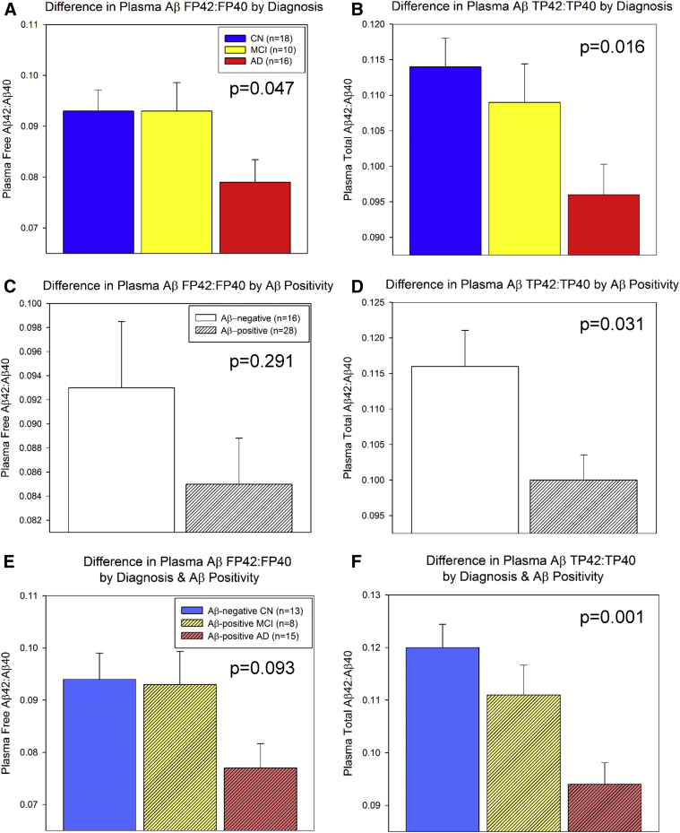
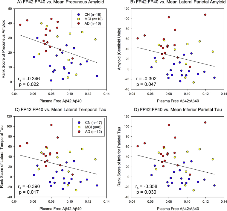
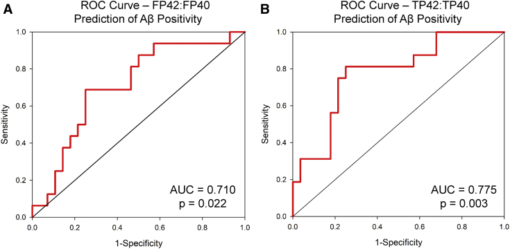
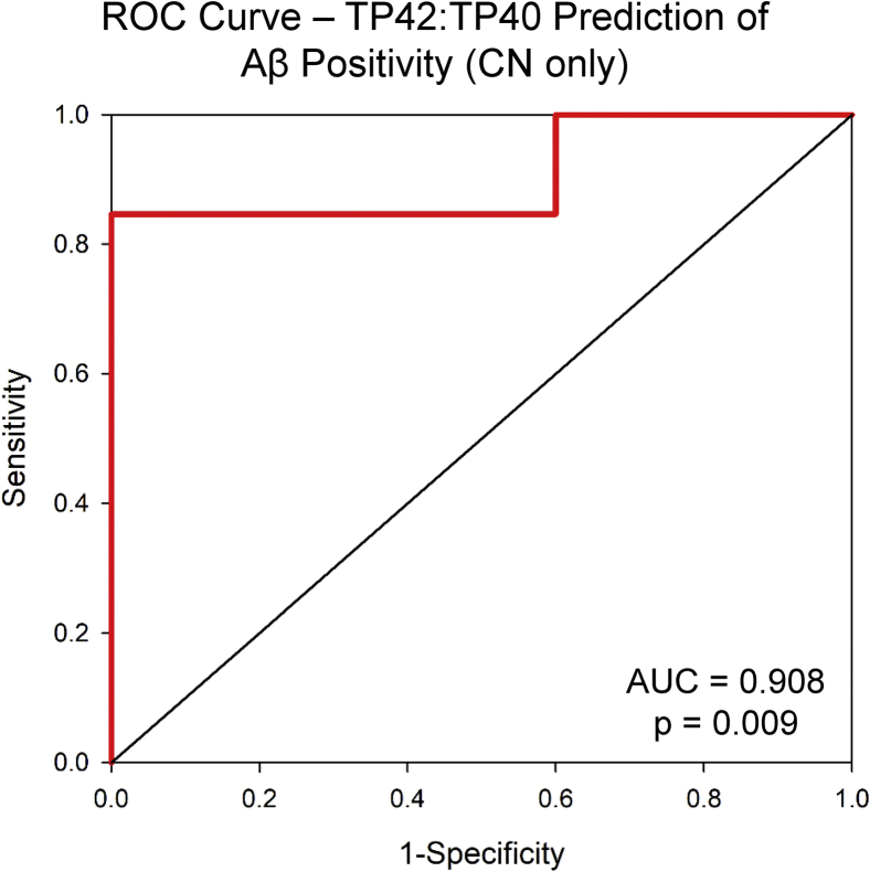
Forty-four participants (18 cognitively normal older adults [CN], 10 mild cognitive impairment, 16 Alzheimer's disease [AD]) underwent amyloid PET and a blood draw. Free and total plasma Aβ40 and Aβ42 were assessed using a validated assay. Thirty-seven participants (17 CN, 8 mild cognitive impairment, 12 AD) also underwent a [F]flortaucipir scan. Scans were preprocessed by standard techniques, and mean global and regional amyloid and tau values were extracted. Free Aβ42/Aβ40 (Aβ F42:F40) and total Aβ42/Aβ40 (Aβ T42:T40) were evaluated for differences by diagnosis and relation to PET Aβ positivity. Relationships between these measures and cerebral Aβ and tau on both regional and voxel-wise basis were also evaluated.
Lower Aβ T42:T40 was associated with diagnosis and PET Aβ positivity. Lower plasma Aβ T42:T40 ratios predicted cerebral Aβ positivity, both across the full sample and in CN only. Finally, lower plasma Aβ T42:T40 ratios were associated with increased cortical Aβ and tau in AD-related regions on both regional and voxel-wise analyses.
Plasma Aβ measures may be useful biomarkers for predicting cerebral Aβ and tau. Additional studies in larger samples are warranted.
我们研究了血浆淀粉样蛋白β(Aβ)与正电子发射断层扫描(PET)上Aβ和tau的脑沉积之间的关系。
44名参与者(18名认知正常的老年人[CN]、10名轻度认知障碍者、16名阿尔茨海默病[AD]患者)接受了淀粉样蛋白PET检查和采血。使用经过验证的检测方法评估游离和总血浆Aβ40和Aβ42。37名参与者(17名CN、8名轻度认知障碍者、12名AD患者)还接受了[F]氟代托卡匹尔扫描。扫描采用标准技术进行预处理,并提取平均全脑和区域淀粉样蛋白及tau值。通过诊断和与PET Aβ阳性的关系评估游离Aβ42/Aβ40(Aβ F42:F40)和总Aβ42/Aβ40(Aβ T42:T40)的差异。还评估了这些指标与区域和体素水平上脑Aβ和tau之间的关系。
较低的Aβ T42:T40与诊断及PET Aβ阳性相关。较低的血浆Aβ T42:T40比值在整个样本以及仅在CN组中均能预测脑Aβ阳性。最后,在区域和体素水平分析中,较低的血浆Aβ T42:T40比值与AD相关区域皮质Aβ和tau增加有关。
血浆Aβ指标可能是预测脑Aβ和tau的有用生物标志物。有必要在更大样本中进行进一步研究。