Department of Immunology and Oncology, and NanoBiomedicine Initiative, Centro Nacional de Biotecnología (CNB)-CSIC, Darwin 3, Cantoblanco, 28049, Madrid, Spain.
J Nanobiotechnology. 2019 Aug 6;17(1):87. doi: 10.1186/s12951-019-0520-0.
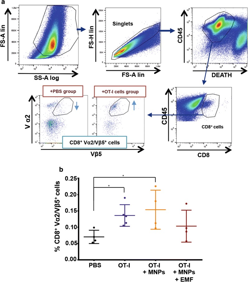
Adoptive T cell-transfer (ATC) therapy is a highly promising cancer-treatment approach. However, in vivo-administered T cells tend to disperse, with only a small proportion reaching the tumour. To remedy this, magnetic targeting of T cells has been recently explored. Magnetic nanoparticles (MNPs) functionalised with antibodies were attached to effector T cells and magnetically recruited to tumour sites under MRI guidance. In this study, we investigated whether 3-aminopropyl-triethoxysilane (APS)-coated MNPs directly attached to CD8 T cell membranes could also magnetically target and accumulate tumour-specific CD8 T cells in solid tumours using an external magnetic field (EMF). As it has been shown that T cells associated with APS-coated MNPs are retained in lymph nodes (LNs), and tumour-draining LNs are the most common sites of solid-tumour metastases, we further evaluated whether magnetic targeting of APS-MNP-loaded CD8 T cells could cause them to accumulate in tumour-draining LNs.
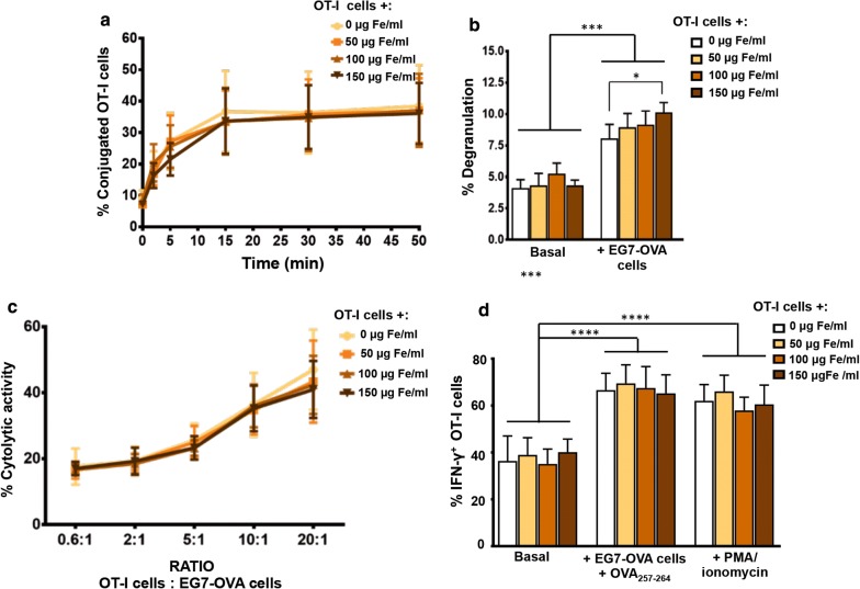
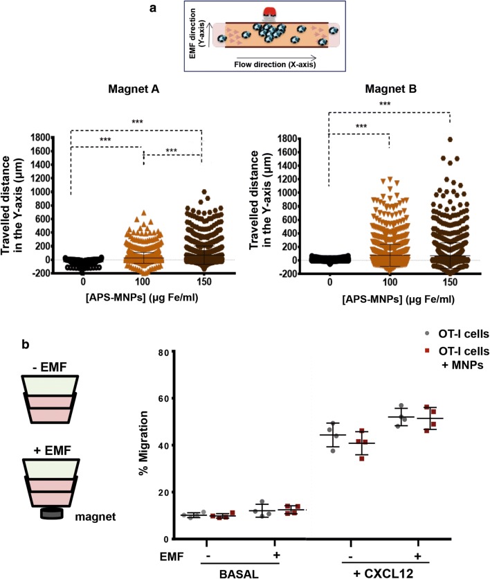
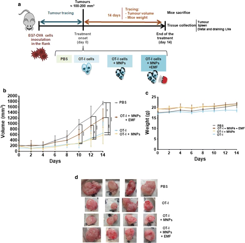
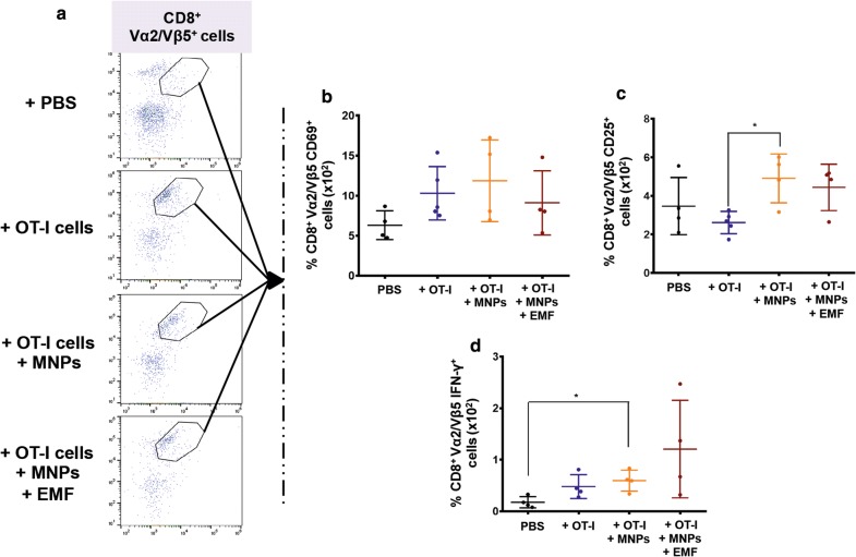
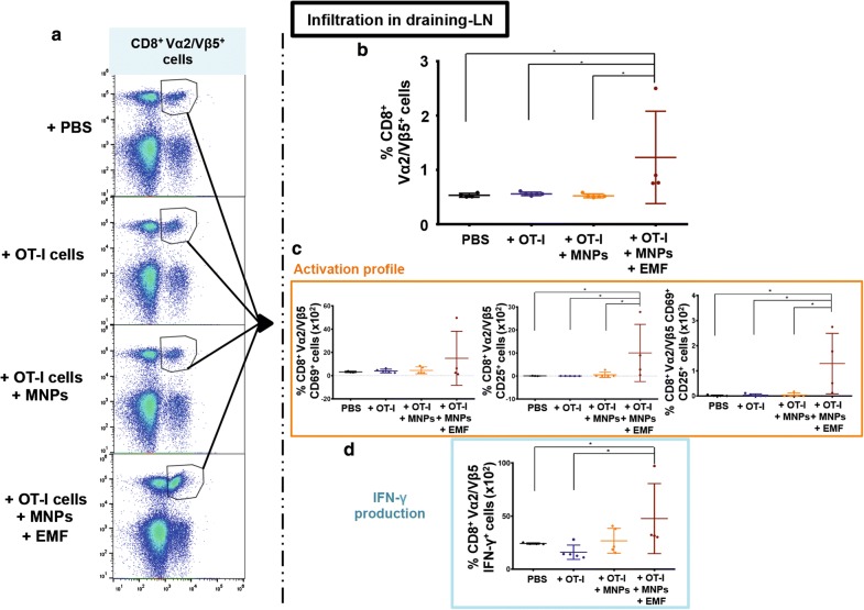
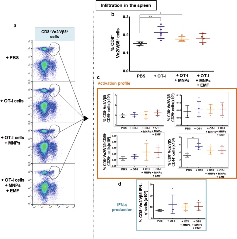
First, we show that antigen-specific CD8 T cells preserve their antitumor activity in vitro when associated with APS-MNPs. Next, we demonstrate that the application of a magnetic field enhanced the retention of APS-MNP-loaded OT-I CD8 T cells under flow conditions in vitro. Using a syngeneic mouse model, we found similar numbers of APS-MNP-loaded OT-I CD8 T cells and OT-I CD8 T cells infiltrating the tumour 14 days after cell transfer. However, when a magnet was placed near the tumour during the transfer of tumour-specific APS-MNP-loaded CD8 T cells to improve tumour infiltration, a reduced percentage of tumour-specific T cells was found infiltrating the tumour 14 days after cell transfer, which was reflected in a smaller reduction in tumour size compared to tumour-specific CD8 T cells transferred with or without MNPs in the absence of a magnetic field. Nonetheless, magnet placement near the tumour site during cell transfer induced infiltration of activated tumour-specific CD8 T cells in tumour-draining LNs, which remained 14 days after cell transfer.
The use of an EMF to improve targeting of tumour-specific T cells modified with APS-MNPs reduced the percentage of these cells infiltrating the tumour, but promoted the retention and the persistence of these cells in the tumour-draining LNs.
过继性 T 细胞转移(ATC)疗法是一种很有前途的癌症治疗方法。然而,体内给予的 T 细胞往往会分散,只有一小部分到达肿瘤部位。为了解决这个问题,最近已经探索了 T 细胞的磁靶向。用抗体功能化的磁性纳米颗粒(MNPs)附着在效应 T 细胞上,并在 MRI 引导下通过磁场将其募集到肿瘤部位。在这项研究中,我们研究了 3-氨基丙基三乙氧基硅烷(APS)涂层的 MNPs 是否可以直接附着在 CD8 T 细胞膜上,通过外加磁场(EMF)靶向和聚集实体瘤中的肿瘤特异性 CD8 T 细胞。因为已经表明与 APS 涂层的 MNPs 相关的 T 细胞保留在淋巴结(LNs)中,并且肿瘤引流 LNs 是实体瘤转移的最常见部位,所以我们进一步评估了磁性靶向负载 APS-MNP 的 CD8 T 细胞是否可以导致它们在肿瘤引流 LNs 中积累。
首先,我们表明,与 APS-MNPs 结合的抗原特异性 CD8 T 细胞在体外保留其抗肿瘤活性。接下来,我们证明,在外加磁场的应用下,体外流动条件下负载 APS-MNP 的 OT-I CD8 T 细胞的保留率增强。使用同种小鼠模型,我们发现细胞转移后 14 天,负载 APS-MNP 的 OT-I CD8 T 细胞和 OT-I CD8 T 细胞浸润肿瘤的数量相似。然而,当在肿瘤特异性 APS-MNP 负载的 CD8 T 细胞转移期间将磁铁放置在肿瘤附近以改善肿瘤浸润时,与没有磁场的情况下转移肿瘤特异性 CD8 T 细胞或不转移 MNPs 相比,发现肿瘤特异性 T 细胞浸润肿瘤的百分比降低,这反映在肿瘤大小的减小与没有磁场的情况下相比。尽管如此,在细胞转移期间将磁铁放置在肿瘤部位附近会诱导肿瘤特异性 CD8 T 细胞在肿瘤引流 LNs 中的浸润,这种浸润在细胞转移后 14 天仍然存在。
使用外加磁场来改善用 APS-MNPs 修饰的肿瘤特异性 T 细胞的靶向作用会降低这些细胞浸润肿瘤的百分比,但会促进这些细胞在肿瘤引流 LNs 中的保留和持久性。