Yovchev Mladen I, Lee Edward J, Rodriguez-Silva Waldemar, Locker Joseph, Oertel Michael
Department of Pathology, Division of Experimental Pathology University of Pittsburgh Pittsburgh PA.
Pittsburgh Liver Research Center University of Pittsburgh Pittsburgh PA.
Hepatol Commun. 2019 May 23;3(8):1137-1150. doi: 10.1002/hep4.1367. eCollection 2019 Aug.
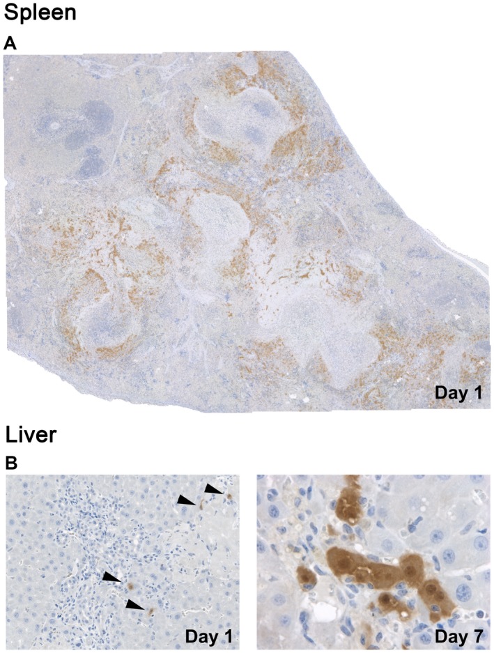
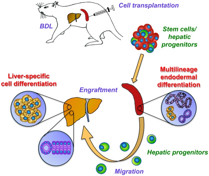
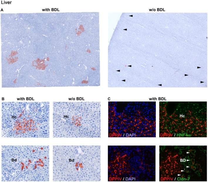
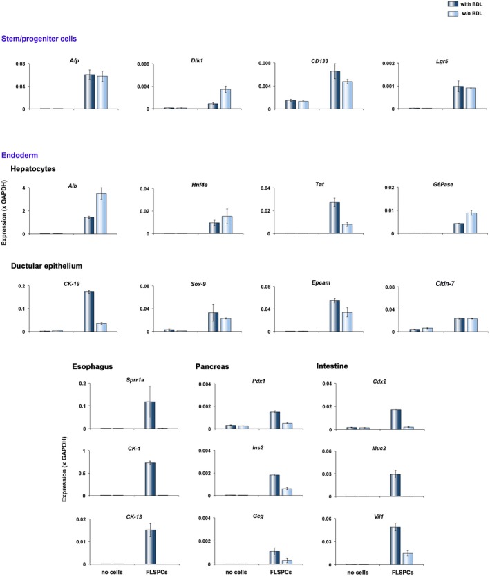
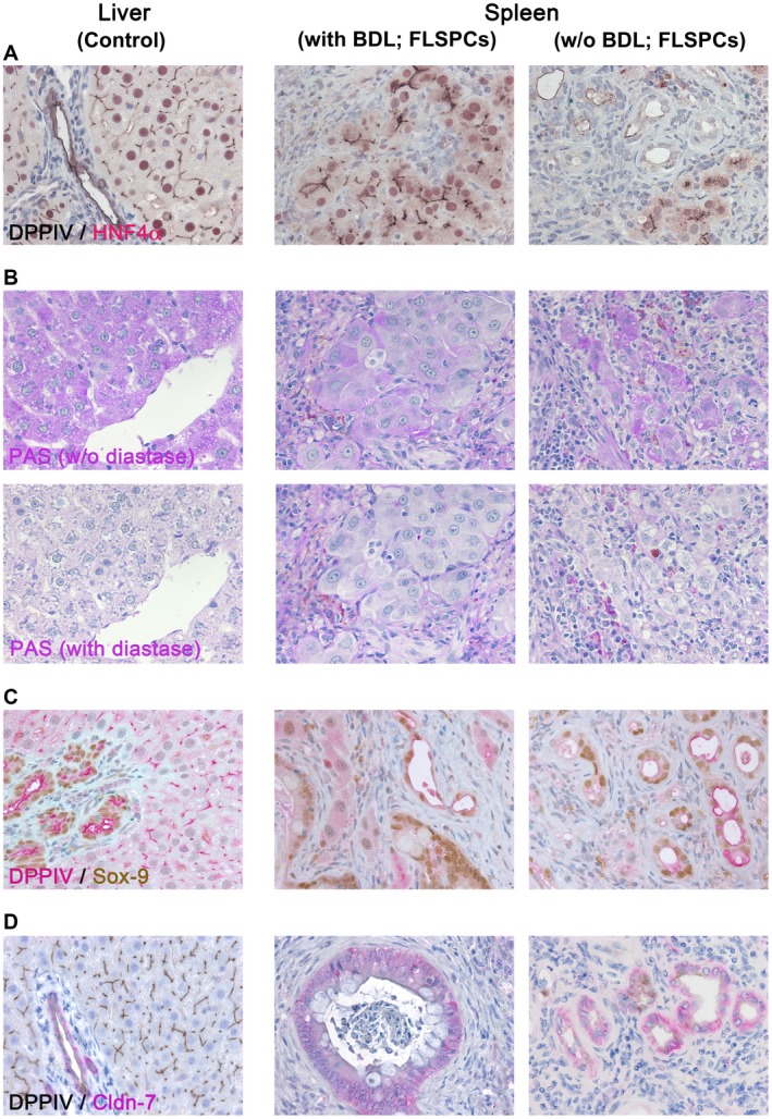
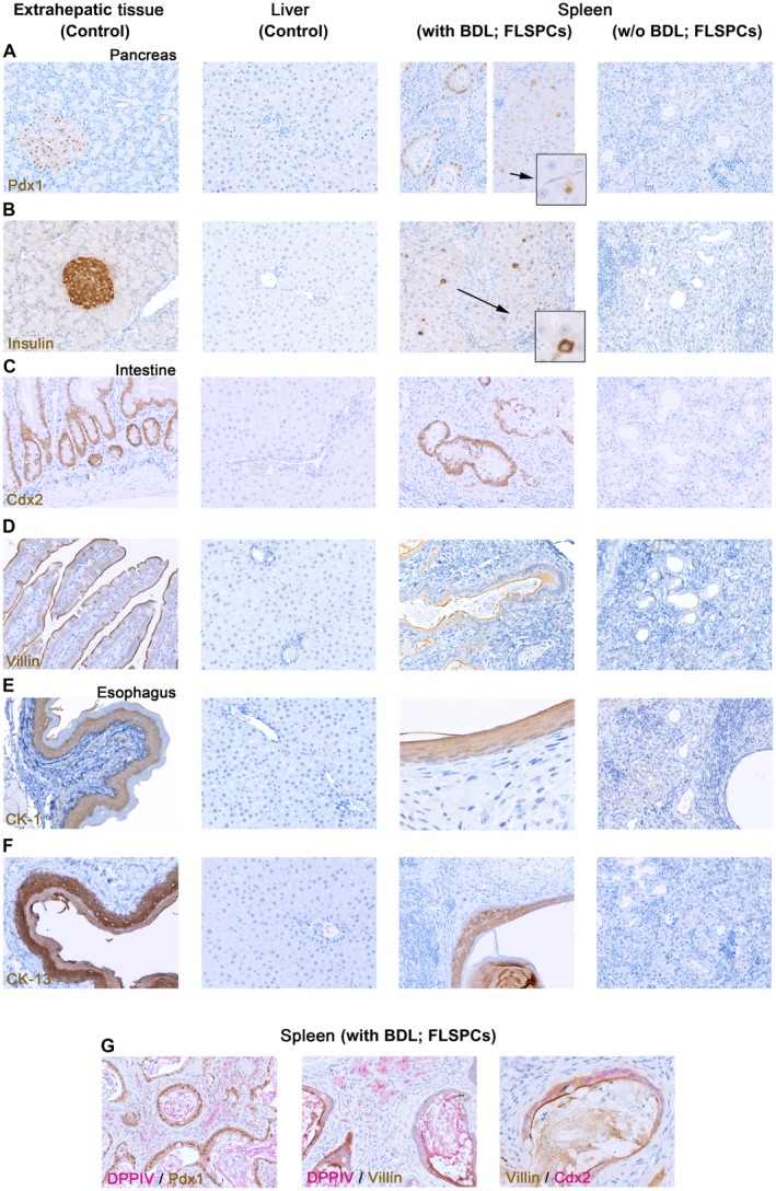
Because of their high regenerative potential, stem cells are an ideal resource for development of therapies that replace injured tissue mass and restore function in patients with end-stage liver diseases. Using a rat model of bile duct ligation (BDL) and biliary fibrosis, we investigated cell engraftment, liver repopulation, and ectopic tissue formation after intrasplenic transplantation of epithelial stem/progenitor cells. Fetal liver cells were infused into the spleens of Fisher 344 rats with progressing biliary fibrosis induced by common BDL or rats without BDL. Cell delivery was well tolerated. After migration to the liver, donor-derived stem/progenitor cells engrafted, differentiated into hepatocytes and cholangiocytes, and formed large cell clusters at 2 months in BDL rats but not controls. Substantial numbers of donor cells were also detected at the splenic injection site where they generated hepatic and nonhepatic tissue. Transplanted cells differentiated into phenotypes other than hepato/cholangiocytic cells only in rats that underwent BDL. Quantitative reverse-transcription polymerase chain reaction analyses demonstrated marked up-regulation of tissue-specific genes of nonhepatic endodermal lineages (e.g., caudal type homeobox 2 [], pancreatic and duodenal homeobox 1 [], keratin 13 []), confirmed by immunohistochemistry. : BDL and its induced fibrosis promote liver repopulation by ectopically transplanted fetal liver-derived cells. These cell fractions contain multipotent stem cells that colonize the spleen of BDL rats and differentiate into multiple gastrointestinal tissues, including liver, pancreas, intestine, and esophagus. The splenic microenvironment, therefore, represents an ideal niche to assess the differentiation of these stem cells, while BDL provides a stimulus that induces their differentiation.
由于干细胞具有很高的再生潜力,它们是开发治疗方法的理想资源,这些治疗方法可替代受损组织并恢复终末期肝病患者的功能。我们使用胆管结扎(BDL)和胆汁性肝纤维化大鼠模型,研究了上皮干细胞/祖细胞脾内移植后的细胞植入、肝脏再填充和异位组织形成。将胎肝细胞注入因普通BDL诱导胆汁性肝纤维化进展的Fisher 344大鼠脾脏或未进行BDL的大鼠脾脏。细胞递送耐受性良好。迁移至肝脏后,供体来源的干细胞/祖细胞植入,分化为肝细胞和胆管细胞,并在BDL大鼠而非对照大鼠中于2个月时形成大细胞簇。在脾注射部位也检测到大量供体细胞,它们在那里形成肝脏和非肝脏组织。仅在接受BDL的大鼠中,移植细胞分化为肝/胆管细胞以外的表型。定量逆转录聚合酶链反应分析显示非肝内胚层谱系的组织特异性基因(如尾型同源框2、胰腺和十二指肠同源框1、角蛋白13)显著上调,免疫组织化学证实了这一点。BDL及其诱导的纤维化通过异位移植的胎肝来源细胞促进肝脏再填充。这些细胞组分包含多能干细胞,它们定植于BDL大鼠的脾脏并分化为多种胃肠组织,包括肝脏、胰腺、肠道和食管。因此,脾脏微环境是评估这些干细胞分化的理想生态位,而BDL提供了诱导其分化的刺激因素。