• 文献检索
  • 文档翻译
  • 深度研究
  • 学术资讯
  • Suppr Zotero 插件Zotero 插件
  • 邀请有礼
  • 套餐&价格
  • 历史记录
应用&插件
Suppr Zotero 插件Zotero 插件浏览器插件Mac 客户端Windows 客户端微信小程序
定价
高级版会员购买积分包购买API积分包
服务
文献检索文档翻译深度研究API 文档MCP 服务
关于我们
关于 Suppr公司介绍联系我们用户协议隐私条款
关注我们

Suppr 超能文献

核心技术专利:CN118964589B侵权必究
粤ICP备2023148730 号-1Suppr @ 2026

文献检索

告别复杂PubMed语法,用中文像聊天一样搜索,搜遍4000万医学文献。AI智能推荐,让科研检索更轻松。

立即免费搜索

文件翻译

保留排版,准确专业,支持PDF/Word/PPT等文件格式,支持 12+语言互译。

免费翻译文档

深度研究

AI帮你快速写综述,25分钟生成高质量综述,智能提取关键信息,辅助科研写作。

立即免费体验

手动多重荧光免疫组织化学的优化、设计与规避陷阱

Optimization, Design and Avoiding Pitfalls in Manual Multiplex Fluorescent Immunohistochemistry.

作者信息

Lazarus Jenny, Akiska Yagiz, Perusina Lanfranca Mirna, Delrosario Lawrence, Sun Lei, Long Daniel, Shi Jiaqi, Crawford Howard, Di Magliano Marina P, Zou Weiping, Frankel Timothy

机构信息

Department of Surgery, University of Michigan.

Department of Molecular and Cellular Physiology, University of Michigan.

出版信息

J Vis Exp. 2019 Jul 26(149). doi: 10.3791/59915.

DOI:10.3791/59915
PMID:31403624
原文链接:https://pmc.ncbi.nlm.nih.gov/articles/PMC6786767/
Abstract

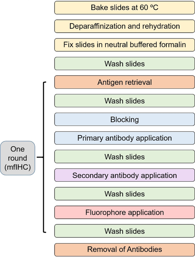
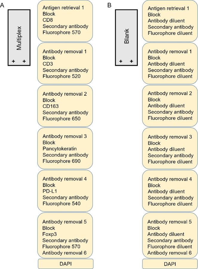
Microenvironment evaluation of intact tissue for analysis of cell infiltration and spatial organization are essential in understanding the complexity of disease processes. The principle techniques used in the past include immunohistochemistry (IHC) and immunofluorescence (IF) which enable visualization of cells as a snapshot in time using between 1 and 4 markers. Both techniques have shortcomings including difficulty staining poorly antigenic targets and limitations related to cross-species reactivity. IHC is reliable and reproducible, but the nature of the chemistry and reliance on the visible light spectrum allows for only a few markers to be used and makes co-localization challenging. Use of IF broadens potential markers but typically relies on frozen tissue due to the extensive tissue autofluorescence following formalin fixation. Flow cytometry, a technique that enables simultaneous labeling of multiple epitopes, abrogates many of the deficiencies of IF and IHC, however, the need to examine cells as a single cell suspension loses the spatial context of cells discarding important biologic relationships. Multiplex fluorescent immunohistochemistry (mfIHC) bridges these technologies allowing for multi-epitope cellular phenotyping in formalin fixed paraffin embedded (FFPE) tissue while preserving the overall microenvironment architecture and spatial relationship of cells within intact undisrupted tissue. High fluorescent intensity fluorophores that covalently bond to the tissue epitope enables multiple applications of primary antibodies without worry of species specific cross-reactivity by secondary antibodies. Although this technology has been proven to produce reliable and accurate images for the study of disease, the process of creating a useful mfIHC staining strategy can be time consuming and exacting due to extensive optimization and design. In order to make robust images that represent accurate cellular interactions in-situ and to mitigate the optimization period for manual analysis, presented here are methods for slide preparation, optimizing antibodies, multiplex design as well as errors commonly encountered during the staining process.

摘要

对完整组织进行微环境评估以分析细胞浸润和空间组织,对于理解疾病过程的复杂性至关重要。过去使用的主要技术包括免疫组织化学(IHC)和免疫荧光(IF),这两种技术能够使用1至4种标记物将细胞可视化为某个时间点的快照。这两种技术都有缺点,包括难以对低抗原性靶点进行染色以及与种间反应性相关的局限性。IHC可靠且可重复,但化学性质和对可见光谱的依赖使得只能使用少数标记物,并且使得共定位具有挑战性。IF的使用拓宽了潜在标记物的范围,但由于福尔马林固定后组织存在广泛的自发荧光,通常依赖于冷冻组织。流式细胞术是一种能够同时标记多个表位的技术,消除了IF和IHC的许多缺陷,然而,将细胞作为单细胞悬液进行检测的需要失去了细胞的空间背景,丢弃了重要的生物学关系。多重荧光免疫组织化学(mfIHC)弥补了这些技术的不足,允许在福尔马林固定石蜡包埋(FFPE)组织中进行多表位细胞表型分析,同时保留完整未破坏组织内细胞的整体微环境结构和空间关系。与组织表位共价结合的高荧光强度荧光团使得能够多次应用一抗,而无需担心二抗的种属特异性交叉反应。尽管该技术已被证明可为疾病研究产生可靠且准确的图像,但由于需要进行广泛的优化和设计,创建有用的mfIHC染色策略的过程可能既耗时又严格。为了制作能够代表原位准确细胞相互作用的稳健图像,并缩短手动分析的优化周期,本文介绍了载玻片制备、抗体优化、多重设计以及染色过程中常见错误的方法。

https://cdn.ncbi.nlm.nih.gov/pmc/blobs/a7dd/6786767/e4ad3b59d029/nihms-1033133-f0005.jpg
https://cdn.ncbi.nlm.nih.gov/pmc/blobs/a7dd/6786767/bdfbd3d5ee82/nihms-1033133-f0001.jpg
https://cdn.ncbi.nlm.nih.gov/pmc/blobs/a7dd/6786767/d67150c7a39f/nihms-1033133-f0002.jpg
https://cdn.ncbi.nlm.nih.gov/pmc/blobs/a7dd/6786767/d0f973986fe8/nihms-1033133-f0003.jpg
https://cdn.ncbi.nlm.nih.gov/pmc/blobs/a7dd/6786767/44fb0e4684b4/nihms-1033133-f0004.jpg
https://cdn.ncbi.nlm.nih.gov/pmc/blobs/a7dd/6786767/e4ad3b59d029/nihms-1033133-f0005.jpg

相似文献

1
Optimization, Design and Avoiding Pitfalls in Manual Multiplex Fluorescent Immunohistochemistry.手动多重荧光免疫组织化学的优化、设计与规避陷阱
J Vis Exp. 2019 Jul 26(149). doi: 10.3791/59915.
2
Multiplex Fluorescent Immunohistochemistry for Preservation of Tumor Microenvironment Architecture and Spatial Relationship of Cells in Tumor Tissues.多重荧光免疫组化技术用于保存肿瘤组织中肿瘤微环境结构和细胞的空间关系。
Methods Mol Biol. 2023;2660:235-246. doi: 10.1007/978-1-0716-3163-8_16.
3
Multiplexed Immunohistochemical Consecutive Staining on Single Slide (MICSSS): Multiplexed Chromogenic IHC Assay for High-Dimensional Tissue Analysis.单玻片多重免疫组化连续染色(MICSSS):用于高维组织分析的多重显色免疫组化检测法
Methods Mol Biol. 2020;2055:497-519. doi: 10.1007/978-1-4939-9773-2_23.
4
Multiple Immunostainings with Different Epitope Retrievals-The FOLGAS Protocol.多种抗原修复的免疫组化染色——FOLGAS 方案。
Int J Mol Sci. 2021 Dec 25;23(1):223. doi: 10.3390/ijms23010223.
5
Automated Multiplex Immunofluorescence Panel for Immuno-oncology Studies on Formalin-fixed Carcinoma Tissue Specimens.用于福尔马林固定癌组织标本免疫肿瘤学研究的自动化多重免疫荧光检测板
J Vis Exp. 2019 Jan 21(143). doi: 10.3791/58390.
6
Multiplex Immunochromogenic Tissue Staining Employing Primary Antibodies from the Same Species.使用来自同一物种的一抗进行多重免疫显色组织染色
Methods Mol Biol. 2023;2593:127-142. doi: 10.1007/978-1-0716-2811-9_8.
7
Using Immunohistochemistry Without Linkers to Determine the Optimum Concentrations of Primary Antibodies for Immunofluorescence Staining of Formalin-fixed Paraffin-embedded Tissue Sections.使用无连接物的免疫组织化学来确定福尔马林固定石蜡包埋组织切片免疫荧光染色的最佳一抗浓度。
Appl Immunohistochem Mol Morphol. 2020 Mar;28(3):249-257. doi: 10.1097/PAI.0000000000000718.
8
Mixed Multiplex Staining: Automated RNAscope™ and OPAL™ for Multiple Targets.多重免疫荧光染色:自动化 RNAscope 和 OPAL 多重靶点检测试剂盒
Methods Mol Biol. 2020;2148:277-298. doi: 10.1007/978-1-0716-0623-0_18.
9
Simultaneous visualization and cell-specific confirmation of RNA and protein in the mouse retina.小鼠视网膜中RNA和蛋白质的同时可视化及细胞特异性确认
Mol Vis. 2014 Sep 21;20:1366-73. eCollection 2014.
10
Fluorescent Immunohistochemistry.荧光免疫组织化学。
Methods Mol Biol. 2022;2422:131-146. doi: 10.1007/978-1-0716-1948-3_9.

引用本文的文献

1
Topological importance of CD8 T-cell enrichment in the tumor microenvironment of classic Hodgkin lymphoma.经典型霍奇金淋巴瘤肿瘤微环境中CD8 T细胞富集的拓扑学重要性。
Ann Hematol. 2025 Feb;104(2):1047-1057. doi: 10.1007/s00277-025-06189-1. Epub 2025 Jan 17.
2
Deep learning application in prediction of cancer molecular alterations based on pathological images: a bibliographic analysis via CiteSpace.深度学习在基于病理图像的癌症分子改变预测中的应用:基于 CiteSpace 的文献分析。
J Cancer Res Clin Oncol. 2024 Oct 18;150(10):467. doi: 10.1007/s00432-024-05992-z.
3
Characterization of undifferentiated carcinomas of the pancreas with and without osteoclast-like giant cells.

本文引用的文献

1
Spatial and phenotypic immune profiling of metastatic colon cancer.转移性结直肠癌的空间和表型免疫特征分析。
JCI Insight. 2018 Nov 15;3(22):121932. doi: 10.1172/jci.insight.121932.
2
Multiplex Immunohistochemistry for Image Analysis of Tertiary Lymphoid Structures in Cancer.用于癌症中三级淋巴结构图像分析的多重免疫组织化学
Methods Mol Biol. 2018;1845:87-98. doi: 10.1007/978-1-4939-8709-2_6.
3
A Functional Spatial Analysis Platform for Discovery of Immunological Interactions Predictive of Low-Grade to High-Grade Transition of Pancreatic Intraductal Papillary Mucinous Neoplasms.
伴有和不伴有破骨细胞样巨细胞的胰腺未分化癌的特征
JNCI Cancer Spectr. 2025 Jan 3;9(1). doi: 10.1093/jncics/pkae097.
4
Development of a Multiplex Immunofluorescence Assay for Tumor Microenvironment Studies of Human and Murine Merkel Cell Carcinoma.开发一种用于人类和鼠类 Merkel 细胞癌肿瘤微环境研究的多重免疫荧光检测方法。
Lab Invest. 2024 Oct;104(10):102128. doi: 10.1016/j.labinv.2024.102128. Epub 2024 Aug 23.
5
CD200 is overexpressed in the pancreatic tumor microenvironment and predictive of overall survival.CD200在胰腺肿瘤微环境中过表达,且可预测总生存期。
Cancer Immunol Immunother. 2024 Apr 15;73(6):96. doi: 10.1007/s00262-024-03678-6.
6
Unique characteristics of the tumor immune microenvironment in young patients with metastatic colorectal cancer.年轻转移性结直肠癌患者肿瘤免疫微环境的独特特征。
Front Immunol. 2023 Dec 13;14:1289402. doi: 10.3389/fimmu.2023.1289402. eCollection 2023.
7
Lineage plasticity enables low-ER luminal tumors to evolve and gain basal-like traits.谱系可塑性使低内质网腔肿瘤能够进化并获得基底样特征。
Breast Cancer Res. 2023 Mar 1;25(1):23. doi: 10.1186/s13058-023-01621-8.
8
Evaluation of paclitaxel-loaded polymeric nanoparticles in 3D tumor model: impact of tumor stroma on penetration and efficacy.载紫杉醇聚合物纳米粒在 3D 肿瘤模型中的评价:肿瘤基质对渗透和疗效的影响。
Drug Deliv Transl Res. 2023 May;13(5):1470-1483. doi: 10.1007/s13346-023-01310-1. Epub 2023 Feb 28.
9
Phenotypic heterogeneity driven by plasticity of the intermediate EMT state governs disease progression and metastasis in breast cancer.表型异质性由中间 EMT 状态的可塑性驱动,控制着乳腺癌的疾病进展和转移。
Sci Adv. 2022 Aug 5;8(31):eabj8002. doi: 10.1126/sciadv.abj8002. Epub 2022 Aug 3.
10
Development of a Multiplex Immunohistochemistry Workflow to Investigate the Immune Microenvironment in Mouse Models of Inflammatory Bowel Disease and Colon Cancer.开发一种多重免疫组化工作流程,以研究炎症性肠病和结肠癌小鼠模型中的免疫微环境。
Int J Mol Sci. 2021 Oct 12;22(20):11001. doi: 10.3390/ijms222011001.
用于发现预测胰腺导管内乳头状黏液性肿瘤从低级别向高级别转变的免疫相互作用的功能空间分析平台。
Cancer Inform. 2018 Jun 28;17:1176935118782880. doi: 10.1177/1176935118782880. eCollection 2018.
4
Multispectral imaging reveals hyper active TGF-β signaling in colorectal cancer.多光谱成像揭示结直肠癌中 TGF-β 信号的过度活跃。
Cancer Biol Ther. 2018 Feb 1;19(2):105-112. doi: 10.1080/15384047.2017.1395116. Epub 2017 Dec 8.
5
Eight-Color Multiplex Immunohistochemistry for Simultaneous Detection of Multiple Immune Checkpoint Molecules within the Tumor Microenvironment.用于同时检测肿瘤微环境中多种免疫检查点分子的八色多重免疫组织化学
J Immunol. 2018 Jan 1;200(1):347-354. doi: 10.4049/jimmunol.1701262. Epub 2017 Nov 15.
6
Validation of multiplex immunofluorescence panels using multispectral microscopy for immune-profiling of formalin-fixed and paraffin-embedded human tumor tissues.利用多光谱显微镜对福尔马林固定石蜡包埋的人类肿瘤组织进行免疫分析的多重免疫荧光试剂盒验证。
Sci Rep. 2017 Oct 17;7(1):13380. doi: 10.1038/s41598-017-13942-8.
7
Topological analysis reveals a PD-L1-associated microenvironmental niche for Reed-Sternberg cells in Hodgkin lymphoma.拓扑分析揭示了霍奇金淋巴瘤中里德-施特恩伯格细胞的一种与程序性死亡配体1相关的微环境生态位。
Blood. 2017 Nov 30;130(22):2420-2430. doi: 10.1182/blood-2017-03-770719. Epub 2017 Sep 11.
8
Fully automated 5-plex fluorescent immunohistochemistry with tyramide signal amplification and same species antibodies.全自动 5 重荧光免疫组化,采用辣根过氧化物酶信号放大技术和同种属抗体。
Lab Invest. 2017 Jul;97(7):873-885. doi: 10.1038/labinvest.2017.37. Epub 2017 May 15.
9
Spatial computation of intratumoral T cells correlates with survival of patients with pancreatic cancer.肿瘤内 T 细胞的空间计算与胰腺癌患者的生存相关。
Nat Commun. 2017 Apr 27;8:15095. doi: 10.1038/ncomms15095.
10
Flow cytometry: basic principles and applications.流式细胞术:基本原理与应用
Crit Rev Biotechnol. 2017 Mar;37(2):163-176. doi: 10.3109/07388551.2015.1128876. Epub 2016 Jan 14.