• 文献检索
  • 文档翻译
  • 深度研究
  • 学术资讯
  • Suppr Zotero 插件Zotero 插件
  • 邀请有礼
  • 套餐&价格
  • 历史记录
应用&插件
Suppr Zotero 插件Zotero 插件浏览器插件Mac 客户端Windows 客户端微信小程序
定价
高级版会员购买积分包购买API积分包
服务
文献检索文档翻译深度研究API 文档MCP 服务
关于我们
关于 Suppr公司介绍联系我们用户协议隐私条款
关注我们

Suppr 超能文献

核心技术专利:CN118964589B侵权必究
粤ICP备2023148730 号-1Suppr @ 2026

文献检索

告别复杂PubMed语法,用中文像聊天一样搜索,搜遍4000万医学文献。AI智能推荐,让科研检索更轻松。

立即免费搜索

文件翻译

保留排版,准确专业,支持PDF/Word/PPT等文件格式,支持 12+语言互译。

免费翻译文档

深度研究

AI帮你快速写综述,25分钟生成高质量综述,智能提取关键信息,辅助科研写作。

立即免费体验

应激诱导的磷酸泛素化导致 parkin 降解。

Stress-induced phospho-ubiquitin formation causes parkin degradation.

机构信息

Department of Biological Sciences, Columbia University, New York, NY, USA.

Departments of Psychiatry, Neurology, and Pharmacology, Columbia University: Division of Molecular Therapeutics, New York State Psychiatric Institute, New York, NY, USA.

出版信息

Sci Rep. 2019 Aug 12;9(1):11682. doi: 10.1038/s41598-019-47952-5.

DOI:10.1038/s41598-019-47952-5
PMID:31406131
原文链接:https://pmc.ncbi.nlm.nih.gov/articles/PMC6690910/
Abstract

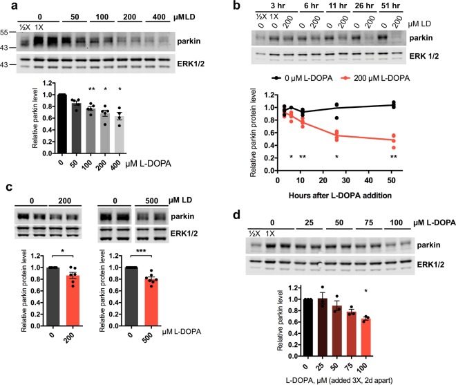
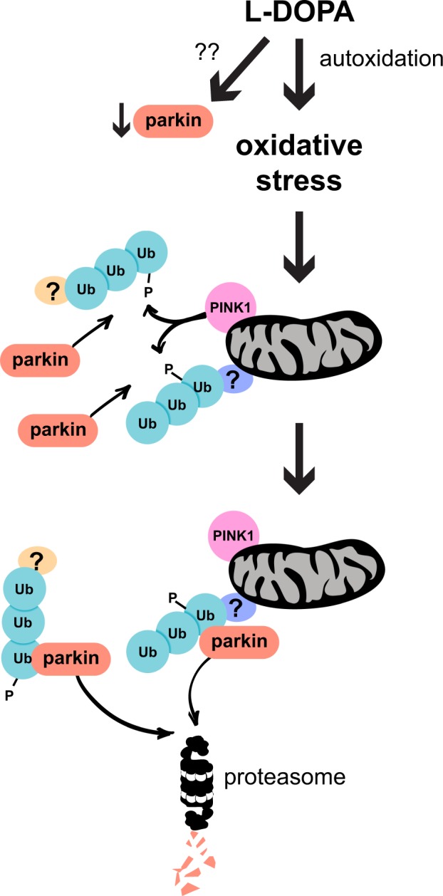
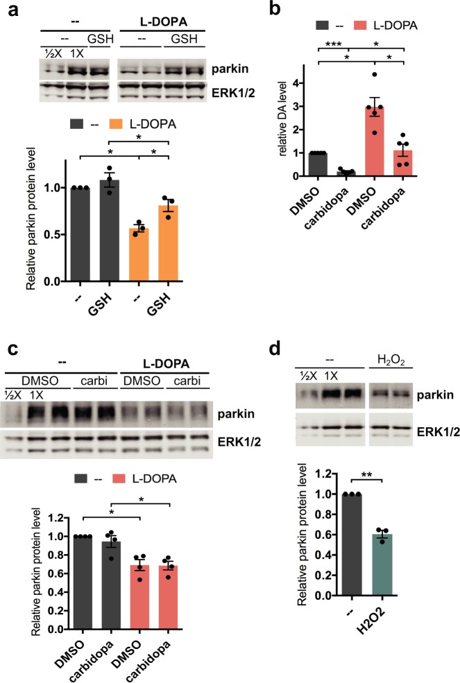
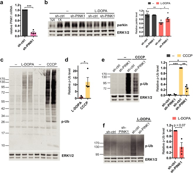
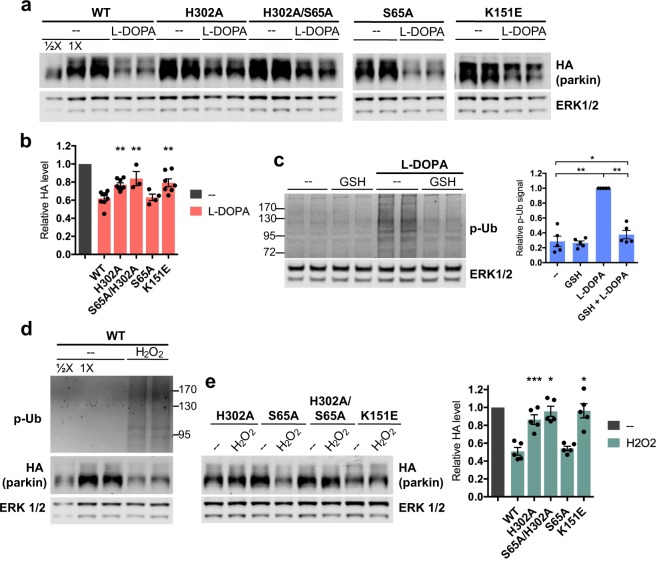
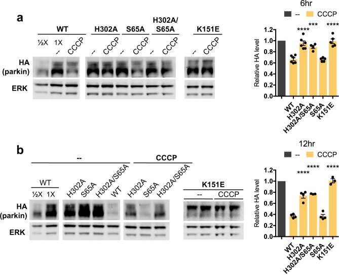
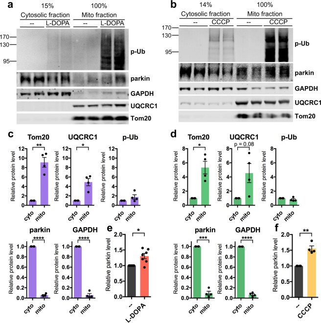
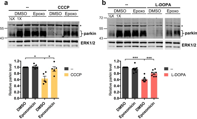
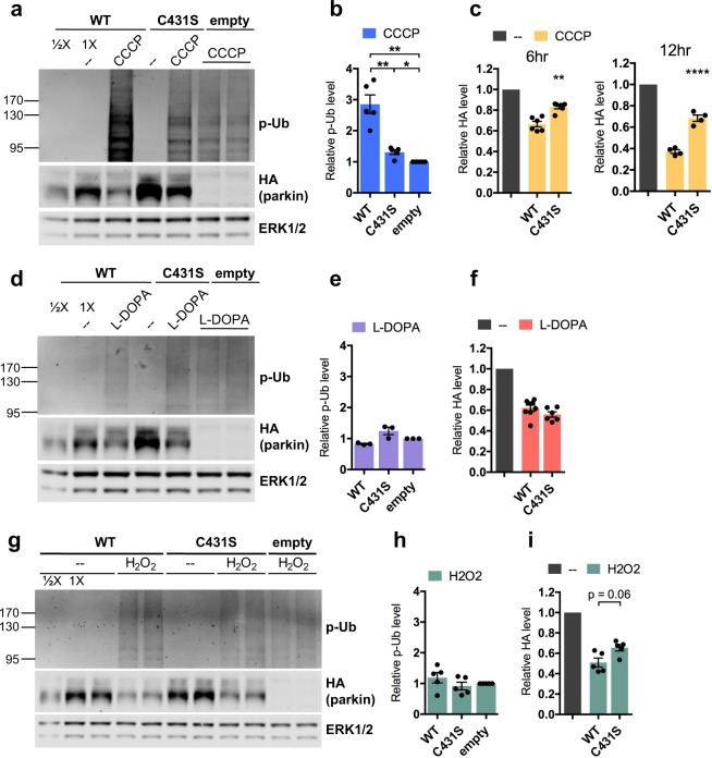
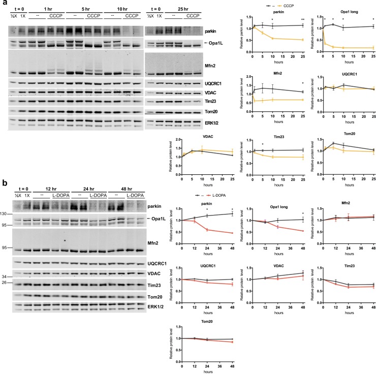
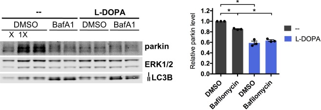
Mutations in the E3 ubiquitin ligase parkin are the most common known cause of autosomal recessive Parkinson's disease (PD), and parkin depletion may play a role in sporadic PD. Here, we sought to elucidate the mechanisms by which stress decreases parkin protein levels using cultured neuronal cells and the PD-relevant stressor, L-DOPA. We find that L-DOPA causes parkin loss through both oxidative stress-independent and oxidative stress-dependent pathways. Characterization of the latter reveals that it requires both the kinase PINK1 and parkin's interaction with phosphorylated ubiquitin (phospho-Ub) and is mediated by proteasomal degradation. Surprisingly, autoubiquitination and mitophagy do not appear to be required for such loss. In response to stress induced by hydrogen peroxide or CCCP, parkin degradation also requires its association with phospho-Ub, indicating that this mechanism is broadly generalizable. As oxidative stress, metabolic dysfunction and phospho-Ub levels are all elevated in PD, we suggest that these changes may contribute to a loss of parkin expression.

摘要

E3 泛素连接酶 parkin 的突变是最常见的常染色体隐性帕金森病 (PD) 的已知原因,而 parkin 的缺失可能在散发性 PD 中发挥作用。在这里,我们试图利用培养的神经元细胞和与 PD 相关的应激源 L-DOPA 来阐明应激降低 parkin 蛋白水平的机制。我们发现,L-DOPA 通过氧化应激独立和依赖的途径导致 parkin 的丢失。对后者的特征分析表明,它需要激酶 PINK1 和 parkin 与磷酸化泛素(磷酸化 Ub)的相互作用,并通过蛋白酶体降解介导。令人惊讶的是,自泛素化和线粒体自噬似乎不是这种丢失所必需的。在过氧化氢或 CCCP 诱导的应激反应中,parkin 的降解也需要其与磷酸化 Ub 的结合,这表明这种机制具有广泛的通用性。由于氧化应激、代谢功能障碍和磷酸化 Ub 水平在 PD 中均升高,我们认为这些变化可能导致 parkin 表达的丧失。

https://cdn.ncbi.nlm.nih.gov/pmc/blobs/4826/6690910/7d2a7471a35b/41598_2019_47952_Fig11_HTML.jpg
https://cdn.ncbi.nlm.nih.gov/pmc/blobs/4826/6690910/e031dcc1a5e3/41598_2019_47952_Fig1_HTML.jpg
https://cdn.ncbi.nlm.nih.gov/pmc/blobs/4826/6690910/e3e70eb3eadf/41598_2019_47952_Fig2_HTML.jpg
https://cdn.ncbi.nlm.nih.gov/pmc/blobs/4826/6690910/229bb57bff53/41598_2019_47952_Fig3_HTML.jpg
https://cdn.ncbi.nlm.nih.gov/pmc/blobs/4826/6690910/51d61b8c4243/41598_2019_47952_Fig4_HTML.jpg
https://cdn.ncbi.nlm.nih.gov/pmc/blobs/4826/6690910/bfc6dff311b7/41598_2019_47952_Fig5_HTML.jpg
https://cdn.ncbi.nlm.nih.gov/pmc/blobs/4826/6690910/735c36820cb0/41598_2019_47952_Fig6_HTML.jpg
https://cdn.ncbi.nlm.nih.gov/pmc/blobs/4826/6690910/79fcd7df2e90/41598_2019_47952_Fig7_HTML.jpg
https://cdn.ncbi.nlm.nih.gov/pmc/blobs/4826/6690910/28cc68d23a3b/41598_2019_47952_Fig8_HTML.jpg
https://cdn.ncbi.nlm.nih.gov/pmc/blobs/4826/6690910/79d73966bf35/41598_2019_47952_Fig9_HTML.jpg
https://cdn.ncbi.nlm.nih.gov/pmc/blobs/4826/6690910/097b4225c5cc/41598_2019_47952_Fig10_HTML.jpg
https://cdn.ncbi.nlm.nih.gov/pmc/blobs/4826/6690910/7d2a7471a35b/41598_2019_47952_Fig11_HTML.jpg

相似文献

1
Stress-induced phospho-ubiquitin formation causes parkin degradation.应激诱导的磷酸泛素化导致 parkin 降解。
Sci Rep. 2019 Aug 12;9(1):11682. doi: 10.1038/s41598-019-47952-5.
2
Evidence that phosphorylated ubiquitin signaling is involved in the etiology of Parkinson's disease.磷酸化泛素信号传导参与帕金森病病因学的证据。
Hum Mol Genet. 2017 Aug 15;26(16):3172-3185. doi: 10.1093/hmg/ddx201.
3
Nix restores mitophagy and mitochondrial function to protect against PINK1/Parkin-related Parkinson's disease.Nix 恢复线粒体自噬和线粒体功能,以预防 PINK1/Parkin 相关帕金森病。
Sci Rep. 2017 Mar 10;7:44373. doi: 10.1038/srep44373.
4
LRRK2 impairs PINK1/Parkin-dependent mitophagy via its kinase activity: pathologic insights into Parkinson's disease.LRRK2 通过其激酶活性损害 PINK1/Parkin 依赖性线粒体自噬:帕金森病的病理见解。
Hum Mol Genet. 2019 May 15;28(10):1645-1660. doi: 10.1093/hmg/ddz004.
5
Binding to serine 65-phosphorylated ubiquitin primes Parkin for optimal PINK1-dependent phosphorylation and activation.与丝氨酸65磷酸化的泛素结合可使帕金蛋白(Parkin)做好准备,以实现最佳的依赖于PTEN诱导激酶1(PINK1)的磷酸化和激活。
EMBO Rep. 2015 Aug;16(8):939-54. doi: 10.15252/embr.201540352. Epub 2015 Jun 25.
6
(Patho-)physiological relevance of PINK1-dependent ubiquitin phosphorylation.PINK1 依赖性泛素磷酸化的(病理)生理相关性。
EMBO Rep. 2015 Sep;16(9):1114-30. doi: 10.15252/embr.201540514. Epub 2015 Jul 10.
7
Mutations in PINK1 and Parkin impair ubiquitination of Mitofusins in human fibroblasts.PINK1 和 Parkin 突变会损害人成纤维细胞中线粒体融合蛋白的泛素化。
PLoS One. 2011 Mar 8;6(3):e16746. doi: 10.1371/journal.pone.0016746.
8
PINK1 phosphorylates ubiquitin to activate Parkin E3 ubiquitin ligase activity.PINK1 通过磷酸化泛素来激活 Parkin E3 泛素连接酶活性。
J Cell Biol. 2014 Apr 28;205(2):143-53. doi: 10.1083/jcb.201402104. Epub 2014 Apr 21.
9
Parkin-catalyzed ubiquitin-ester transfer is triggered by PINK1-dependent phosphorylation.Parkin 催化的泛素酯转移是由 PINK1 依赖性磷酸化触发的。
J Biol Chem. 2013 Jul 26;288(30):22019-32. doi: 10.1074/jbc.M113.467530. Epub 2013 Jun 10.
10
The roles of PINK1, parkin, and mitochondrial fidelity in Parkinson's disease.PINK1、parkin 和线粒体保真度在帕金森病中的作用。
Neuron. 2015 Jan 21;85(2):257-73. doi: 10.1016/j.neuron.2014.12.007.

引用本文的文献

1
PRKN-linked familial Parkinson's disease: cellular and molecular mechanisms of disease-linked variants.PRKN 相关家族性帕金森病:疾病相关变异的细胞和分子机制。
Cell Mol Life Sci. 2024 May 20;81(1):223. doi: 10.1007/s00018-024-05262-8.
2
Basal activity of PINK1 and PRKN in cell models and rodent brain.PINK1 和 PRKN 在细胞模型和啮齿动物大脑中的基础活性。
Autophagy. 2024 May;20(5):1147-1158. doi: 10.1080/15548627.2023.2286414. Epub 2023 Dec 2.
3
Mitochondrial Hydrogen Peroxide Activates PTEN and Inactivates Akt Leading to Autophagy Inhibition-Dependent Cell Death in Neuronal Models of Parkinson's Disease.

本文引用的文献

1
Mechanism of parkin activation by PINK1.Parkin 的激活机制由 PINK1 介导。
Nature. 2018 Jul;559(7714):410-414. doi: 10.1038/s41586-018-0224-x. Epub 2018 Jun 6.
2
Mechanism of parkin activation by phosphorylation.Parkin 通过磷酸化激活的机制。
Nat Struct Mol Biol. 2018 Jul;25(7):623-630. doi: 10.1038/s41594-018-0088-7. Epub 2018 Jul 2.
3
Age- and disease-dependent increase of the mitophagy marker phospho-ubiquitin in normal aging and Lewy body disease.年龄和疾病依赖性磷酸泛素化标志在正常衰老和路易体病中的增加。
线粒体过氧化氢激活 PTEN 并失活 Akt,导致帕金森病神经元模型中自噬抑制依赖性细胞死亡。
Mol Neurobiol. 2023 Jun;60(6):3345-3364. doi: 10.1007/s12035-023-03286-y. Epub 2023 Feb 28.
4
Core transcription programs controlling injury-induced neurodegeneration of retinal ganglion cells.核心转录程序控制视网膜神经节细胞损伤诱导的神经变性。
Neuron. 2022 Aug 17;110(16):2607-2624.e8. doi: 10.1016/j.neuron.2022.06.003. Epub 2022 Jun 28.
5
Parkinson's Disease Phenotypes in Patient Neuronal Cultures and Brain Organoids Improved by 2-Hydroxypropyl-β-Cyclodextrin Treatment.2-羟丙基-β-环糊精治疗可改善患者神经元培养物和脑类器官中的帕金森病表型。
Mov Disord. 2022 Jan;37(1):80-94. doi: 10.1002/mds.28810. Epub 2021 Oct 12.
6
Mitochondrial quality control: from molecule to organelle.线粒体质量控制:从分子到细胞器。
Cell Mol Life Sci. 2021 Apr;78(8):3853-3866. doi: 10.1007/s00018-021-03775-0. Epub 2021 Mar 29.
Autophagy. 2018;14(8):1404-1418. doi: 10.1080/15548627.2018.1461294. Epub 2018 Jul 28.
4
PTEN-L is a novel protein phosphatase for ubiquitin dephosphorylation to inhibit PINK1-Parkin-mediated mitophagy.PTEN-L 是一种新型的泛素去磷酸化蛋白磷酸酶,可抑制 PINK1-Parkin 介导的线粒体自噬。
Cell Res. 2018 Aug;28(8):787-802. doi: 10.1038/s41422-018-0056-0. Epub 2018 Jun 22.
5
Ari-1 Regulates Myonuclear Organization Together with Parkin and Is Associated with Aortic Aneurysms.Ari-1 通过与 Parkin 共同调节肌核组织,与主动脉瘤相关。
Dev Cell. 2018 Apr 23;45(2):226-244.e8. doi: 10.1016/j.devcel.2018.03.020.
6
Mfn2 ubiquitination by PINK1/parkin gates the p97-dependent release of ER from mitochondria to drive mitophagy.Mfn2 通过 PINK1/parkin 的泛素化将 ER 从线粒体中释放出来,从而驱动线粒体自噬。
Elife. 2018 Apr 20;7:e32866. doi: 10.7554/eLife.32866.
7
Building and decoding ubiquitin chains for mitophagy.构建和解析用于线粒体自噬的泛素链。
Nat Rev Mol Cell Biol. 2018 Jan 23;19(2):93-108. doi: 10.1038/nrm.2017.129.
8
Genome-wide CRISPR screen for PARKIN regulators reveals transcriptional repression as a determinant of mitophagy.全基因组 CRISPR 筛选 PARKIN 调控因子揭示转录抑制是调控线粒体自噬的决定因素。
Proc Natl Acad Sci U S A. 2018 Jan 9;115(2):E180-E189. doi: 10.1073/pnas.1711023115. Epub 2017 Dec 21.
9
PARKIN overexpression in human mesenchymal stromal cells from Wharton's jelly suppresses 6-hydroxydopamine-induced apoptosis: Potential therapeutic strategy in Parkinson's disease.人脐带华通氏胶间充质基质细胞中 PARKIN 的过表达抑制 6-羟多巴胺诱导的细胞凋亡:帕金森病的潜在治疗策略。
Cytotherapy. 2018 Jan;20(1):45-61. doi: 10.1016/j.jcyt.2017.09.011. Epub 2017 Nov 2.
10
Dopamine induces soluble α-synuclein oligomers and nigrostriatal degeneration.多巴胺诱导可溶性α-突触核蛋白寡聚体和黑质纹状体变性。
Nat Neurosci. 2017 Nov;20(11):1560-1568. doi: 10.1038/nn.4641. Epub 2017 Sep 18.