Agarwal Nisha Rani, Dowlatshahi Pour Masoumeh, Vandikas Maria Siekkeri, Neittaanmäki Noora, Osmancevic Amra, Malmberg Per
Department of Biology and Biological Engineering, Chalmers University of Technology, Gothenburg, Sweden.
Department of Chemistry and Chemical Engineering, Chalmers University of Technology, Gothenburg, Sweden.
Psoriasis (Auckl). 2019 Jul 17;9:43-57. doi: 10.2147/PTT.S200366. eCollection 2019.
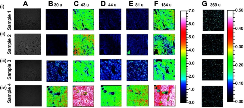
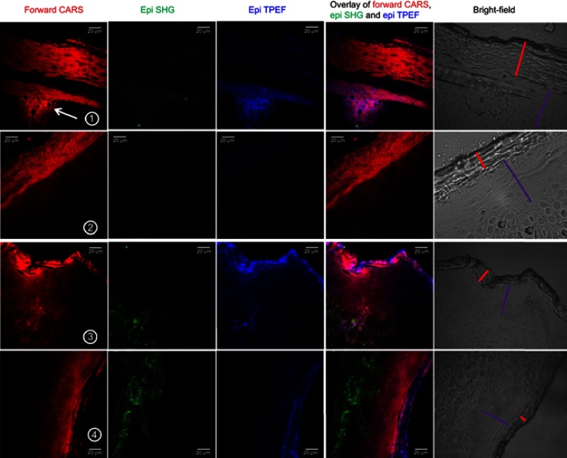
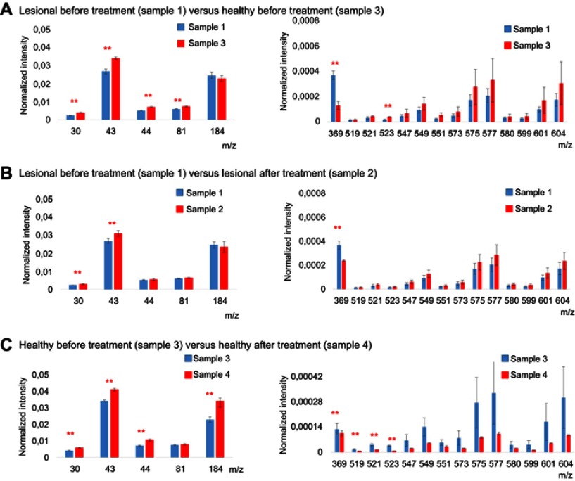
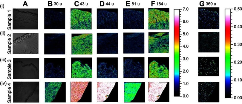
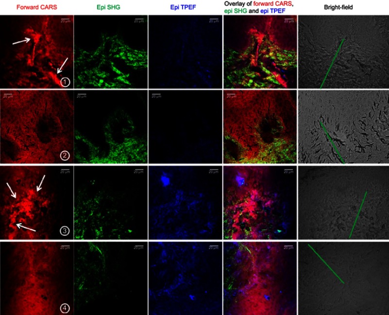
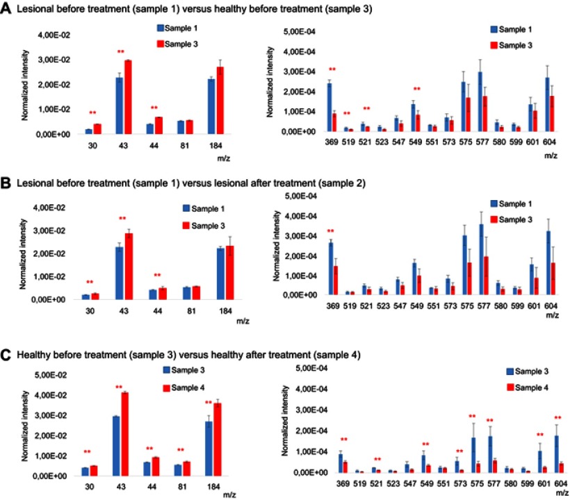
Psoriasis is a systemic inflammatory disease characterized by epidermal proliferation in the skin. Altered lipid metabolism is considered to be a central factor in the psoriatic etiopathogenesis. Thus, it is necessary to visualize chemical specificity of the samples for better medical diagnosis and treatment. Here, we investigate its role in the development of psoriatic lesions, before and after ultraviolet phototherapy, in a case study. The distribution and morphology of different lipids and fibrous proteins in psoriatic (lesional) tissues were visualized by two complementary label-free imaging techniques: 1) non-linear microscopy (NLM), providing images of lipids/proteins throughout the skin layers at submicrometer resolution; and 2) mass spectrometry imaging (MSI), offering high chemical specificity and hence the detection of different lipid species in the epidermal and dermal regions. A conventional method of histological evaluation was performed on the tissues, with no direct comparison with NLM and MSI. Psoriatic tissues had a higher lipid content, mainly in cholesterol, in both the epidermal and dermal regions, compared to healthy tissues. Moreover, the collagen and elastin fibers in the psoriatic tissues had a tendency to assemble as larger bundles, while healthy tissues showed smaller fibers more homogeneously spread. Although phototherapy significantly reduced the cholesterol content, it also increased the amounts of collagen in both lesional and non-lesional tissues. This study introduces NLM and MSI as two complementary techniques which are chemical specific and can be used to assess and visualize the distribution of lipids, collagen, and elastin in a non-invasive and label-free manner.
银屑病是一种全身性炎症性疾病,其特征为皮肤表皮增生。脂质代谢改变被认为是银屑病发病机制的核心因素。因此,为了更好地进行医学诊断和治疗,有必要可视化样本的化学特异性。在此,我们通过一个病例研究,调查了其在紫外线光疗前后银屑病皮损发展过程中的作用。采用两种互补的无标记成像技术,对银屑病(皮损)组织中不同脂质和纤维蛋白的分布及形态进行了可视化:1)非线性显微镜(NLM),以亚微米分辨率提供整个皮肤层脂质/蛋白质的图像;2)质谱成像(MSI),具有高化学特异性,从而能够检测表皮和真皮区域的不同脂质种类。对组织进行了传统的组织学评估,但未与NLM和MSI进行直接比较。与健康组织相比,银屑病组织在表皮和真皮区域均具有较高的脂质含量,主要为胆固醇。此外,银屑病组织中的胶原纤维和弹性纤维倾向于聚集成更大的束状,而健康组织显示较小的纤维分布更均匀。尽管光疗显著降低了胆固醇含量,但它也增加了皮损组织和非皮损组织中的胶原含量。本研究引入NLM和MSI作为两种互补技术,它们具有化学特异性,可用于以非侵入性和无标记的方式评估和可视化脂质、胶原和弹性蛋白的分布。