Department of Orthopaedic Surgery, Center for Cellular and Molecular Engineering, University of Pittsburgh School of Medicine, 450 Technology Drive, Pittsburgh, PA, 15219, USA.
McGowan Institute for Regenerative Medicine, University of Pittsburgh School of Medicine, Pittsburgh, USA.
Stem Cell Res Ther. 2019 Aug 14;10(1):254. doi: 10.1186/s13287-019-1350-6.
Stem cell-based bone tissue engineering shows promise for bone repair but faces some challenges, such as insufficient osteogenesis and limited architecture flexibility of the cell-delivery scaffold.
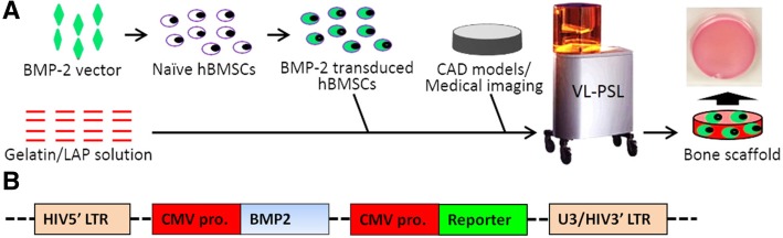
In this study, we first used lentiviral constructs to transduce ex vivo human bone marrow-derived stem cells with human bone morphogenetic protein-2 (BMP-2) gene (BMP-hBMSCs). We then introduced these cells into a hydrogel scaffold using an advanced visible light-based projection stereolithography (VL-PSL) technology, which is compatible with concomitant cell encapsulation and amenable to computer-aided architectural design, to fabricate scaffolds fitting local physical and structural variations in different bones and defects.
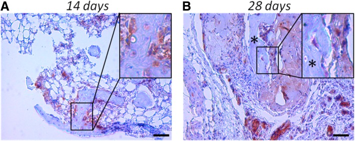
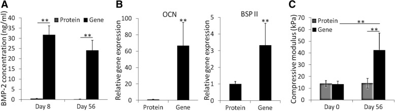
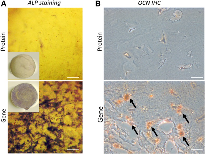
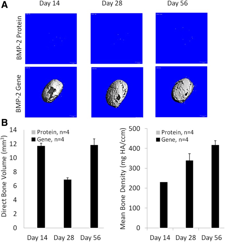
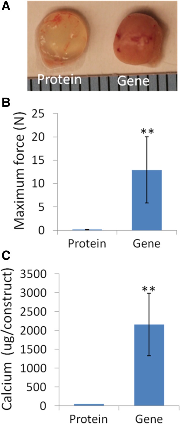
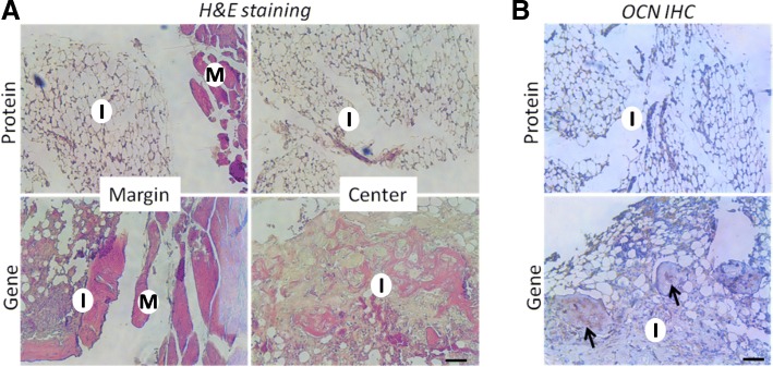
The results showed that the BMP-hBMSCs encapsulated within the scaffolds had high viability with sustained BMP-2 gene expression and differentiated toward an osteogenic lineage without the supplement of additional BMP-2 protein. In vivo bone formation efficacy was further assessed using an intramuscular implantation model in severe combined immunodeficiency (SCID) mice. Microcomputed tomography (micro-CT) imaging indicated rapid bone formation by the BMP-hBMSC-laden constructs as early as 14 days post-implantation. Histological examination revealed a mature trabecular bone structure with considerable vascularization. Through tracking of the implanted cells, we also found that BMP-hBMSC were directly involved in the new bone formation.
The robust, self-driven osteogenic capability and computer-designed architecture of the construct developed in this study should have potential applications for customized clinical repair of large bone defects or non-unions.
基于干细胞的骨组织工程在骨修复方面显示出巨大的潜力,但也面临一些挑战,例如成骨能力不足和细胞输送支架结构灵活性有限。
在本研究中,我们首先使用慢病毒构建体将人骨形态发生蛋白-2(BMP-2)基因(BMP-hBMSCs)转导到体外培养的人骨髓源性干细胞中。然后,我们使用先进的基于可见光的投影立体光刻(VL-PSL)技术将这些细胞引入水凝胶支架中,该技术与同时进行细胞包封兼容,并适用于计算机辅助建筑设计,以制造适合不同骨骼和缺陷的局部物理和结构变化的支架。
结果表明,支架内包封的 BMP-hBMSCs 具有高活力,持续表达 BMP-2 基因,并向成骨谱系分化,而无需补充额外的 BMP-2 蛋白。进一步使用严重联合免疫缺陷(SCID)小鼠的肌肉内植入模型评估体内骨形成效果。微计算机断层扫描(micro-CT)成像表明,BMP-hBMSC 负载构建物在植入后 14 天即可快速形成骨。组织学检查显示出具有相当大血管化的成熟小梁骨结构。通过对植入细胞的追踪,我们还发现 BMP-hBMSC 直接参与了新骨形成。
本研究中开发的具有强大的、自我驱动的成骨能力和计算机设计的支架结构,应该具有用于定制化临床修复大骨缺损或骨不连的潜在应用。