• 文献检索
  • 文档翻译
  • 深度研究
  • 学术资讯
  • Suppr Zotero 插件Zotero 插件
  • 邀请有礼
  • 套餐&价格
  • 历史记录
应用&插件
Suppr Zotero 插件Zotero 插件浏览器插件Mac 客户端Windows 客户端微信小程序
定价
高级版会员购买积分包购买API积分包
服务
文献检索文档翻译深度研究API 文档MCP 服务
关于我们
关于 Suppr公司介绍联系我们用户协议隐私条款
关注我们

Suppr 超能文献

核心技术专利:CN118964589B侵权必究
粤ICP备2023148730 号-1Suppr @ 2026

文献检索

告别复杂PubMed语法,用中文像聊天一样搜索,搜遍4000万医学文献。AI智能推荐,让科研检索更轻松。

立即免费搜索

文件翻译

保留排版,准确专业,支持PDF/Word/PPT等文件格式,支持 12+语言互译。

免费翻译文档

深度研究

AI帮你快速写综述,25分钟生成高质量综述,智能提取关键信息,辅助科研写作。

立即免费体验

CT 和超声心动图在主动脉瓣狭窄严重程度方面的不一致性:使用计算流体动力学研究其机制差异的患病率和见解。

Inconsistency in aortic stenosis severity between CT and echocardiography: prevalence and insights into mechanistic differences using computational fluid dynamics.

机构信息

Heart Assessment, Royal Brompton and Harefield NHS Foundation Trust, London, Middlesex, UK.

Faculty of Medicine, NHLI, Imperial College London, London, United Kingdom.

出版信息

Open Heart. 2019 Jul 29;6(2):e001044. doi: 10.1136/openhrt-2019-001044. eCollection 2019.

DOI:10.1136/openhrt-2019-001044
PMID:31413845
原文链接:https://pmc.ncbi.nlm.nih.gov/articles/PMC6667934/
Abstract

OBJECTIVES

The aims of this study were to evaluate the inconsistency of aortic stenosis (AS) severity between CT aortic valve area (CT-AVA) and echocardiographic Doppler parameters, and to investigate potential underlying mechanisms using computational fluid dynamics (CFD).

METHODS

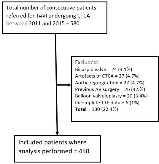
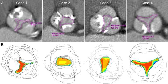
A total of 450 consecutive eligible patients undergoing transcatheter AV implantation assessment underwent CT cardiac angiography (CTCA) following echocardiography. CT-AVA derived by direct planimetry and echocardiographic parameters were used to assess severity. CFD simulation was performed in 46 CTCA cases to evaluate velocity profiles.

RESULTS

A CT-AVA>1 cm was present in 23% of patients with echocardiographic peak velocity≥4 m/s (r=-0.33) and in 15% patients with mean Doppler gradient≥40 mm Hg (r=-0.39). Patients with inconsistent severity grading between CT and echocardiography had higher stroke volume index (43 vs 38 mL/m, p<0.003) and left ventricular outflow tract (LVOT) flow rate (235 vs 192 cm/s, p<0.001). CFD simulation revealed high flow, either in isolation (p=0.01), or when associated with a skewed velocity profile (p=0.007), as the main cause for inconsistency between CT and echocardiography.

CONCLUSION

Severe AS by Doppler criteria may be associated with a CT-AVA>1 cm in up to a quarter of patients. CFD demonstrates that haemodynamic severity may be exaggerated on Doppler analysis due to high LVOT flow rates, with or without skewed velocity profiles, across the valve orifice. These factors should be considered before making a firm diagnosis of severe AS and evaluation with CT can be helpful.

摘要

目的

本研究旨在评估 CT 主动脉瓣口面积(CT-AVA)与超声心动图多普勒参数评估主动脉瓣狭窄(AS)严重程度的不一致性,并通过计算流体动力学(CFD)研究潜在的机制。

方法

对 450 例连续的经导管主动脉瓣植入术评估患者在超声心动图检查后进行 CT 心脏血管造影(CTCA)。采用直接平面测量法获得 CT-AVA,并采用超声心动图参数评估严重程度。对 46 例 CTCA 病例进行 CFD 模拟,以评估速度剖面。

结果

超声心动图峰值速度≥4 m/s(r=-0.33)的患者中,有 23%的 CT-AVA>1 cm;平均多普勒梯度≥40 mm Hg(r=-0.39)的患者中有 15%的 CT-AVA>1 cm。CT 和超声心动图严重程度分级不一致的患者的每搏量指数(43 比 38 mL/m,p<0.003)和左心室流出道(LVOT)流速(235 比 192 cm/s,p<0.001)更高。CFD 模拟显示,高流量(单独存在或与速度剖面偏斜相关)是导致 CT 和超声心动图之间不一致的主要原因(p=0.01 或 p=0.007)。

结论

多普勒标准评估的严重 AS 患者中,多达四分之一的患者 CT-AVA>1 cm。CFD 表明,由于 LVOT 流速较高,无论是否存在速度剖面偏斜,瓣口的血流动力学严重程度可能在多普勒分析中被夸大。在做出严重 AS 的明确诊断之前,应考虑这些因素,而 CT 评估可能会有所帮助。

https://cdn.ncbi.nlm.nih.gov/pmc/blobs/4dca/6667934/e0aba08d27d6/openhrt-2019-001044f03.jpg
https://cdn.ncbi.nlm.nih.gov/pmc/blobs/4dca/6667934/3849e085e633/openhrt-2019-001044f01.jpg
https://cdn.ncbi.nlm.nih.gov/pmc/blobs/4dca/6667934/8dec0d85953e/openhrt-2019-001044f02.jpg
https://cdn.ncbi.nlm.nih.gov/pmc/blobs/4dca/6667934/e0aba08d27d6/openhrt-2019-001044f03.jpg
https://cdn.ncbi.nlm.nih.gov/pmc/blobs/4dca/6667934/3849e085e633/openhrt-2019-001044f01.jpg
https://cdn.ncbi.nlm.nih.gov/pmc/blobs/4dca/6667934/8dec0d85953e/openhrt-2019-001044f02.jpg
https://cdn.ncbi.nlm.nih.gov/pmc/blobs/4dca/6667934/e0aba08d27d6/openhrt-2019-001044f03.jpg

相似文献

1
Inconsistency in aortic stenosis severity between CT and echocardiography: prevalence and insights into mechanistic differences using computational fluid dynamics.CT 和超声心动图在主动脉瓣狭窄严重程度方面的不一致性:使用计算流体动力学研究其机制差异的患病率和见解。
Open Heart. 2019 Jul 29;6(2):e001044. doi: 10.1136/openhrt-2019-001044. eCollection 2019.
2
The complex nature of discordant severe calcified aortic valve disease grading: new insights from combined Doppler echocardiographic and computed tomographic study.不和谐严重钙化主动脉瓣疾病分级的复杂性:联合多普勒超声心动图和计算机断层扫描研究的新见解。
J Am Coll Cardiol. 2013 Dec 17;62(24):2329-38. doi: 10.1016/j.jacc.2013.08.1621. Epub 2013 Sep 24.
3
Anatomic estimation of aortic stenosis severity vs "fusion" of data from computed tomography and Doppler echocardiography.主动脉瓣狭窄严重程度的解剖学评估与计算机断层扫描和多普勒超声心动图数据的“融合”
Echocardiography. 2018 Jun;35(6):777-784. doi: 10.1111/echo.13855. Epub 2018 Mar 9.
4
Inconsistent echocardiographic grading of aortic stenosis: is the left ventricular outflow tract important?主动脉瓣狭窄的超声心动图分级不一致:左心室流出道重要吗?
Heart. 2013 Jul;99(13):921-31. doi: 10.1136/heartjnl-2012-302881. Epub 2013 Jan 24.
5
Multimodality assessment of high- vs. low-gradient aortic stenosis using echocardiography and cardiac CT.超声心动图和心脏 CT 评估主动脉瓣狭窄的高梯度与低梯度
J Cardiovasc Comput Tomogr. 2023 Nov-Dec;17(6):421-428. doi: 10.1016/j.jcct.2023.09.002. Epub 2023 Oct 6.
6
Inconsistent grading of aortic valve stenosis by current guidelines: haemodynamic studies in patients with apparently normal left ventricular function.现行指南对主动脉瓣狭窄分级不一致:左心室功能正常的患者的血液动力学研究。
Heart. 2010 Sep;96(18):1463-8. doi: 10.1136/hrt.2009.181982.
7
Differences in aortic valve area measured with CT planimetry and echocardiography (continuity equation) are related to divergent estimates of left ventricular outflow tract area.通过CT平面测量法和超声心动图(连续方程)测量的主动脉瓣面积差异与左心室流出道面积的不同估计值相关。
AJR Am J Roentgenol. 2009 Jun;192(6):1668-73. doi: 10.2214/AJR.08.1986.
8
Discordant Grading of Aortic Stenosis Severity: Echocardiographic Predictors of Survival Benefit Associated With Aortic Valve Replacement.主动脉瓣狭窄严重程度的不一致分级:超声心动图预测主动脉瓣置换术相关生存获益的指标。
JACC Cardiovasc Imaging. 2016 Jul;9(7):797-805. doi: 10.1016/j.jcmg.2015.09.026. Epub 2016 May 18.
9
Effect of the ellipsoid shape of the left ventricular outflow tract on the echocardiographic assessment of aortic valve area in aortic stenosis.左心室流出道椭圆形形态对主动脉瓣狭窄时主动脉瓣面积超声心动图评估的影响。
J Cardiovasc Comput Tomogr. 2014 Jan-Feb;8(1):52-7. doi: 10.1016/j.jcct.2013.12.006. Epub 2014 Jan 11.
10
Evaluation of aortic stenosis severity: role of contrast echocardiography in comparison with conventional echocardiography and cardiac catheterization.主动脉瓣狭窄严重程度的评估:对比超声心动图与传统超声心动图及心导管检查相比的作用。
Rev Port Cardiol. 2002 May;21(5):555-72.

引用本文的文献

1
Performance of Computed Tomographic Angiography-Based Aortic Valve Area for Assessment of Aortic Stenosis.基于计算机断层扫描血管造影的主动脉瓣口面积评估主动脉瓣狭窄的性能。
J Am Heart Assoc. 2023 Aug 15;12(16):e029973. doi: 10.1161/JAHA.123.029973. Epub 2023 Aug 10.
2
Computed Tomography-Based Assessment of Transvalvular Pressure Gradient in Aortic Stenosis.基于计算机断层扫描的主动脉瓣狭窄跨瓣压差评估
Front Cardiovasc Med. 2021 Sep 8;8:706628. doi: 10.3389/fcvm.2021.706628. eCollection 2021.

本文引用的文献

1
Should we forget about valve area when assessing aortic stenosis?在评估主动脉瓣狭窄时,我们应该忽略瓣口面积吗?
Heart. 2019 Jan;105(2):92-93. doi: 10.1136/heartjnl-2018-313666. Epub 2018 Aug 3.
2
2017 ESC/EACTS Guidelines for the management of valvular heart disease.2017年欧洲心脏病学会/欧洲心胸外科学会瓣膜性心脏病管理指南。
Eur Heart J. 2017 Sep 21;38(36):2739-2791. doi: 10.1093/eurheartj/ehx391.
3
Impact of stroke volume assessment by integrating multi-detector computed tomography and Doppler data on the classification of aortic stenosis.
多排螺旋 CT 与多普勒数据整合评估心搏量对主动脉瓣狭窄分类的影响。
Int J Cardiol. 2017 Nov 1;246:80-86. doi: 10.1016/j.ijcard.2017.03.112.
4
Recommendations on the echocardiographic assessment of aortic valve stenosis: a focused update from the European Association of Cardiovascular Imaging and the American Society of Echocardiography.主动脉瓣狭窄的超声心动图评估建议:欧洲心血管影像学会和美国超声心动图学会的重点更新
Eur Heart J Cardiovasc Imaging. 2017 Mar 1;18(3):254-275. doi: 10.1093/ehjci/jew335.
5
Computational medical imaging and hemodynamics framework for functional analysis and assessment of cardiovascular structures.用于心血管结构功能分析和评估的计算医学成像与血流动力学框架。
Biomed Eng Online. 2017 Mar 21;16(1):35. doi: 10.1186/s12938-017-0326-y.
6
Beyond Bernoulli: Improving the Accuracy and Precision of Noninvasive Estimation of Peak Pressure Drops.超越伯努利原理:提高无创估计峰值压力降的准确性和精度。
Circ Cardiovasc Imaging. 2017 Jan;10(1):e005207. doi: 10.1161/CIRCIMAGING.116.005207.
7
Echocardiographic predictors of outcomes in adults with aortic stenosis.成人主动脉瓣狭窄患者预后的超声心动图预测指标
Heart. 2016 Jun 15;102(12):934-42. doi: 10.1136/heartjnl-2015-308742. Epub 2016 Apr 5.
8
Computational fluid dynamics modelling in cardiovascular medicine.心血管医学中的计算流体动力学建模
Heart. 2016 Jan;102(1):18-28. doi: 10.1136/heartjnl-2015-308044. Epub 2015 Oct 28.
9
STARD 2015: an updated list of essential items for reporting diagnostic accuracy studies.STARD 2015:报告诊断准确性研究的必备项目更新清单。
BMJ. 2015 Oct 28;351:h5527. doi: 10.1136/bmj.h5527.
10
Inconsistency in hemodynamic characterization of severe aortic stenosis.重度主动脉瓣狭窄血流动力学特征的不一致性。
Int J Cardiol. 2015 Oct 15;197:309-11. doi: 10.1016/j.ijcard.2015.06.102. Epub 2015 Jun 30.