Orthopaedic Biomechanics, Department of Biomedical Engineering, Eindhoven University of Technology, PO Box 513, 5600 MB, Eindhoven, The Netherlands.
Institute for Complex Molecular Systems, Eindhoven University of Technology, PO Box 513, 5600 MB, Eindhoven, The Netherlands.
J Mater Sci Mater Med. 2019 Aug 14;30(8):94. doi: 10.1007/s10856-019-6295-x.
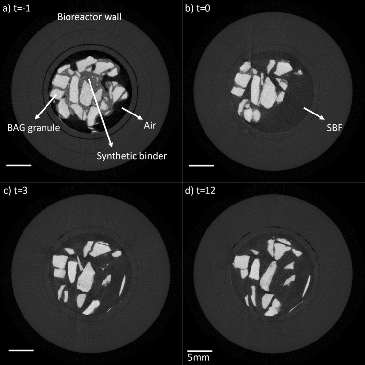
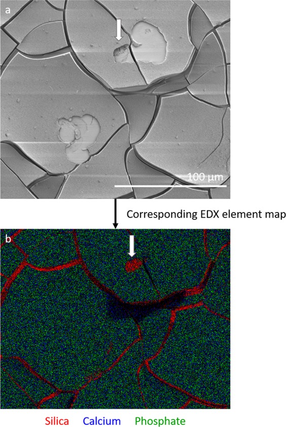
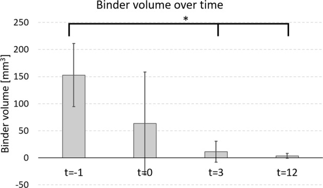
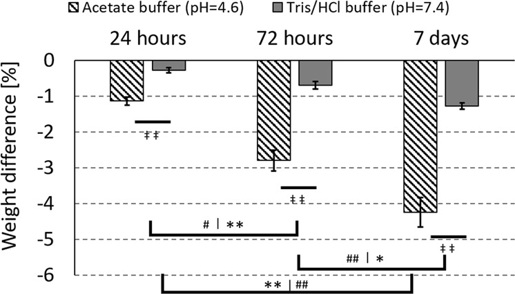
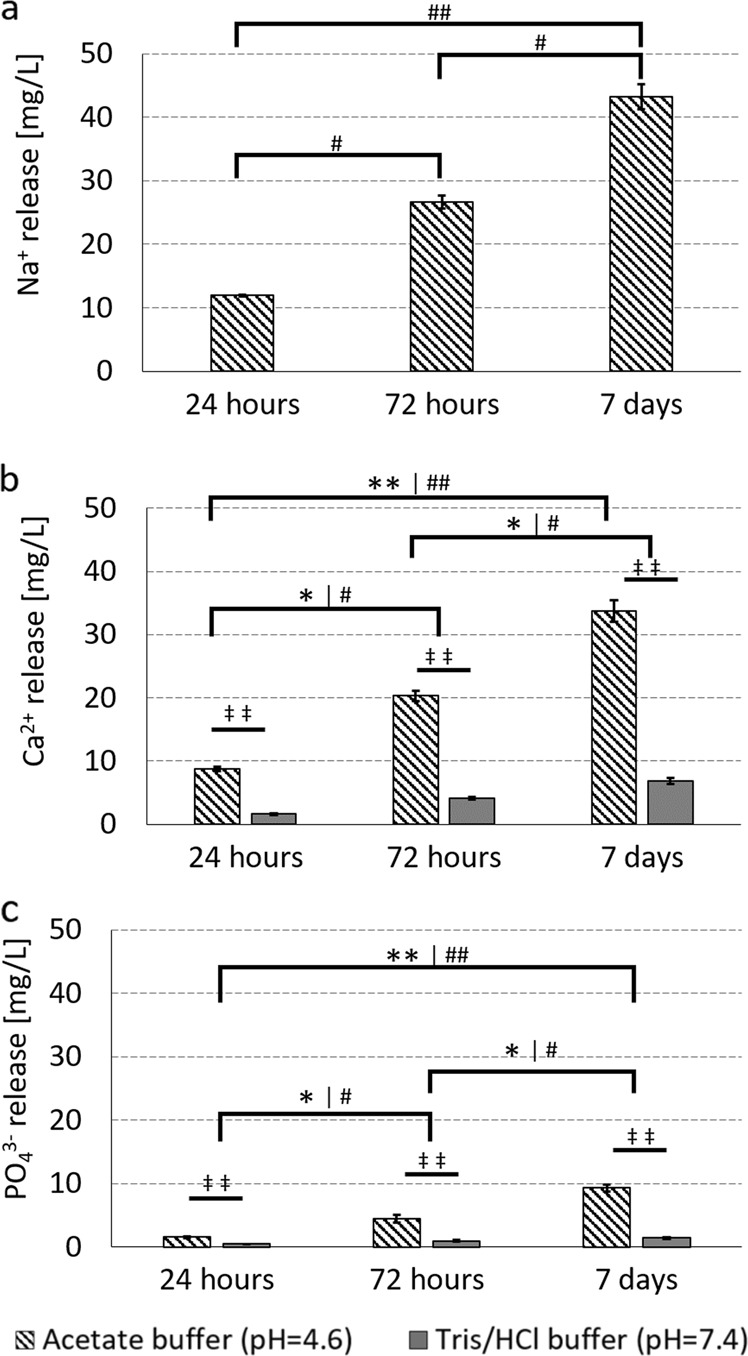
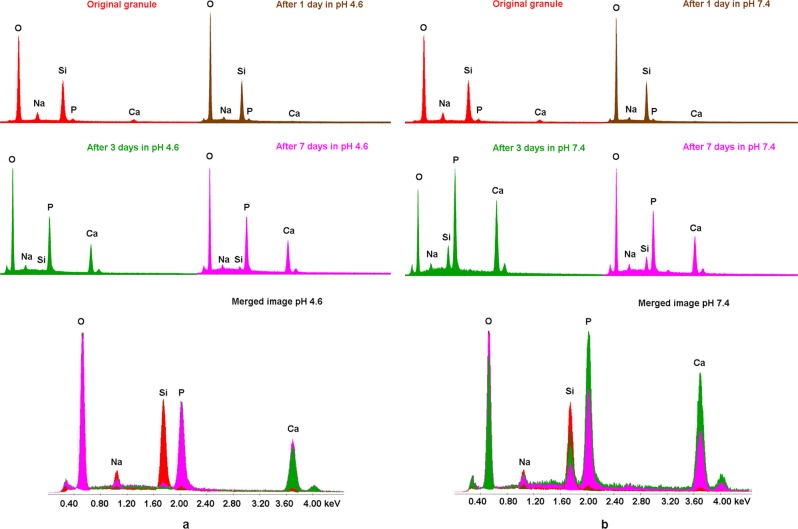
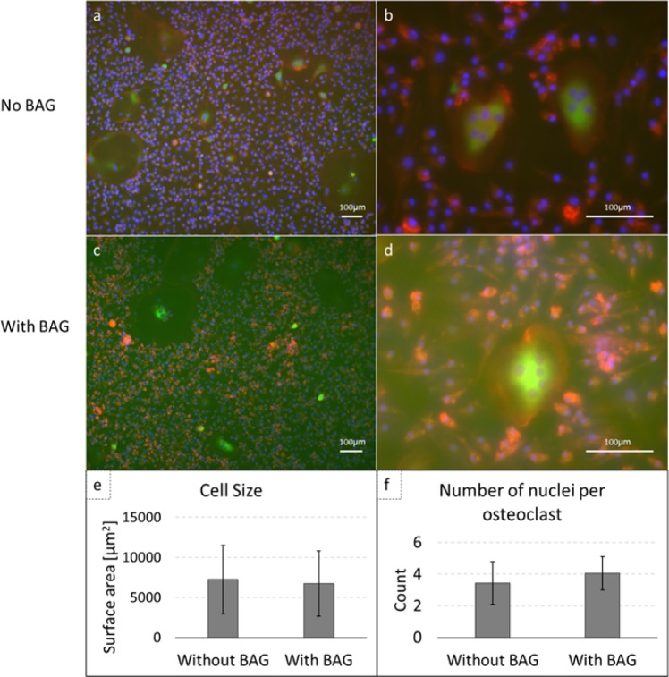
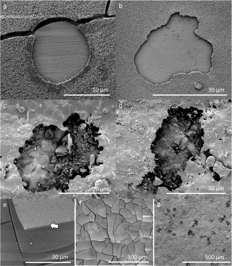
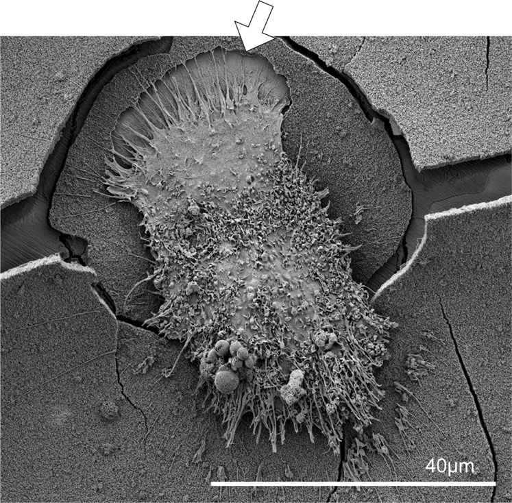
Clinically, S53P4 bioactive glass (BAG) has shown very promising results in bone infection treatment, but it is also known to degrade very slowly in vivo. To evaluate which mechanisms (cellular or dissolution) can play a role in the degradation of S53P4 BAG and S53P4 BAG putty, in vitro degradation experiments at different pH (7.4 and 4.6) were performed. Micro computed tomography showed a rapid dissolution of the synthetic binder in the putty formulation, within 12 h is simulated body fluid (pH = 7.4), leaving behind only loose granules. Therefore the degradation of the loose granules was investigated further. Significant weight loss was observed and ion chromatography showed that Ca, Na and PO ions were released from S54P4 BAG granules in the two fluids. It was observed that the weight loss and ion release were increased when the pH of the fluid was decreased to 4.6. Osteoclasts are known to create such a low pH when resorbing bone and therefore their capacity to degrade S53P4 surfaces were studied as well. Scanning electron microscopy and energy-dispersive X-ray spectroscopy confirmed that osteoclasts were able to create resorption pits in the calcium phosphate layer on S53P4 BAG surfaces. The silica of the BAG, located underneath the calcium phosphate, seemed to hinder further osteclastic resorption of the material. To our knowledge we were the first to observe actively resorbing osteoclasts on S53P4 bioactive glass surfaces, in vitro. Future research is needed to define the specific role osteoclasts play in the degradation of BAG in vivo.
临床上,S53P4 生物活性玻璃(BAG)在治疗骨感染方面显示出非常有前景的结果,但也已知其在体内降解非常缓慢。为了评估哪些机制(细胞或溶解)可以在 S53P4 BAG 和 S53P4 BAG 油灰的体外降解中发挥作用,在不同 pH 值(7.4 和 4.6)下进行了体外降解实验。微计算机断层扫描显示,在模拟体液中(pH=7.4),合成粘合剂在油灰配方中迅速溶解,在 12 小时内仅留下松散的颗粒。因此,进一步研究了松散颗粒的降解。观察到明显的重量损失,离子色谱法表明 Ca、Na 和 PO 离子从 S54P4 BAG 颗粒在两种流体中释放。当流体的 pH 值降低到 4.6 时,观察到重量损失和离子释放增加。破骨细胞在吸收骨时会产生这种低 pH 值,因此研究了它们降解 S53P4 表面的能力。扫描电子显微镜和能谱分析证实,破骨细胞能够在 S53P4 BAG 表面的磷酸钙层上形成吸收陷窝。位于磷酸钙下的 BAG 中的二氧化硅似乎阻碍了材料的进一步破骨细胞吸收。据我们所知,我们是第一个在体外观察到 S53P4 生物活性玻璃表面主动吸收的破骨细胞。需要进一步的研究来确定破骨细胞在体内 BAG 降解中的具体作用。