Department of Otorhinolaryngology, National Medical Center, Seoul 04564, Republic of Korea.
Department of Otorhinolaryngology‑Head and Neck Surgery, Chung‑Ang University College of Medicine, Seoul 06974, Republic of Korea.
Int J Mol Med. 2019 Oct;44(4):1473-1483. doi: 10.3892/ijmm.2019.4316. Epub 2019 Aug 19.
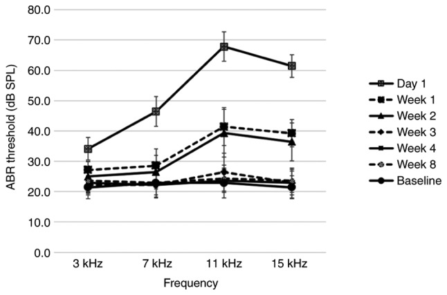
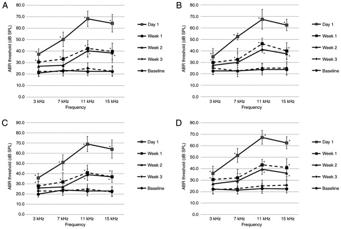
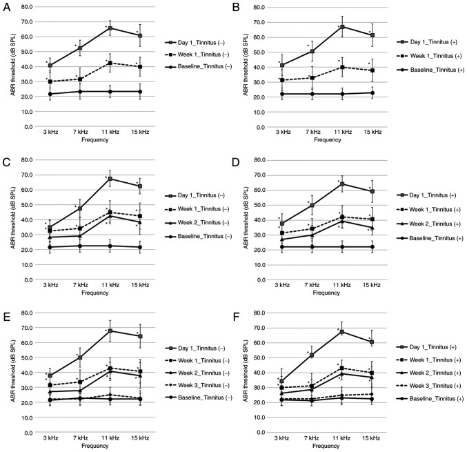
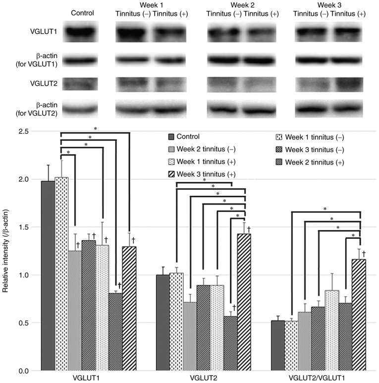
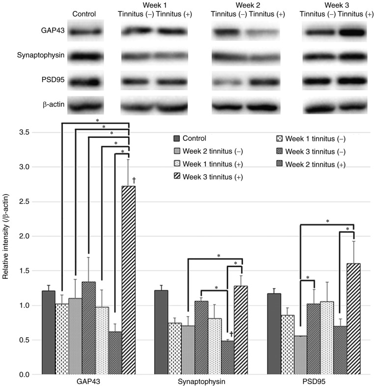
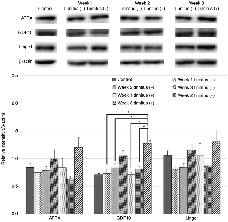
One of the primary theories of the pathogenesis of tinnitus involves maladaptive auditory‑somatosensory plasticity in the dorsal cochlear nucleus (DCN), which is assumed to be due to axonal sprouting. Although a disrupted balance between auditory and somatosensory inputs may occur following hearing damage and may induce tinnitus, examination of this phenomenon employed a model of hearing damage that does not account for the causal relationship between these changes and tinnitus. The present study aimed to investigate changes in auditory‑somatosensory innervation and the role that axonal sprouting serves in this process by comparing results between animals with and without tinnitus. Rats were exposed to a noise‑inducing temporary threshold shift and were subsequently divided into tinnitus and non‑tinnitus groups based on the results of gap prepulse inhibition of the acoustic startle reflex. DCNs were collected from rats divided into three sub‑groups according to the number of weeks (1, 2 or 3) following noise exposure, and the protein levels of vesicular glutamate transporter 1 (VGLUT1), which is associated with auditory input to the DCN, and VGLUT2, which is in turn primarily associated with somatosensory inputs, were assessed. In addition, factors related to axonal sprouting, including growth‑associated protein 43 (GAP43), postsynaptic density protein 95, synaptophysin, α‑thalassemia/mental retardation syndrome X‑linked homolog (ATRX), growth differentiation factor 10 (GDF10), and leucine‑rich repeat and immunoglobulin domain‑containing 1, were measured by western blot analyses. Compared to the non‑tinnitus group, the tinnitus group exhibited a significant decrease in VGLUT1 at 1 week and a significant increase in VGLUT2 at 3 weeks post‑exposure. In addition, rats in the tinnitus group exhibited significant increases in GAP43 and GDF10 protein expression levels in their DCN at 3 weeks following noise exposure. Results from the present study provided further evidence that changes in the neural input distribution to the DCN may cause tinnitus and that axonal sprouting underlies these alterations.
耳鸣发病机制的主要理论之一涉及背侧耳蜗核 (DCN) 中的听觉-躯体感觉可塑性失调,这被认为是由于轴突发芽所致。尽管听觉损伤后可能会出现听觉和躯体感觉输入之间的平衡失调,并可能导致耳鸣,但对这种现象的检查采用了一种听力损伤模型,该模型不能说明这些变化与耳鸣之间的因果关系。本研究旨在通过比较有耳鸣和无耳鸣动物的结果,研究听觉-躯体感觉传入的变化以及轴突发芽在此过程中的作用。将大鼠暴露于致聋的短暂阈移噪声中,然后根据声反射的声刺激潜伏期间隙抑制的结果将其分为耳鸣和非耳鸣组。根据噪声暴露后 1、2 或 3 周的时间,将大鼠分为三组,收集 DCN,并评估与 DCN 听觉传入相关的囊泡谷氨酸转运体 1 (VGLUT1) 和主要与躯体感觉传入相关的 VGLUT2 的蛋白水平。此外,还通过 Western blot 分析测量了与轴突发芽相关的因子,包括生长相关蛋白 43 (GAP43)、突触后密度蛋白 95、突触小体蛋白、X 连锁α-地中海贫血/智力低下综合征同源物 (ATRX)、生长分化因子 10 (GDF10) 和富含亮氨酸重复和免疫球蛋白域包含蛋白 1。与非耳鸣组相比,耳鸣组在暴露后 1 周时 VGLUT1 显著降低,在暴露后 3 周时 VGLUT2 显著升高。此外,耳鸣组大鼠在噪声暴露后 3 周时 DCN 中的 GAP43 和 GDF10 蛋白表达水平显著增加。本研究的结果进一步证明,DCN 神经传入分布的变化可能导致耳鸣,而轴突发芽是这些变化的基础。