Hearing and Neuronal activity Lab, Brain Institute, Federal University of Rio Grande do Norte, Natal, Brazil.
Institute for Analysis and Scientific Computing, Vienna University of Technology, Vienna, Austria.
BMC Biol. 2022 May 12;20(1):102. doi: 10.1186/s12915-022-01288-1.
The dorsal cochlear nucleus (DCN) is a region known to integrate somatosensory and auditory inputs and is identified as a potential key structure in the generation of phantom sound perception, especially noise-induced tinnitus. Yet, how altered homeostatic plasticity of the DCN induces and maintains the sensation of tinnitus is not clear. Here, we chemogenetically decrease activity of a subgroup of DCN neurons, Ca/Calmodulin kinase 2 α (CaMKII α)-positive DCN neurons, using Gi-coupled human M4 Designer Receptors Exclusively Activated by Designer Drugs (hM4Di DREADDs), to investigate their role in noise-induced tinnitus.
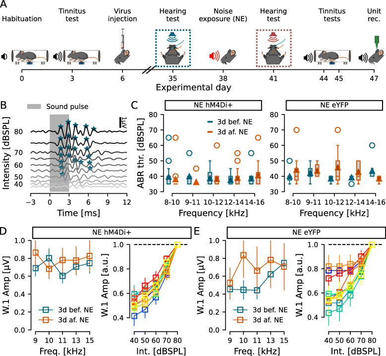
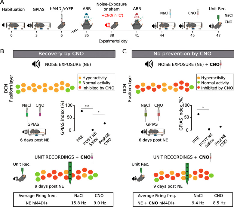
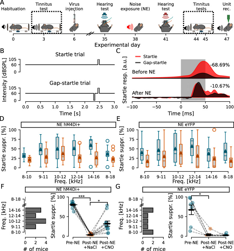
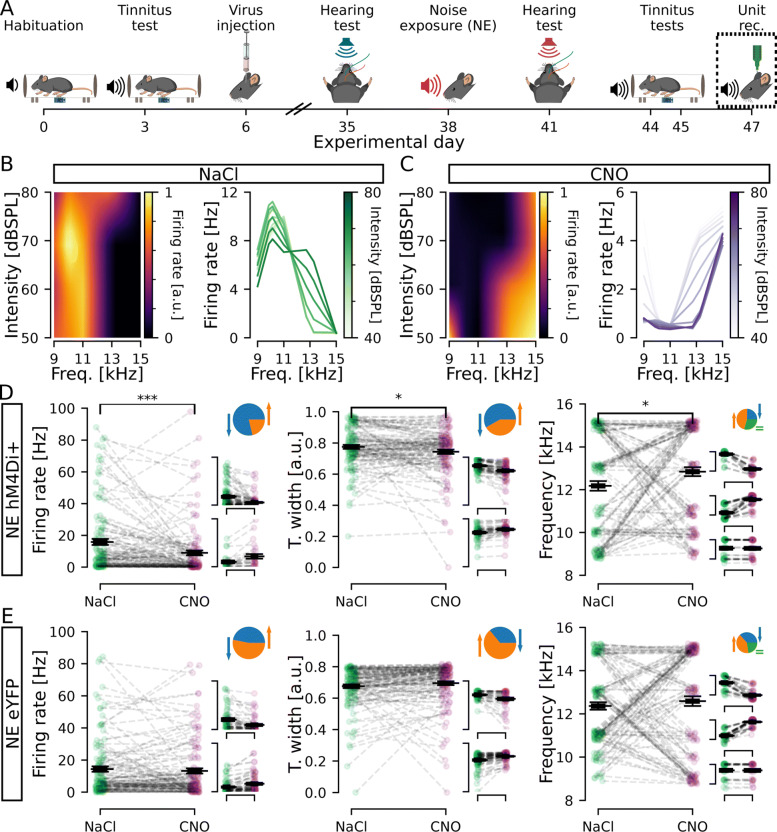
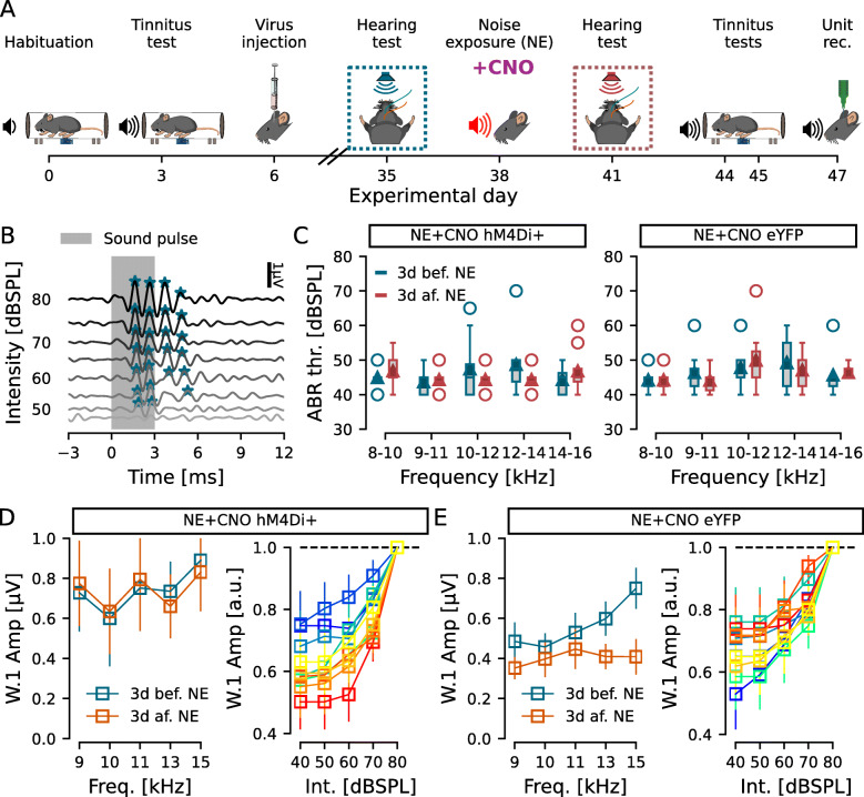
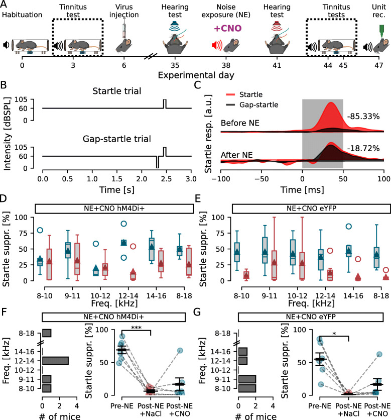
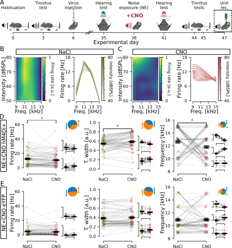
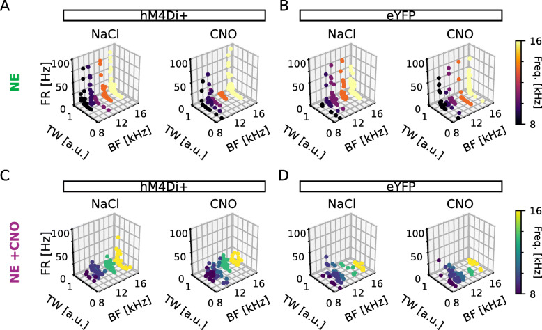
Mice were exposed to loud noise (9-11kHz, 90dBSPL, 1h, followed by 2h of silence), and auditory brainstem responses (ABRs) and gap prepulse inhibition of acoustic startle (GPIAS) were recorded 2 days before and 2 weeks after noise exposure to identify animals with a significantly decreased inhibition of startle, indicating tinnitus but without permanent hearing loss. Neuronal activity of CaMKII α+ neurons expressing hM4Di in the DCN was lowered by administration of clozapine-N-oxide (CNO). We found that acutely decreasing firing rate of CaMKII α+ DCN units decrease tinnitus-like responses (p = 3e -3, n = 11 mice), compared to the control group that showed no improvement in GPIAS (control virus; CaMKII α-YFP + CNO, p = 0.696, n = 7 mice). Extracellular recordings confirmed CNO to decrease unit firing frequency of CaMKII α-hM4Di+ mice and alter best frequency and tuning width of response to sound. However, these effects were not seen if CNO had been previously administered during the noise exposure (n = 6 experimental and 6 control mice).
We found that lowering DCN activity in mice displaying tinnitus-related behavior reduces tinnitus, but lowering DCN activity during noise exposure does not prevent noise-induced tinnitus. Our results suggest that CaMKII α-positive cells in the DCN are not crucial for tinnitus induction but play a significant role in maintaining tinnitus perception in mice.
背侧耳蜗核(DCN)是一个整合躯体感觉和听觉输入的区域,被认为是产生幻听感知的潜在关键结构,特别是噪声诱导的耳鸣。然而,DCN 的稳态可塑性如何改变导致并维持耳鸣的感觉尚不清楚。在这里,我们使用 Gi 偶联的人源 M4 Designer Receptors Exclusively Activated by Designer Drugs(hM4Di DREADDs),化学遗传学地降低一组 DCN 神经元,即钙/钙调蛋白激酶 2α(CaMKIIα)阳性 DCN 神经元的活性,以研究它们在噪声诱导的耳鸣中的作用。
小鼠暴露于强噪声(9-11kHz,90dBSPL,1h,随后 2h 安静),在噪声暴露前 2 天和后 2 周记录听觉脑干反应(ABR)和声刺激的潜伏期预脉冲抑制(GPIAS),以识别出明显抑制起始惊跳反应降低的动物,表明有耳鸣但无永久性听力损失。在 DCN 中表达 hM4Di 的 CaMKIIα+神经元的神经元活性通过氯氮平-N-氧化物(CNO)降低。我们发现,急性降低 CaMKIIα+DCN 单位的放电率可降低耳鸣样反应(p=3e-3,n=11 只小鼠),而对照组 GPIAS 没有改善(对照病毒;CaMKIIα-YFP+CNO,p=0.696,n=7 只小鼠)。细胞外记录证实 CNO 降低 CaMKIIα-hM4Di+小鼠的单位放电频率,并改变对声音的最佳频率和调谐宽度的反应。然而,如果在噪声暴露期间已经给予 CNO,则不会出现这些效果(n=6 只实验和 6 只对照小鼠)。
我们发现,在表现出与耳鸣相关行为的小鼠中降低 DCN 活性可降低耳鸣,但在噪声暴露期间降低 DCN 活性并不能预防噪声诱导的耳鸣。我们的结果表明,DCN 中的 CaMKIIα 阳性细胞对于耳鸣的诱导不是至关重要的,但在维持小鼠的耳鸣感知中起着重要作用。