Department II of Internal Medicine and Center for Molecular Medicine Cologne, University of Cologne, Faculty of Medicine and University Hospital Cologne, Cologne, Germany.
CECAD, University of Cologne, Faculty of Medicine and University Hospital Cologne, Cologne, Germany.
BMC Nephrol. 2019 Aug 22;20(1):326. doi: 10.1186/s12882-019-1478-8.
Podocyte infolding glomerulopathy (PIG) is a rare histopathologic finding with global infolding of the podocytes into the glomerular basement membrane (GBM), accompanied by microstructures underneath. Described in 2002 for the first time, PIG was proposed as a new pathological entity in 2008 based on the largest case series so far. Yet all of the described cases derive from Asian countries. We report a case from Germany fulfilling the diagnostic criteria of PIG. Considering the scarcity of data on this entity especially in Western countries, collecting cases like ours and multicentric meta-analyses will be crucial to obtain a better understanding of PIG, its causes, clinical course and potential treatment options.
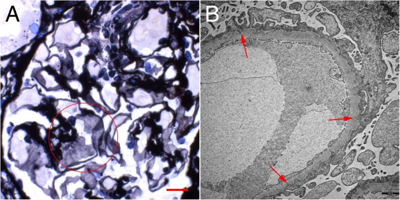
A 56-year-old Caucasian woman with a history of rheumatoid arthritis (RA), no other comorbidities and no known renal disease was admitted to the hospital with acute kidney injury (AKI) and nephrotic syndrome. Physical examination was unremarkable except for anasarca. Renal ultrasound revealed no abnormalities. Laboratory and urine analyses were consistent with the nephrotic syndrome and renal failure. Serological studies regarding ANA, ANCA, anti-PLA2R autoantibodies, complement, virus infections, immunofixation and quantitative light chain analysis were unremarkable. A renal biopsy was performed. Light microscopic examination showed flattened tubular epithelium consistent with acute tubular damage, no infiltrates and unremarkable glomeruli except diffuse and global holes in the GBM (Fig. 1a) and negative staining for immunoglobulin heavy-chains, light-chains and complement split products. Electron microscopy revealed a rare correlate for these holes: global peculiar infolding of podocyte cytoplasm into the GBM. Most of these infoldings were accompanied by condensation of the GBM underneath. No such condensation or electron dense deposits were found without these infoldings or outside the GBM.
Here we report the first case of PIG outside of Asia. Since there are only few reports about this specific finding, we feel there is a need to share information in an attempt to accumulate knowledge about this possible new entity and potential treatment options.
足细胞内褶肾小球病(PIG)是一种罕见的组织病理学表现,其特征为足细胞整体内褶入肾小球基底膜(GBM),伴有其下的微观结构。该病于 2002 年首次描述,2008 年基于迄今为止最大的病例系列,将其提出为一种新的病理实体。然而,所有描述的病例均来自亚洲国家。我们报告了一例来自德国的病例,符合 PIG 的诊断标准。鉴于该实体在西方国家的数据稀缺,特别是在西方国家,收集像我们这样的病例和多中心荟萃分析将是理解 PIG、其病因、临床过程和潜在治疗选择的关键。
一名 56 岁白人女性,患有类风湿关节炎(RA),无其他合并症,无已知的肾脏疾病,因急性肾损伤(AKI)和肾病综合征住院。体格检查无异常,仅出现水肿。肾脏超声无异常。实验室和尿液分析与肾病综合征和肾衰竭一致。血清学研究抗核抗体(ANA)、抗中性粒细胞胞浆抗体(ANCA)、抗磷脂酶 A2 受体自身抗体、补体、病毒感染、免疫固定电泳和定量轻链分析均无异常。进行了肾活检。光镜检查显示扁平的肾小管上皮细胞符合急性肾小管损伤,无浸润,肾小球无明显异常,除 GBM 弥漫性和全层孔(图 1a)外,免疫球蛋白重链、轻链和补体裂解产物染色均为阴性。电子显微镜显示这些孔的罕见相关物:足细胞细胞质整体内褶入 GBM。这些内褶大多伴有 GBM 下的凝聚。在没有这些内褶或不在 GBM 外的情况下,没有发现这种凝聚或电子致密沉积物。
我们报告了首例亚洲以外的 PIG 病例。由于关于这种特殊表现的报道很少,我们认为有必要分享信息,试图积累有关这种可能的新实体和潜在治疗选择的知识。