Department of Immunology, Veterinary Research Institute, Brno, Czechia.
Faculty of Veterinary Medicine, University of Veterinary and Pharmaceutical Sciences, Brno, Czechia.
Front Immunol. 2019 Aug 6;10:1689. doi: 10.3389/fimmu.2019.01689. eCollection 2019.
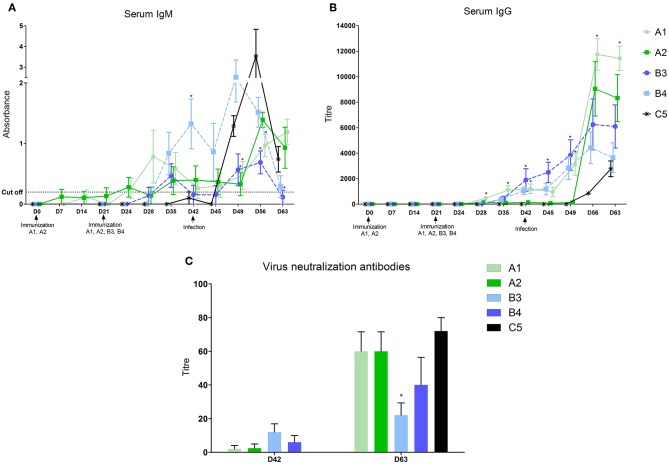
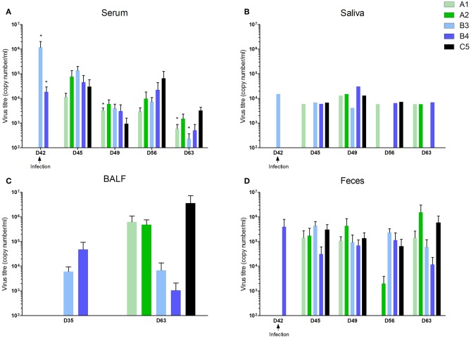
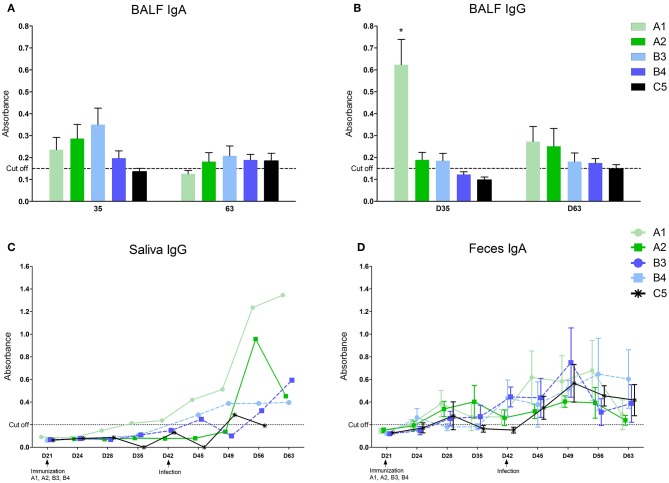
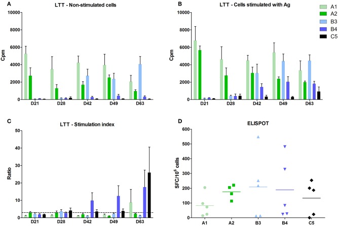
The goals of our study were to compare the immune response to different killed and modified live vaccines against PRRS virus and to monitor the antibody production and the cell mediated immunity both at the systemic and local level. In the experiment, we immunized four groups of piglets with two commercial inactivated (A1-Progressis, A2-Suivac) and two modified live vaccines (B3-Amervac, B4-Porcilis). Twenty-one days after the final vaccination, all piglets, including the control non-immunized group (C5), were i.n., infected with the Lelystad strain of PRRS virus. The serum antibody response (IgM and IgG) was the strongest in group A1 followed by two MLV (B3 and B4) groups. Locally, we demonstrated the highest level of IgG antibodies in bronchoalveolar lavages (BALF), and saliva in group A1, whereas low IgA antibody responses in BALF and feces were detected in all groups. We have found virus neutralization antibody at DPV 21 (days post vaccination) and higher levels in all groups including the control at DPI 21 (days post infection). Positive antigen specific cell-mediated response in lymphocyte transformation test (LTT) was observed in groups B3 and B4 at DPV 7 and in group B4 at DPV 21 and in all intervals after infection. The IFN-γ producing lymphocytes after antigen stimulation were found in CD4CD8 and CD4CD8 subsets of all immunized groups 7 days after infection. After infection, there were obvious differences in virus excretion. The virus was detected in all groups of piglets in serum, saliva, and occasionally in feces at DPI 3. Significantly lower virus load was found in groups A1 and B3 at DPI 21. Negative samples appeared at DPI 21 in B3 group in saliva. It can be concluded that antibodies after immunization and infection, and the virus after infection can be detected in all the compartments monitored. Immunization with inactivated vaccine A1-Progressis induces high levels of antibodies produced both systemically and locally. Immunization with MLV-vaccines (Amervac and Porcilis) produces sufficient antibody levels and also cell-mediated immunity. After infection virus secretion gradually decreases in group B3, indicating tendency to induce sterile immunity.
我们的研究目的是比较针对 PRRS 病毒的不同灭活和改良活疫苗的免疫反应,并监测系统和局部水平的抗体产生和细胞介导免疫。在实验中,我们用两种商业灭活疫苗(A1-Progressis、A2-Suivac)和两种改良活疫苗(B3-Amervac、B4-Porcilis)免疫四组仔猪。最后一次接种后 21 天,所有仔猪,包括非免疫对照组(C5),经鼻内感染 Lelystad 株 PRRS 病毒。血清抗体反应(IgM 和 IgG)在 A1 组最强,其次是两个 MLV(B3 和 B4)组。局部,我们在 A1 组的支气管肺泡灌洗液(BALF)和唾液中检测到最高水平的 IgG 抗体,而在所有组中 BALF 和粪便中的 IgA 抗体反应均较低。我们在 DPV 21(接种后天数)时发现了病毒中和抗体,并且在包括对照组在内的所有组中都发现了更高水平的病毒中和抗体。在 DPV 7 时,B3 和 B4 组和 DPV 21 时以及感染后的所有时间间隔内,在淋巴细胞转化试验(LTT)中均观察到了针对抗原的阳性特异性细胞介导反应。在感染后 7 天,在所有免疫组的 CD4CD8 和 CD4CD8 亚群中均发现了经抗原刺激后产生 IFN-γ的淋巴细胞。感染后,病毒的排泄有明显的差异。在 DPI 3 时,在所有仔猪血清、唾液中均检测到病毒,偶尔在粪便中也检测到病毒。在 DPI 21 时,在 A1 和 B3 组中发现了显著较低的病毒载量。在 B3 组的唾液中,在 DPI 21 时出现了阴性样本。可以得出结论,在所有监测的隔室中,都可以检测到免疫接种和感染后的抗体以及感染后的病毒。用灭活疫苗 A1-Progressis 免疫诱导了全身性和局部高水平的抗体产生。用 MLV 疫苗(Amervac 和 Porcilis)免疫可产生足够的抗体水平,也可诱导细胞介导免疫。在 B3 组中,病毒分泌逐渐减少,表明有诱导无菌免疫的趋势。