Wen Qiuting, Mustafi Sourajit M, Li Junjie, Risacher Shannon L, Tallman Eileen, Brown Steven A, West John D, Harezlak Jaroslaw, Farlow Martin R, Unverzagt Frederick W, Gao Sujuan, Apostolova Liana G, Saykin Andrew J, Wu Yu-Chien
Department of Radiology and Imaging Sciences, Indiana University School of Medicine, Indianapolis, IN, USA.
Indiana Alzheimer Disease Center, Indiana University School of Medicine, Indianapolis, IN, USA.
Alzheimers Dement (Amst). 2019 Aug 21;11:576-587. doi: 10.1016/j.dadm.2019.06.003. eCollection 2019 Dec.
Diffusion magnetic resonance imaging may allow for microscopic characterization of white matter degeneration in early stages of Alzheimer's disease.
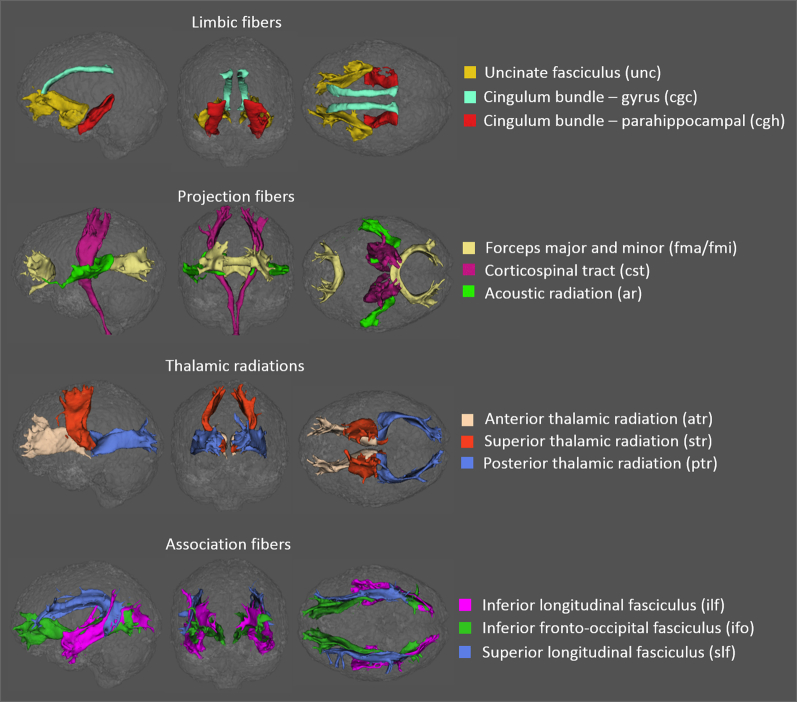
Multishell Diffusion magnetic resonance imaging data were acquired from 100 participants (40 cognitively normal, 38 with subjective cognitive decline, and 22 with mild cognitive impairment [MCI]). White matter microscopic degeneration in 27 major tracts of interest was assessed using diffusion tensor imaging (DTI), neurite orientation dispersion and density imaging, and q-space imaging.
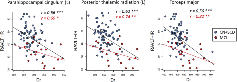
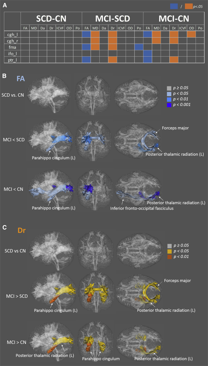
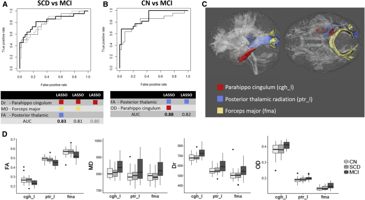
Lower DTI fractional anisotropy and higher radial diffusivity were observed in the cingulum, thalamic radiation, and forceps major of participants with MCI. These tracts of interest also had the highest predictive power to discriminate groups. Diffusion metrics were associated with cognitive performance, particularly Rey Auditory Verbal Learning Test immediate recall, with the highest association observed in participants with MCI.
While DTI was the most sensitive, neurite orientation dispersion and density imaging and q-space imaging complementarily characterized reduced axonal density accompanied with dispersed and less restricted white matter microstructures.
扩散磁共振成像可能有助于在阿尔茨海默病早期对白质退变进行微观特征描述。
从100名参与者(40名认知正常者、38名主观认知衰退者和22名轻度认知障碍[MCI]者)获取多壳层扩散磁共振成像数据。使用扩散张量成像(DTI)、神经突方向离散度和密度成像以及q空间成像评估27条主要感兴趣脑区白质的微观退变情况。
在MCI参与者的扣带束、丘脑辐射和胼胝体压部观察到较低的DTI分数各向异性和较高的径向扩散率。这些感兴趣脑区在区分不同组时也具有最高的预测能力。扩散指标与认知表现相关,尤其是雷伊听觉词语学习测验即时回忆,在MCI参与者中观察到的相关性最高。
虽然DTI最敏感,但神经突方向离散度和密度成像以及q空间成像互补地描述了轴突密度降低,同时伴有白质微观结构的离散和受限程度降低。