Developmental Neurogenomics Unit, Human Genetics Branch, National Institute of Mental Health, Bethesda, MD, 20892, USA.
Department of Medical Biophysics, University of Toronto, Toronto, ON, M5T 1R8, Canada; Neurosciences and Mental Health, the Hospital for Sick Children, Toronto, ON, M5T 3H7, Canada.
Neuroimage. 2020 Jan 1;204:116122. doi: 10.1016/j.neuroimage.2019.116122. Epub 2019 Aug 27.
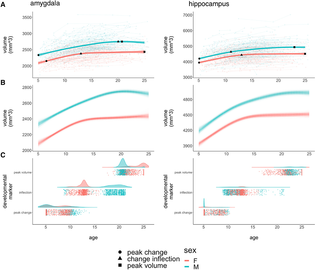
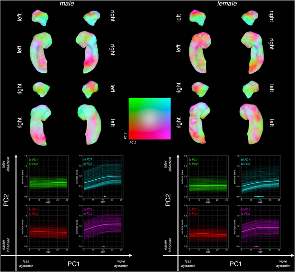
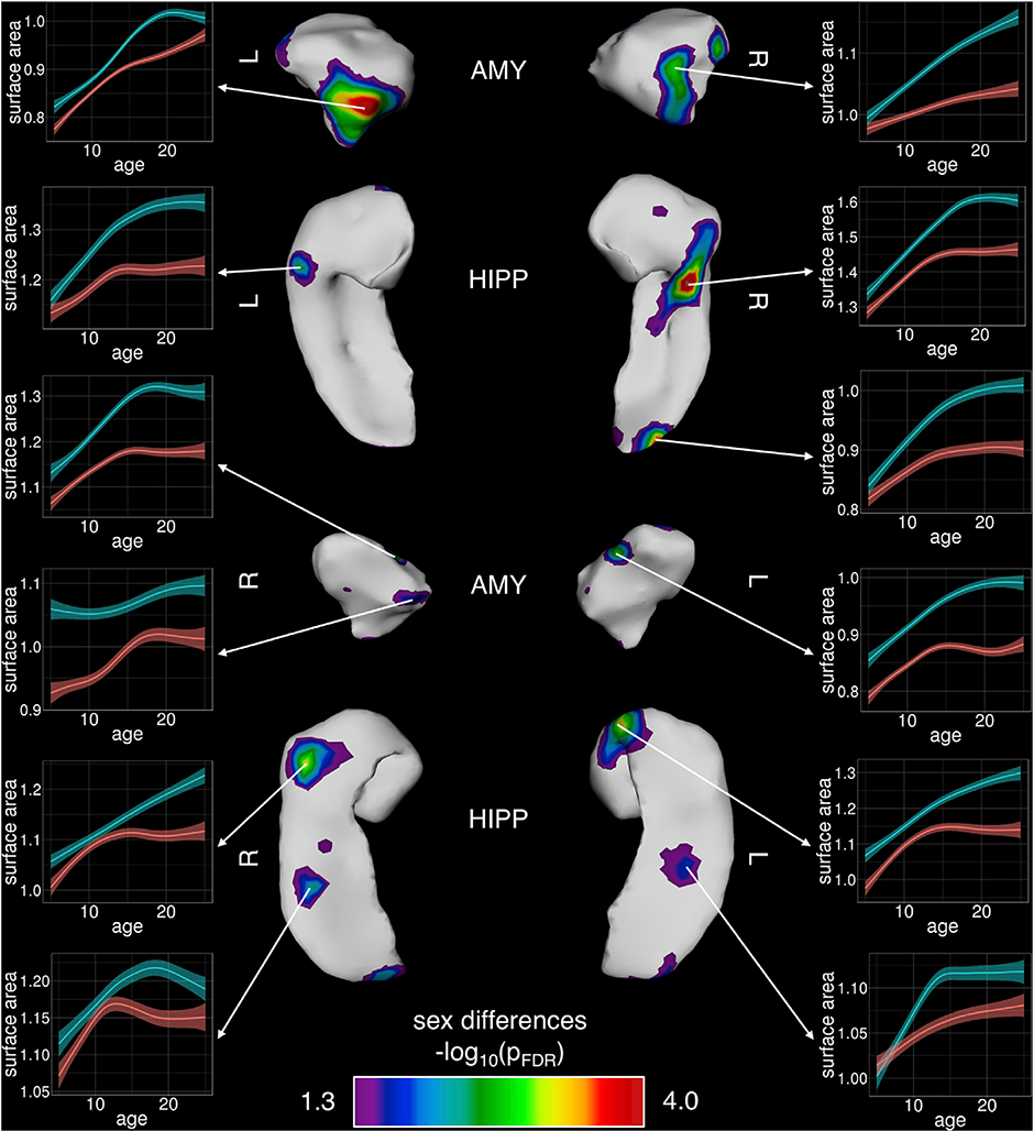
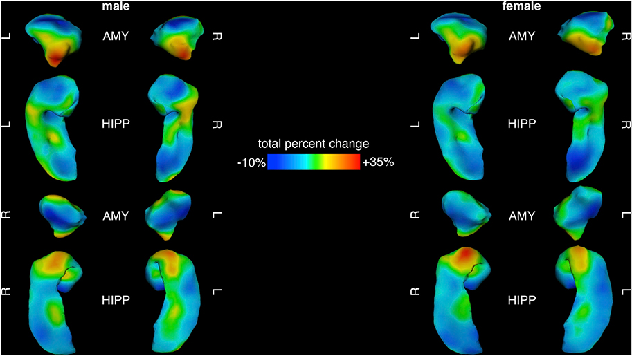
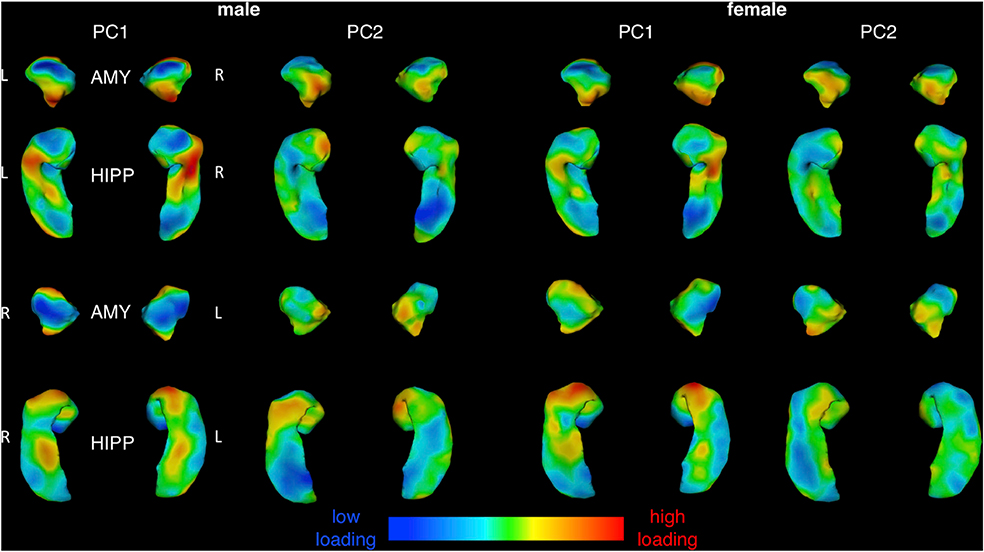
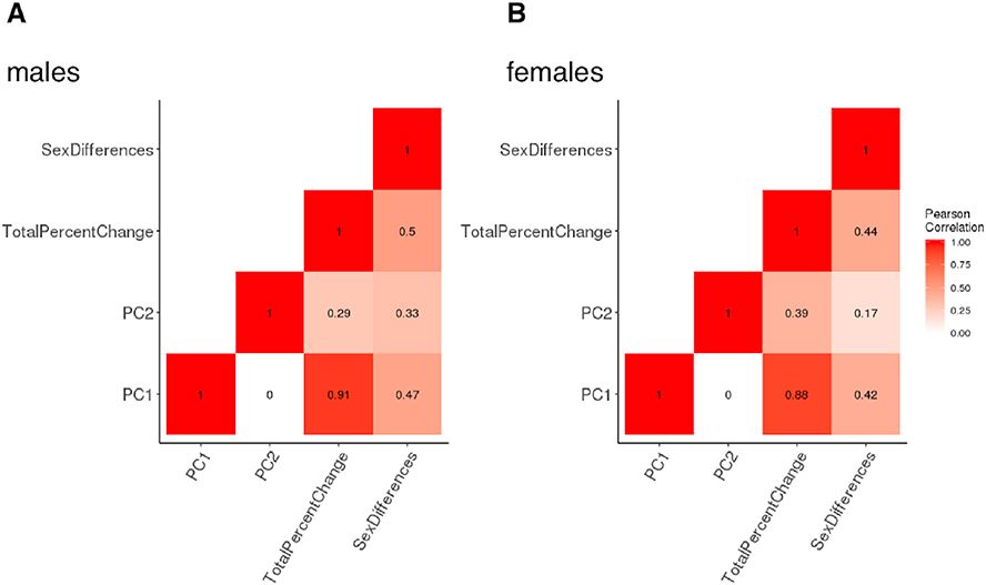
The amygdala and hippocampus are two adjacent allocortical structures implicated in sex-biased and developmentally-emergent psychopathology. However, the spatiotemporal dynamics of amygdalo-hippocampal development remain poorly understood in healthy humans. The current study defined trajectories of volume and shape change for the amygdala and hippocampus by applying a multi-atlas segmentation pipeline (MAGeT-Brain) and semi-parametric mixed-effects spline modeling to 1,529 longitudinally-acquired structural MRI brain scans from a large, single-center cohort of 792 youth (403 males, 389 females) between the ages of 5 and 25 years old. We found that amygdala and hippocampus volumes both follow curvilinear and sexually dimorphic growth trajectories. These sex-biases were particularly striking in the amygdala: males showed a significantly later and slower adolescent deceleration in volume expansion (at age 20 years) than females (age 13 years). Shape analysis localized significant hot-spots of sex-biased anatomical development in sub-regional territories overlying rostral and caudal extremes of the CA1/2 in the hippocampus, and the centromedial nuclear group of the amygdala. In both sexes, principal components analysis revealed close integration of amygdala and hippocampus shape change along two main topographically-organized axes - low vs. high areal expansion, and early vs. late growth deceleration. These results (i) bring greater resolution to our spatiotemporal understanding of amygdalo-hippocampal development in healthy males and females, and (ii) uncover focal sex-differences in the structural maturation of the brain components that may contribute to differences in behavior and psychopathology that emerge during adolescence.
杏仁核和海马体是两个相邻的皮质下结构,与性别偏向和发育出现的精神病理学有关。然而,健康人群中杏仁核-海马体发育的时空动态仍知之甚少。本研究通过应用多图谱分割管道(MAGeT-Brain)和半参数混合效应样条建模,对 792 名 5 至 25 岁的青少年(403 名男性,389 名女性)的 1529 次纵向结构 MRI 脑扫描进行分析,确定了杏仁核和海马体的体积和形状变化轨迹。我们发现杏仁核和海马体的体积都遵循曲线和性别二态性的生长轨迹。这些性别差异在杏仁核中尤为明显:男性的体积扩张青春期减速明显晚且慢(20 岁),而女性(13 岁)则更早。形态分析定位于海马体 CA1/2 头侧和尾侧以及杏仁核中央核团的亚区域内的显著性别偏侧化的解剖发育热点。在两性中,主成分分析揭示了杏仁核和海马体形状变化沿着两个主要的地形组织轴的紧密整合——区域扩张的高低,以及生长减速的早晚。这些结果(i)使我们对健康男性和女性的杏仁核-海马体发育的时空理解更加深入,(ii)揭示了大脑成分结构成熟中的焦点性别差异,这些差异可能导致青春期出现的行为和精神病理学差异。