Sabry Dina, Mohamed Abbas, Monir Manar, Ibrahim Heba A
Department of Medical Biochemistry and Molecular Biology, Faculty of Medicine, Cairo University, Giza, Egypt.
Department of Pathology, Faculty of Medicine, Cairo University, Giza, Egypt.
Int J Stem Cells. 2019 Nov 30;12(3):400-409. doi: 10.15283/ijsc18143.
The release of microvesicles (MVs) from mesenchymal stem cells (MSCs) has been implicated in intercellular communication, and may contribute to beneficial paracrine effects of stem cell-based therapies. We investigated the effect of administration of MSC-MVs on the therapeutic potential of carbon tetrachloride (CCL) induced liver fibrosis in rats.
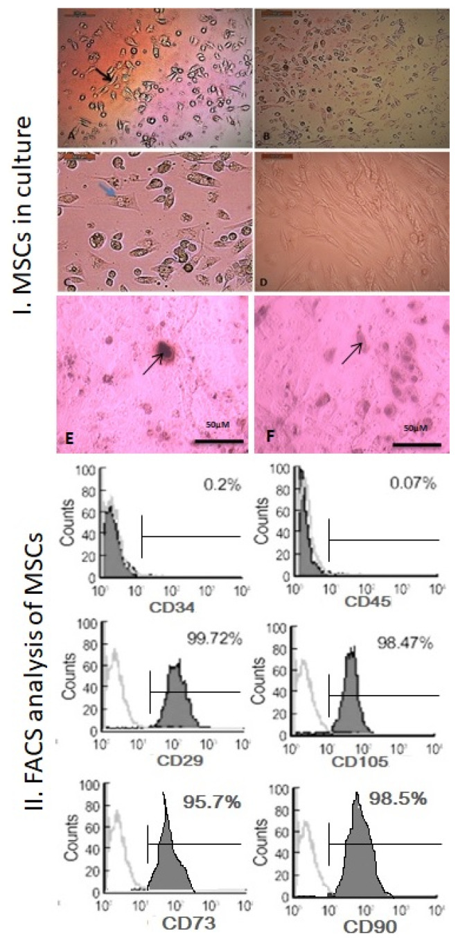
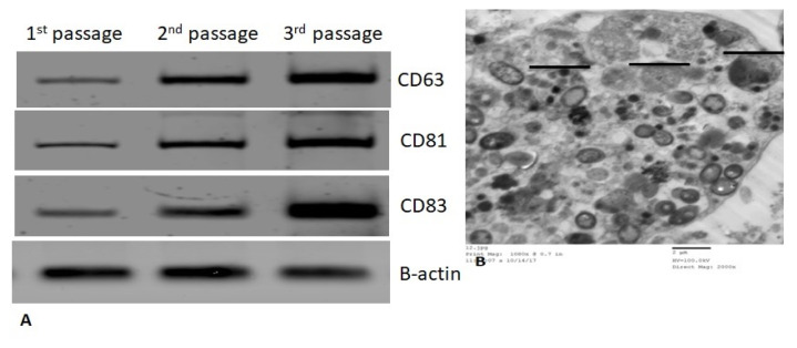
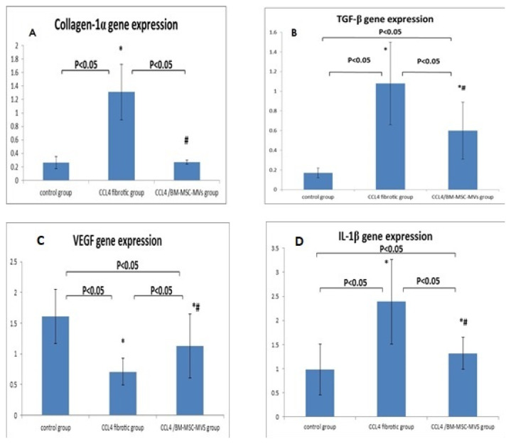
Our work included: isolation and further identification of bone marrow MSC-MVs by transmission electron microscopy (TEM). Liver fibrosis was induced in rats by CCl4 followed by injection of prepared MSC-MVs in injured rats. The effects of MSC-MVs were evaluated by biochemical analysis of liver functions, RNA gene expression quantitation for collagen-1, transforming growth factor (TGF-), interleukin-1 (IL-1), vascular endothelial growth factor (VEGF) by real time reverse transcription PCR (RT-PCR) techniques. Finally histopathological examination of the liver tissues was assessed for all studied groups.
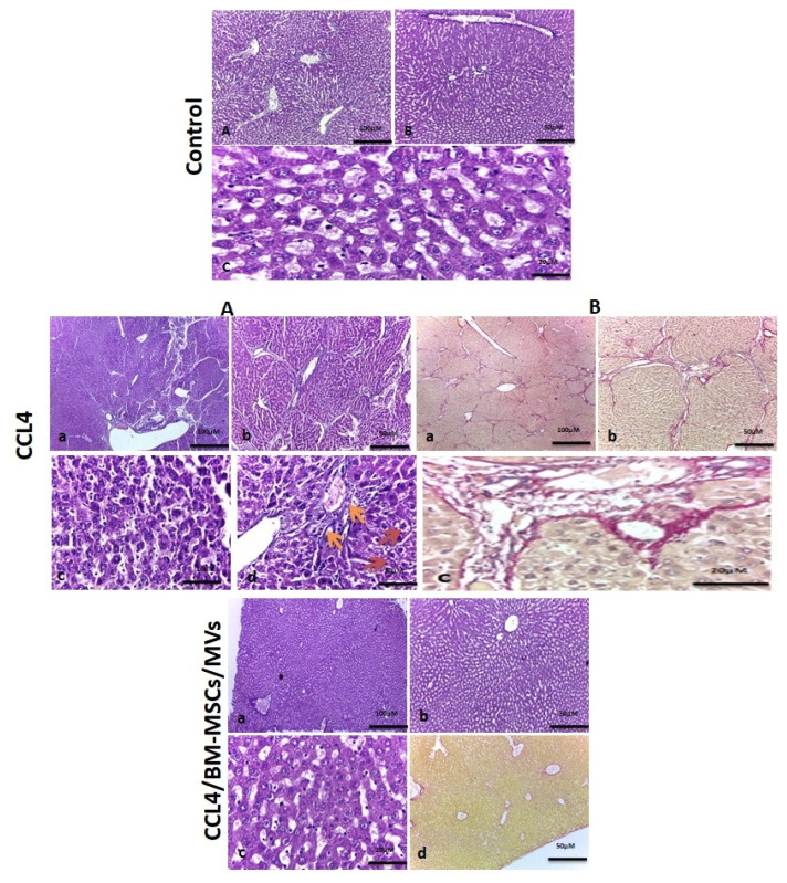
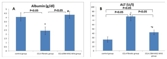
BM-MSC-MVs treated group showed significant increase in serum albumin levels, VEGF quantitative gene expression (p<0.05), while it showed a significant decrease in serum alanine transaminase (ALT) enzyme levels, quantitative gene expression of TGF-, collagen-1, IL-1 compared to CCL fibrotic group (p<0.05). Additionally, the histopathological assessment of the liver tissues of BM-MSC-MVs treated group showed marked decrease in the collagen deposition & improvement of histopathological picture in comparison with CCL fibrotic group.
Our study demonstrates that BM-MSC-MVs possess anti-fibrotic, anti-inflammatory, and pro-angiogenic properties which can promote the resolution of CCL induced liver fibrosis in rats.
间充质干细胞(MSC)释放的微泡(MV)参与细胞间通讯,可能有助于基于干细胞的治疗产生有益的旁分泌效应。我们研究了给予MSC-MV对四氯化碳(CCL)诱导的大鼠肝纤维化治疗潜力的影响。
我们的工作包括:通过透射电子显微镜(TEM)分离并进一步鉴定骨髓MSC-MV。用CCl4诱导大鼠肝纤维化,然后将制备好的MSC-MV注射到受损大鼠体内。通过肝功能生化分析、实时逆转录PCR(RT-PCR)技术对胶原蛋白-1、转化生长因子(TGF-)、白细胞介素-1(IL-1)、血管内皮生长因子(VEGF)进行RNA基因表达定量,评估MSC-MV的作用。最后对所有研究组的肝组织进行组织病理学检查。
与CCL纤维化组相比,BM-MSC-MV治疗组血清白蛋白水平、VEGF定量基因表达显著增加(p<0.05),而血清丙氨酸转氨酶(ALT)酶水平、TGF-、胶原蛋白-1、IL-1的定量基因表达显著降低(p<0.05)。此外,与CCL纤维化组相比,BM-MSC-MV治疗组肝组织的组织病理学评估显示胶原沉积明显减少,组织病理学图像有所改善。
我们的研究表明,BM-MSC-MV具有抗纤维化、抗炎和促血管生成特性,可促进CCL诱导的大鼠肝纤维化的消退。