• 文献检索
  • 文档翻译
  • 深度研究
  • 学术资讯
  • Suppr Zotero 插件Zotero 插件
  • 邀请有礼
  • 套餐&价格
  • 历史记录
应用&插件
Suppr Zotero 插件Zotero 插件浏览器插件Mac 客户端Windows 客户端微信小程序
定价
高级版会员购买积分包购买API积分包
服务
文献检索文档翻译深度研究API 文档MCP 服务
关于我们
关于 Suppr公司介绍联系我们用户协议隐私条款
关注我们

Suppr 超能文献

核心技术专利:CN118964589B侵权必究
粤ICP备2023148730 号-1Suppr @ 2026

文献检索

告别复杂PubMed语法,用中文像聊天一样搜索,搜遍4000万医学文献。AI智能推荐,让科研检索更轻松。

立即免费搜索

文件翻译

保留排版,准确专业,支持PDF/Word/PPT等文件格式,支持 12+语言互译。

免费翻译文档

深度研究

AI帮你快速写综述,25分钟生成高质量综述,智能提取关键信息,辅助科研写作。

立即免费体验

褪黑素预处理骨髓间充质干细胞和脂肪间充质干细胞对肝纤维化的再生和治疗作用比较。

Comparison between the Regenerative and Therapeutic Impacts of Bone Marrow Mesenchymal Stem Cells and Adipose Mesenchymal Stem Cells Pre-Treated with Melatonin on Liver Fibrosis.

机构信息

Department of Anatomy and Histology, College of Medicine, Qassim University, Buraydah 51452, Saudi Arabia.

Department of Anatomy and Embryology, College of Medicine, Cairo University, Cairo 11956, Egypt.

出版信息

Biomolecules. 2024 Mar 1;14(3):297. doi: 10.3390/biom14030297.

DOI:10.3390/biom14030297
PMID:38540717
原文链接:https://pmc.ncbi.nlm.nih.gov/articles/PMC10968153/
Abstract

BACKGROUND

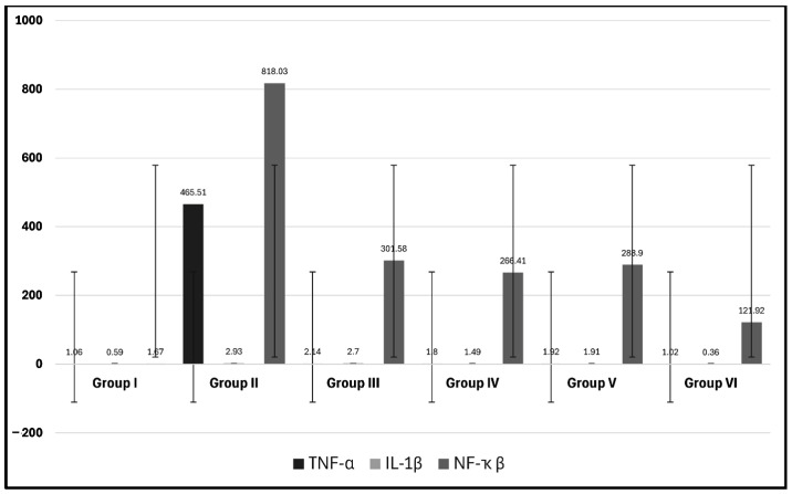
The distinctive feature of liver fibrosis is the progressive replacement of healthy hepatic cells by the extracellular matrix protein, which is abundant in collagen I and III, with impaired matrix remodeling. The activation of myofibroblastic cells enhances the fibrogenic response of complex interactions of hepatic stellate cells, fibroblasts, and inflammatory cells to produce the excessive deposition of the extracellular protein matrix. This process is activated by multiple fibrogenic mediators and cytokines, such as TNF-α and IL-1β, accompanied with a decrease in the anti-fibrogenic factor NF-κβ. Mesenchymal stem cells (MSCs) represent a promising therapy for liver fibrosis, allowing for a more advanced regenerative influence when cultured with extrinsic or intrinsic proliferative factors, cytokines, antioxidants, growth factors, and hormones such as melatonin (MT). However, previous studies showed conflicting findings concerning the therapeutic effects of adipose (AD) and bone marrow (BM) MSCs; therefore, the present work aimed to conduct a comparative and comprehensive study investigating the impact of MT pre-treatment on the immunomodulatory, anti-inflammatory, and anti-apoptotic effects of AD- and BM-MSCs and to critically analyze whether MT-pre-treated AD-MSCs and BM-MSCs reveal equal or different therapeutic and regenerative potentials in a CCl4-injured liver experimental rat model.

MATERIALS AND METHODS

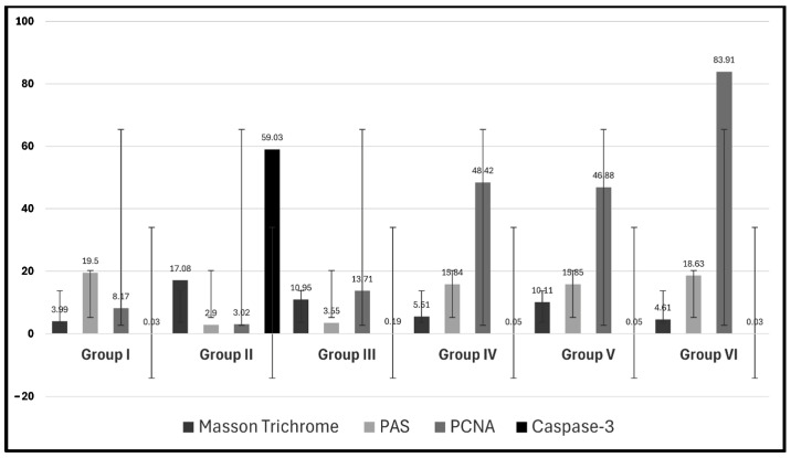
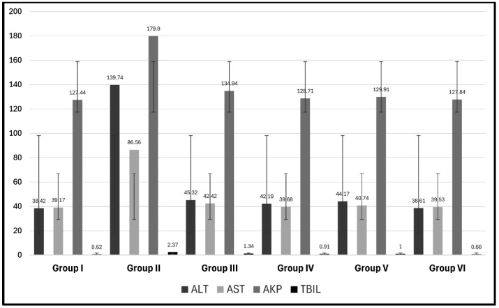
Six groups of experimental rats were used, with ten rats in each group: group I (control group), group II (CCl4-treated group), group III (CCl4- and BM-MSC-treated group), group IV (CCl4 and MT-pre-treated BM-MSC group), group V (CCl4- and AD-MSC-treated group), and group VI (CCl4 and MT-pre-treated AD-MSC group). Liver function tests and the gene expression of inflammatory, fibrogenic, apoptotic, and proliferative factors were analyzed. Histological and immunohistochemical changes were assessed.

RESULTS

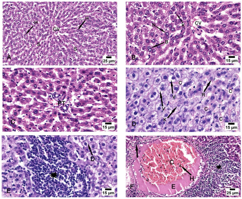
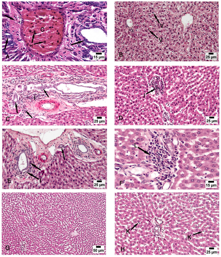
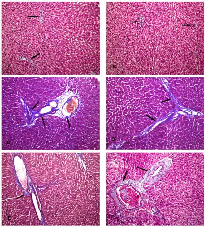
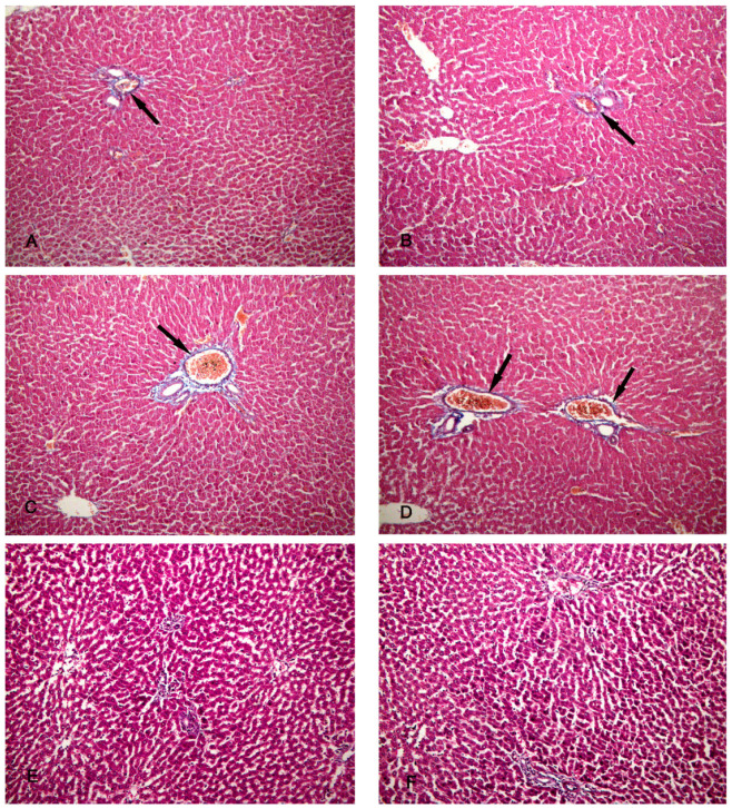
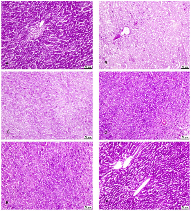
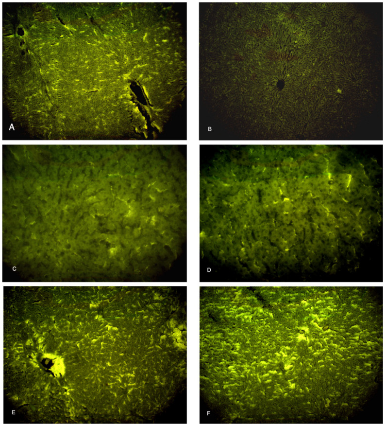
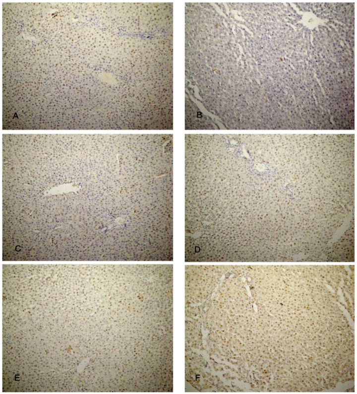
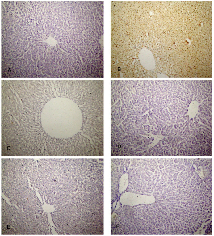
The present study compared the ability of AD- and BM-MSCs, with and without MT pre-treatment, to reduce hepatic fibrosis. Both types of MSCs improved hepatocyte function by reducing the serum levels of ALT, aspartate aminotransferase (AST), alkaline phosphatase (AKP), and total bilirubin (TBIL). In addition, the changes in the hepatocellular architecture, including the hepatocytes, liver sinusoids, central veins, portal veins, biliary ducts, and hepatic arteries, showed a decrease in hepatocyte injury and cholestasis with a reduction in inflammation, apoptosis, and necrosis of the hepatic cells, together with an inhibition of liver tissue fibrosis. These results were augmented by an analysis of the expression of the pro-inflammatory cytokines TNFα and IL-1β, the anti-fibrogenic factor NF-κβ, the apoptotic factor caspase-3, and the proliferative indicators antigen Ki-67 and proliferating cell nuclear antigen (PCNA). These findings were found to be statistically significant, with the restoration of normal parameters in the rats that received AD-MSCs pre-treated with MT, denoting optimal regenerative and therapeutic effects.

CONCLUSIONS

AD-MSCs pre-treated with MT are the preferred choice in improving hepatic fibrosis and promoting the therapeutic and regenerative ability of liver tissue. They represent a very significant tool for future stem cell use in the tissue regeneration strategy for the treatment of liver diseases.

https://cdn.ncbi.nlm.nih.gov/pmc/blobs/a5b8/10968153/8f0e748d3e80/biomolecules-14-00297-g011.jpg
https://cdn.ncbi.nlm.nih.gov/pmc/blobs/a5b8/10968153/42236555d643/biomolecules-14-00297-g001.jpg
https://cdn.ncbi.nlm.nih.gov/pmc/blobs/a5b8/10968153/e96fc294e1a3/biomolecules-14-00297-g002.jpg
https://cdn.ncbi.nlm.nih.gov/pmc/blobs/a5b8/10968153/66ee081880a9/biomolecules-14-00297-g003.jpg
https://cdn.ncbi.nlm.nih.gov/pmc/blobs/a5b8/10968153/3526917d40b3/biomolecules-14-00297-g004.jpg
https://cdn.ncbi.nlm.nih.gov/pmc/blobs/a5b8/10968153/d1d7e2f058a3/biomolecules-14-00297-g005.jpg
https://cdn.ncbi.nlm.nih.gov/pmc/blobs/a5b8/10968153/35b08e706f19/biomolecules-14-00297-g006.jpg
https://cdn.ncbi.nlm.nih.gov/pmc/blobs/a5b8/10968153/1f49dd185671/biomolecules-14-00297-g007.jpg
https://cdn.ncbi.nlm.nih.gov/pmc/blobs/a5b8/10968153/366cc102196d/biomolecules-14-00297-g008.jpg
https://cdn.ncbi.nlm.nih.gov/pmc/blobs/a5b8/10968153/5dd76fe86a28/biomolecules-14-00297-g009.jpg
https://cdn.ncbi.nlm.nih.gov/pmc/blobs/a5b8/10968153/dcb4e585602c/biomolecules-14-00297-g010.jpg
https://cdn.ncbi.nlm.nih.gov/pmc/blobs/a5b8/10968153/8f0e748d3e80/biomolecules-14-00297-g011.jpg
摘要

背景

肝纤维化的显著特征是健康的肝细胞被细胞外基质蛋白逐渐取代,细胞外基质蛋白富含 I 型和 III 型胶原,基质重塑受损。肌成纤维细胞的激活增强了肝星状细胞、成纤维细胞和炎症细胞之间复杂相互作用的纤维生成反应,导致细胞外蛋白基质的过度沉积。这一过程被多种纤维生成介质和细胞因子激活,如 TNF-α 和 IL-1β,并伴有抗纤维生成因子 NF-κβ 的减少。间充质干细胞(MSCs)是肝纤维化有前途的治疗方法,当与外源性或内源性增殖因子、细胞因子、抗氧化剂、生长因子和激素(如褪黑素(MT))一起培养时,允许更先进的再生影响。然而,先前的研究对脂肪(AD)和骨髓(BM)MSC 的治疗效果存在相互矛盾的发现;因此,本研究旨在进行一项比较和综合研究,调查 MT 预处理对 AD 和 BM-MSCs 的免疫调节、抗炎和抗细胞凋亡作用的影响,并批判性分析 MT 预处理的 AD-MSCs 和 BM-MSCs 是否在 CCl4 损伤的大鼠实验模型中显示出相同或不同的治疗和再生潜力。

材料和方法

使用六组实验大鼠,每组 10 只:第 I 组(对照组)、第 II 组(CCl4 处理组)、第 III 组(CCl4 和 BM-MSC 处理组)、第 IV 组(CCl4 和 MT 预处理 BM-MSC 组)、第 V 组(CCl4 和 AD-MSC 处理组)和第 VI 组(CCl4 和 MT 预处理 AD-MSC 组)。分析肝功能试验和炎症、纤维生成、细胞凋亡和增殖因子的基因表达。评估组织学和免疫组织化学变化。

结果

本研究比较了 AD 和 BM-MSCs,以及未经 MT 预处理的 MSC,减少肝纤维化的能力。两种类型的 MSC 都通过降低 ALT、天冬氨酸转氨酶(AST)、碱性磷酸酶(AKP)和总胆红素(TBIL)的血清水平来改善肝细胞功能。此外,肝细胞、肝窦、中央静脉、门静脉、胆管和肝动脉的肝组织结构变化表明,通过减少炎症、细胞凋亡和坏死以及抑制肝组织纤维化,肝细胞损伤和胆汁淤积减少。这种变化通过分析促炎细胞因子 TNFα 和 IL-1β、抗纤维生成因子 NF-κβ、凋亡因子 caspase-3 以及增殖指标抗原 Ki-67 和增殖细胞核抗原(PCNA)的表达得到了增强。这些发现具有统计学意义,接受 MT 预处理的 AD-MSCs 恢复了正常参数,表明具有最佳的再生和治疗效果。

结论

MT 预处理的 AD-MSCs 是改善肝纤维化和促进肝组织治疗和再生能力的首选方法。它们为未来干细胞在肝脏疾病组织再生策略中的应用提供了非常重要的工具。

相似文献

1
Comparison between the Regenerative and Therapeutic Impacts of Bone Marrow Mesenchymal Stem Cells and Adipose Mesenchymal Stem Cells Pre-Treated with Melatonin on Liver Fibrosis.褪黑素预处理骨髓间充质干细胞和脂肪间充质干细胞对肝纤维化的再生和治疗作用比较。
Biomolecules. 2024 Mar 1;14(3):297. doi: 10.3390/biom14030297.
2
The role of the adipose tissue-derived mesenchymal stem cells enriched with melatonin in pancreatic cellular regeneration.富含褪黑素的脂肪组织来源间充质干细胞在胰腺细胞再生中的作用。
Folia Morphol (Warsz). 2022;81(4):931-941. doi: 10.5603/FM.a2021.0093. Epub 2021 Sep 21.
3
Comparison of bone marrow-vs. adipose tissue-derived mesenchymal stem cells for attenuating liver fibrosis.骨髓源性与脂肪组织源性间充质干细胞对减轻肝纤维化作用的比较
Exp Ther Med. 2017 Dec;14(6):5956-5964. doi: 10.3892/etm.2017.5333. Epub 2017 Oct 18.
4
Preconditioning with melatonin improves therapeutic outcomes of bone marrow-derived mesenchymal stem cells in targeting liver fibrosis induced by CCl4.褪黑素预处理可改善骨髓间充质干细胞靶向 CCl4 诱导肝纤维化的治疗效果。
Cell Tissue Res. 2017 Aug;369(2):303-312. doi: 10.1007/s00441-017-2604-1. Epub 2017 Apr 17.
5
The Role of Mesenchymal Stem Cells with Ascorbic Acid and N-Acetylcysteine on TNF-, IL 1, and NF- Expressions in Acute Pancreatitis in Albino Rats.维生素 C 和 N-乙酰半胱氨酸对骨髓间充质干细胞在急性胰腺炎中 TNF-α、IL-1 和 NF-κB 表达的作用。
J Diabetes Res. 2021 Oct 16;2021:6229460. doi: 10.1155/2021/6229460. eCollection 2021.
6
The therapeutic effects of bone marrow-derived mesenchymal stem cells and simvastatin in a rat model of liver fibrosis.骨髓间充质干细胞和辛伐他汀对肝纤维化大鼠模型的治疗作用。
Cell Biochem Biophys. 2014 Jan;68(1):111-25. doi: 10.1007/s12013-013-9698-1.
7
Extracellular vesicles derived from human embryonic stem cell-MSCs ameliorate cirrhosis in thioacetamide-induced chronic liver injury.人胚胎干细胞-MSCs 来源的细胞外囊泡改善硫代乙酰胺诱导的慢性肝损伤性肝硬化。
J Cell Physiol. 2018 Dec;233(12):9330-9344. doi: 10.1002/jcp.26413. Epub 2018 Jun 19.
8
Treatment Efficiency of Different Routes of Bone Marrow-Derived Mesenchymal Stem Cell Injection in Rat Liver Fibrosis Model.不同途径注射骨髓间充质干细胞对大鼠肝纤维化模型的治疗效果
Cell Physiol Biochem. 2018;48(5):2161-2171. doi: 10.1159/000492558. Epub 2018 Aug 16.
9
Human umbilical cord-derived mesenchymal stem cells attenuate hepatic stellate cells activation and liver fibrosis.人脐带间充质干细胞可减轻肝星状细胞的活化和肝纤维化。
Mol Biol Rep. 2024 Jun 14;51(1):734. doi: 10.1007/s11033-024-09664-6.
10
Transplantation of bone marrow mesenchymal stromal cells attenuates liver fibrosis in mice by regulating macrophage subtypes.骨髓间充质基质细胞移植通过调节巨噬细胞亚型减轻小鼠肝纤维化。
Stem Cell Res Ther. 2019 Jan 11;10(1):16. doi: 10.1186/s13287-018-1122-8.

引用本文的文献

1
Extracellular vesicles from melatonin-preconditioned mesenchymal stromal cells protect human umbilical vein endothelial cells against hypoxia/reoxygenation detected by UHPLC-QE-MS/MS untargeted metabolic profiling.褪黑素预处理的间充质基质细胞分泌的细胞外囊泡可保护人脐静脉内皮细胞免受缺氧/复氧损伤,这是通过超高效液相色谱-四极杆静电场轨道阱高分辨质谱非靶向代谢谱检测得出的。
Cell Transplant. 2025 Jan-Dec;34:9636897251347389. doi: 10.1177/09636897251347389. Epub 2025 Jun 13.
2
Mesenchymal Stem Cells Loaded in Injectable Alginate Hydrogels Promote Liver Growth and Attenuate Liver Fibrosis in Cirrhotic Rats.负载于可注射藻酸盐水凝胶中的间充质干细胞促进肝硬化大鼠肝脏生长并减轻肝纤维化
Gels. 2025 Mar 27;11(4):250. doi: 10.3390/gels11040250.
3

本文引用的文献

1
Inflammation and aging: signaling pathways and intervention therapies.炎症与衰老:信号通路与干预治疗。
Signal Transduct Target Ther. 2023 Jun 8;8(1):239. doi: 10.1038/s41392-023-01502-8.
2
Comparative effectiveness of adipose-derived mesenchymal stromal cells in the management of knee osteoarthritis: A meta-analysis.脂肪来源间充质基质细胞治疗膝关节骨关节炎的比较疗效:一项荟萃分析。
World J Orthop. 2023 Jan 18;14(1):23-41. doi: 10.5312/wjo.v14.i1.23.
3
The Molecular Mechanisms of Liver Fibrosis and Its Potential Therapy in Application.
Reduced glutathione enhances adipose tissue-derived mesenchymal stem cell engraftment efficiency for liver fibrosis by targeting TGFβ1/SMAD3/NOX4 pathway.
还原型谷胱甘肽通过靶向TGFβ1/SMAD3/NOX4信号通路提高脂肪组织来源间充质干细胞对肝纤维化的植入效率。
Bioeng Transl Med. 2024 Dec 10;10(2):e10735. doi: 10.1002/btm2.10735. eCollection 2025 Mar.
4
Hotspots and trends in stem cell therapy for liver fibrosis and cirrhosis: A bibliometric analysis.肝纤维化和肝硬化干细胞治疗的热点与趋势:一项文献计量分析
World J Hepatol. 2025 Jan 27;17(1):96105. doi: 10.4254/wjh.v17.i1.96105.
5
Clinical outcomes of autologous adipose-derived mesenchymal stem cell combined with high tibial osteotomy for knee osteoarthritis are correlated with stem cell stemness and senescence.自体脂肪来源间充质干细胞联合胫骨高位截骨术治疗膝骨关节炎的临床疗效与干细胞干性和衰老相关。
J Transl Med. 2024 Nov 18;22(1):1039. doi: 10.1186/s12967-024-05814-3.
肝脏纤维化的分子机制及其在应用中的潜在治疗方法。
Int J Mol Sci. 2022 Oct 20;23(20):12572. doi: 10.3390/ijms232012572.
4
Mesenchymal stem cells improve liver fibrosis and protect hepatocytes by promoting microRNA-148a-5p-mediated inhibition of Notch signaling pathway.间充质干细胞通过促进 microRNA-148a-5p 介导的 Notch 信号通路抑制来改善肝纤维化并保护肝细胞。
Stem Cell Res Ther. 2022 Jul 26;13(1):354. doi: 10.1186/s13287-022-03030-8.
5
Stem cells for treatment of liver fibrosis/cirrhosis: clinical progress and therapeutic potential.用于治疗肝纤维化/肝硬化的干细胞:临床进展和治疗潜力。
Stem Cell Res Ther. 2022 Jul 26;13(1):356. doi: 10.1186/s13287-022-03041-5.
6
Combined effect of hydrogen sulfide and mesenchymal stem cells on mitigating liver fibrosis induced by bile duct ligation: Role of anti-inflammatory, anti-oxidant, anti-apoptotic, and anti-fibrotic biomarkers.硫化氢与间充质干细胞联合减轻胆管结扎诱导的肝纤维化的作用:抗炎、抗氧化、抗凋亡和抗纤维化生物标志物的作用
Iran J Basic Med Sci. 2021 Dec;24(12):1753-1762. doi: 10.22038/IJBMS.2021.56477.12604.
7
Mesenchymal stem cell-originated exosomal circDIDO1 suppresses hepatic stellate cell activation by miR-141-3p/PTEN/AKT pathway in human liver fibrosis.间质干细胞来源的外泌体 circDIDO1 通过 miR-141-3p/PTEN/AKT 通路抑制人肝纤维化中肝星状细胞的激活。
Drug Deliv. 2022 Dec;29(1):440-453. doi: 10.1080/10717544.2022.2030428.
8
Targeting the Wnt Signaling Pathway in Liver Fibrosis for Drug Options: An Update.针对肝纤维化中Wnt信号通路的药物选择:最新进展
J Clin Transl Hepatol. 2021 Dec 28;9(6):960-971. doi: 10.14218/JCTH.2021.00065. Epub 2021 Sep 13.
9
Mesenchymal stem cell therapy in decompensated liver cirrhosis: a long-term follow-up analysis of the randomized controlled clinical trial.间质干细胞治疗失代偿期肝硬化:一项随机对照临床试验的长期随访分析。
Hepatol Int. 2021 Dec;15(6):1431-1441. doi: 10.1007/s12072-021-10199-2. Epub 2021 Nov 29.
10
The Role of Mesenchymal Stem Cells with Ascorbic Acid and N-Acetylcysteine on TNF-, IL 1, and NF- Expressions in Acute Pancreatitis in Albino Rats.维生素 C 和 N-乙酰半胱氨酸对骨髓间充质干细胞在急性胰腺炎中 TNF-α、IL-1 和 NF-κB 表达的作用。
J Diabetes Res. 2021 Oct 16;2021:6229460. doi: 10.1155/2021/6229460. eCollection 2021.