Wang Xin, Feng Hao, Wang Yu, Zhou Jian, Zhao Xingquan
Department of Neurology, Beijing Tiantan Hospital, Capital Medical University, Beijing, China.
Department of Radiology, Beijing Tiantan Hospital, Capital Medical University, Beijing, China.
Front Neurol. 2019 Aug 14;10:881. doi: 10.3389/fneur.2019.00881. eCollection 2019.
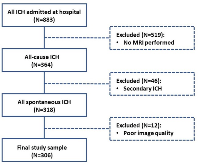
Cerebral small vessel disease (SVD) is associated with cognitive decline, depression, increased mortality, and disability in stroke patients. MRI-visible perivascular spaces (PVS) are a sensitive neuroimaging marker of SVD. We aimed to explore the risk factors and associations with other SVD markers of PVS in two topographical regions (in the basal ganglia [BG] and centrum semiovale [CS]) in a cohort of spontaneous intracerebral hemorrhage (ICH) patients. We included 306 consecutive patients from a prospective spontaneous ICH cohort. We rated PVS, white matter hyperintensities (WMH), cerebral microbleeds (CMB), and lacunes with validated visual rating scale. We collected clinical information using standardized forms. We predefined severe PVS as score > 2 and examined associations between PVS in both BG and CS regions and clinical and imaging markers of SVD by logistic regression. In the multivariable logistic regression, increasing age (OR = 1.075; 95% CI = 1.038-1.113, < 0.001), high CS PVS degrees (OR = 6.906; 95% CI = 3.024-15.774, < 0.001), extensive periventricular WMH (OR = 2.878; 95% CI = 1.298-6.379, = 0.009), and the presence of CMB (OR = 4.073, 95% CI = 1.869-8.877, < 0.001) were independently associated with BG PVS severity. Alcohol-drinking habit (OR = 2.805; 95% CI = 1.451-5.422, = 0.002), hyperlipidemia history (OR = 3.782; 95% CI = 1.582-8.783, = 0.003), high BG PVS degrees (OR = 6.293; 95% CI = 2.755-14.371, < 0.001) and the presence of strictly lobar CMB (OR = 2.556, 95% CI = 1.285-5.085, = 0.008) were independent predictors of increased CS PVS severity. MRI-visible PVS in BG and CS regions are inter-related and have different risk factors in spontaneous ICH patients. Further studies are needed to explore the mechanism and clinical importance of PVS, with possible implications for cerebrovascular disease prevention and effective treatments.
脑小血管病(SVD)与认知功能下降、抑郁、死亡率增加以及卒中患者的残疾有关。磁共振成像(MRI)可见的血管周围间隙(PVS)是SVD的一种敏感神经影像学标志物。我们旨在探讨一组自发性脑出血(ICH)患者两个解剖区域(基底节[BG]和半卵圆中心[CS])中PVS的危险因素及其与其他SVD标志物的关联。我们纳入了来自一个前瞻性自发性ICH队列的306例连续患者。我们使用经过验证的视觉评分量表对PVS、白质高信号(WMH)、脑微出血(CMB)和腔隙进行评分。我们使用标准化表格收集临床信息。我们将严重PVS预先定义为评分>2,并通过逻辑回归分析BG和CS区域的PVS与SVD的临床和影像学标志物之间的关联。在多变量逻辑回归中,年龄增加(比值比[OR]=1.075;95%置信区间[CI]=1.038 - 1.113,P<0.001)、CS区PVS程度高(OR = 6.906;95% CI = 3.024 - 15.774,P<0.001)、广泛的脑室周围WMH(OR = 2.878;95% CI = 1.298 - 6.379,P = 0.009)以及存在CMB(OR = 4.073,95% CI = 1.869 - 8.877,P<0.001)与BG区PVS严重程度独立相关。饮酒习惯(OR = 2.805;95% CI = 1.451 - 5.422,P = 0.002)、高脂血症病史(OR = 3.782;95% CI = 1.582 - 8.783,P = 0.003)、BG区PVS程度高(OR = 6.293;95% CI = 2.755 - 14.371,P<0.001)以及存在严格的叶性CMB(OR = 2.556,95% CI = 1.285 - 5.085,P = 0.008)是CS区PVS严重程度增加的独立预测因素。BG和CS区域中MRI可见的PVS相互关联,且在自发性ICH患者中有不同的危险因素。需要进一步研究以探索PVS的机制和临床重要性,这可能对脑血管疾病的预防和有效治疗具有启示意义。