Potter Gillian M, Chappell Francesca M, Morris Zoe, Wardlaw Joanna M
Brain Imaging Research Centre, University of Edinburgh, Edinburgh, UK.
Cerebrovasc Dis. 2015;39(3-4):224-31. doi: 10.1159/000375153. Epub 2015 Mar 19.
Perivascular spaces (PVS) are an important component of cerebral small vessel disease (SVD), several inflammatory disorders, hypertension and blood-brain barrier breakdown, but are difficult to quantify. A recent international collaboration of SVD experts has highlighted the need for a robust, easy-to-use PVS rating scale for the effective investigation of the diagnostic and prognostic significance of PVS. The purpose of the current study was to develop and extend existing PVS scales to provide a more comprehensive scale for the measurement of PVS in the basal ganglia, centrum semiovale and midbrain, and to test its intra- and inter-rater agreement, assessing reasons for discrepancy.
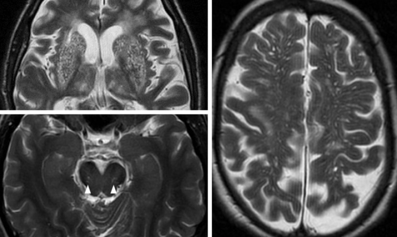
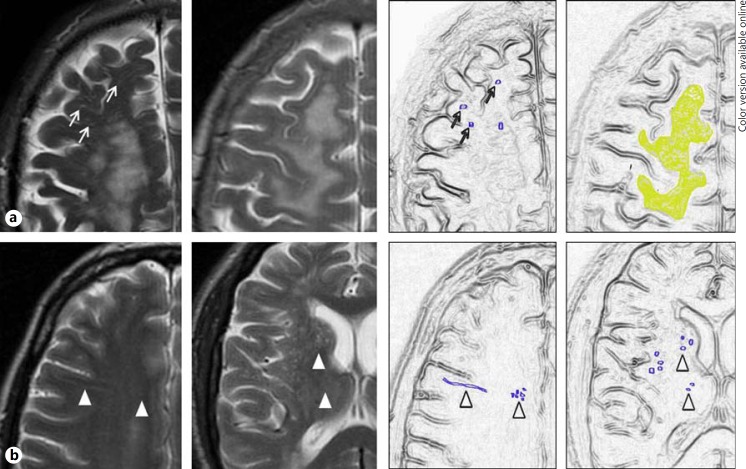
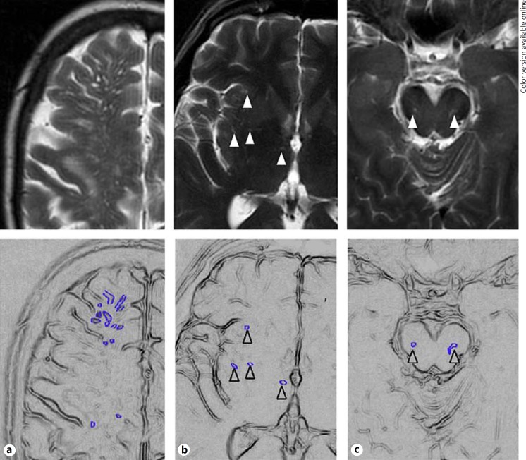
We reviewed previously published PVS scales, including site of PVS assessed, rating method, and size and morphological criteria. Retaining key features, we devised a more comprehensive scale in order to improve the reliability of PVS rating. Two neuroradiologists tested the new scale in MRI brain scans of 60 patients from two studies (stroke, ageing population), chosen to represent a full range of PVS, and demonstrating concomitant features of SVD such as lacunes and white matter hyperintensities. We rated basal ganglia, centrum semiovale, and midbrain PVS. Basal ganglia and centrum semiovale PVS were rated 0 (none), 1 (1-10), 2 (11-20), 3 (21-40) and 4 (>40), and midbrain PVS were rated 0 (none visible) or 1 (visible). We calculated kappa statistics for rating, assessed consistency in use of PVS categories (Bhapkar test) and reviewed sources of discrepancy.
Intra- and inter-rater kappa statistics were highest for basal ganglia PVS (range 0.76-0.87 and 0.8-0.9, respectively) than for centrum semiovale PVS (range 0.68-0.75 and 0.61-0.8, respectively) or midbrain PVS (inter-rater range 0.51-0.52). Inter-rater consistency was better for basal ganglia compared to centrum semiovale PVS (Bhapkar statistic 2.49-3.72, compared to 6.79-21.08, respectively). Most inter-rater disagreements were due to very faint PVS, coexisting extensive white matter hyperintensities (WMH) or the presence of lacunes.
We developed a more inclusive and robust visual PVS rating scale allowing rating of all grades of PVS severity on structural brain imaging. The revised PVS rating scale has good observer reliability for basal ganglia and centrum semiovale PVS, best for basal ganglia PVS, and moderate reliability for midbrain PVS. Agreement is influenced by PVS severity and the presence of background features of SVD. The current scale can be used in further studies to assess the clinical implications of PVS.
脑周血管间隙(PVS)是脑小血管病(SVD)、多种炎症性疾病、高血压和血脑屏障破坏的重要组成部分,但难以量化。近期SVD专家的一项国际合作强调,需要一种强大且易于使用的PVS评分量表,以有效研究PVS的诊断和预后意义。本研究的目的是开发并扩展现有的PVS量表,以提供一个更全面的量表来测量基底节、半卵圆中心和中脑的PVS,并测试其评分者内和评分者间的一致性,评估差异原因。
我们回顾了先前发表的PVS量表,包括评估的PVS部位、评分方法以及大小和形态学标准。保留关键特征后,我们设计了一个更全面的量表,以提高PVS评分的可靠性。两名神经放射科医生在两项研究(中风、老年人群)的60例患者的脑部MRI扫描中测试了新量表,这些患者被选来代表各种程度的PVS,并显示出SVD的伴随特征,如腔隙和白质高信号。我们对基底节、半卵圆中心和中脑的PVS进行评分。基底节和半卵圆中心的PVS评分为0(无)、1(1 - 10)、2(11 - 20)、3(21 - 40)和4(>40),中脑的PVS评分为0(不可见)或1(可见)。我们计算评分的kappa统计量,评估PVS类别使用的一致性(Bhapkar检验)并审查差异来源。
基底节PVS的评分者内和评分者间kappa统计量(分别为0.76 - 0.87和0.8 - 0.9)高于半卵圆中心PVS(分别为0.68 - 0.75和0.61 - 0.8)或中脑PVS(评分者间范围为0.51 - 0.52)。与半卵圆中心PVS相比,基底节的评分者间一致性更好(Bhapkar统计量分别为2.49 - 3.72和6.79 - 21.08)。大多数评分者间的分歧是由于非常微弱的PVS、共存的广泛白质高信号(WMH)或腔隙的存在。
我们开发了一种更具包容性和稳健性的视觉PVS评分量表,可在结构性脑成像上对所有等级的PVS严重程度进行评分。修订后的PVS评分量表对基底节和半卵圆中心PVS具有良好的观察者可靠性,对基底节PVS最佳,对中脑PVS具有中等可靠性。一致性受PVS严重程度和SVD背景特征的影响。当前量表可用于进一步研究以评估PVS的临床意义。