• 文献检索
  • 文档翻译
  • 深度研究
  • 学术资讯
  • Suppr Zotero 插件Zotero 插件
  • 邀请有礼
  • 套餐&价格
  • 历史记录
应用&插件
Suppr Zotero 插件Zotero 插件浏览器插件Mac 客户端Windows 客户端微信小程序
定价
高级版会员购买积分包购买API积分包
服务
文献检索文档翻译深度研究API 文档MCP 服务
关于我们
关于 Suppr公司介绍联系我们用户协议隐私条款
关注我们

Suppr 超能文献

核心技术专利:CN118964589B侵权必究
粤ICP备2023148730 号-1Suppr @ 2026

文献检索

告别复杂PubMed语法,用中文像聊天一样搜索,搜遍4000万医学文献。AI智能推荐,让科研检索更轻松。

立即免费搜索

文件翻译

保留排版,准确专业,支持PDF/Word/PPT等文件格式,支持 12+语言互译。

免费翻译文档

深度研究

AI帮你快速写综述,25分钟生成高质量综述,智能提取关键信息,辅助科研写作。

立即免费体验

线粒体通过一种被细胞质 Ca 拮抗的机制向 ER 供应 ATP。

Mitochondria supply ATP to the ER through a mechanism antagonized by cytosolic Ca.

机构信息

Degenerative Diseases Program, SBP Medical Discovery Institute, La Jolla, United States.

Molecular Biology and Biochemistry, Gottfried Schatz Research Center, Medical University of Graz, Graz, Austria.

出版信息

Elife. 2019 Sep 9;8:e49682. doi: 10.7554/eLife.49682.

DOI:10.7554/eLife.49682
PMID:31498082
原文链接:https://pmc.ncbi.nlm.nih.gov/articles/PMC6763289/
Abstract

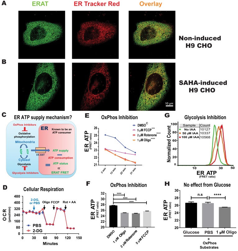
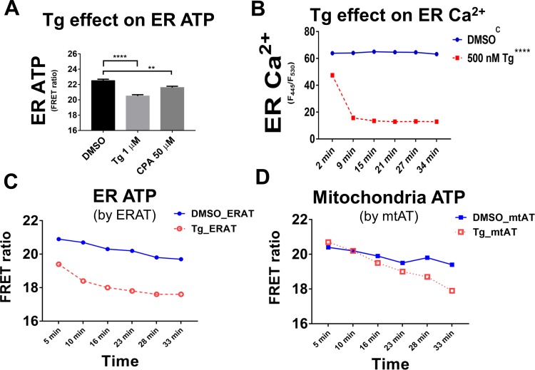
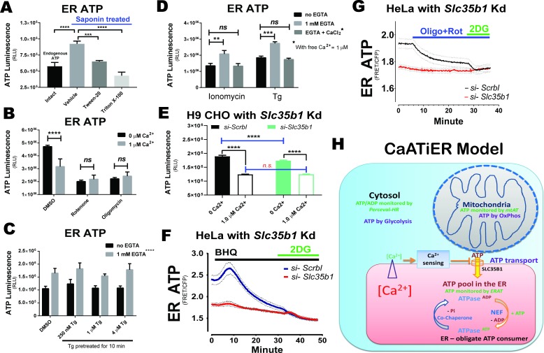
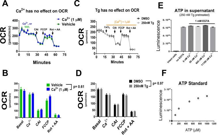
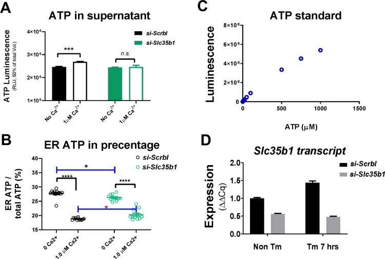
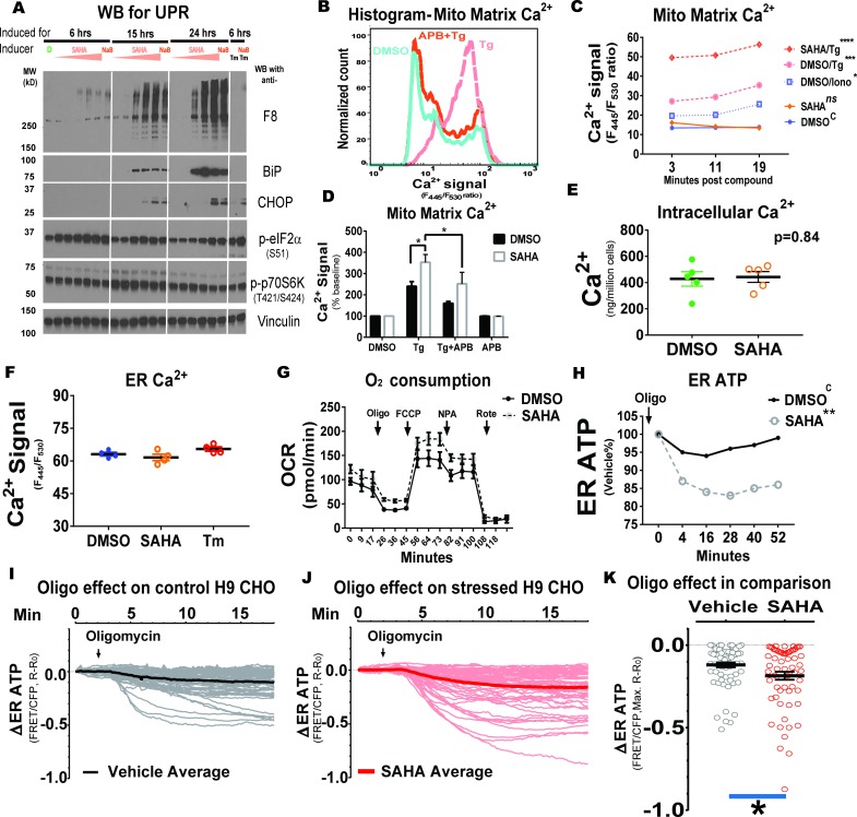
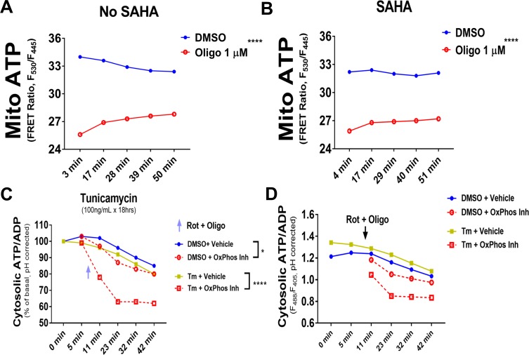
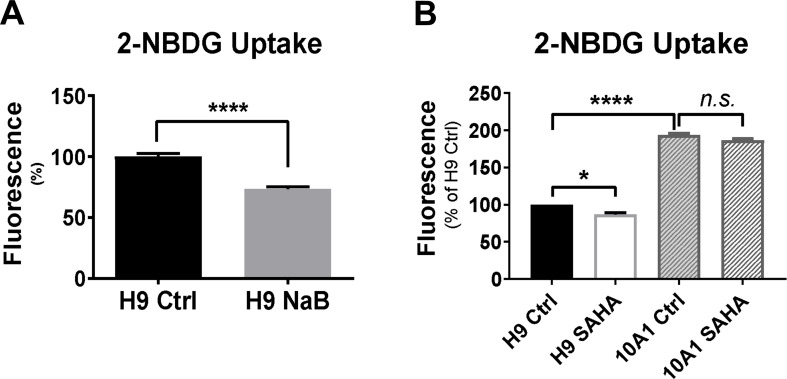
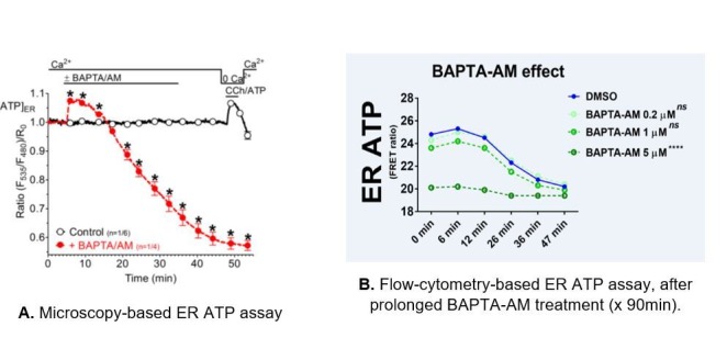
The endoplasmic reticulum () imports ATP and uses energy from ATP hydrolysis for protein folding and trafficking. However, little is known about how this vital ATP transport occurs across the ER membrane. Here, using three commonly used cell lines (CHO, INS1 and HeLa), we report that ATP enters the ER lumen through a cytosolic Ca-antagonized mechanism, or (-ntagonized ransport nto ). Significantly, we show that mitochondria supply ATP to the ER and a SERCA-dependent Ca gradient across the ER membrane is necessary for ATP transport into the ER, through SLC35B1/AXER. We propose that under physiological conditions, increases in cytosolic Ca inhibit ATP import into the ER lumen to limit ER ATP consumption. Furthermore, the ATP level in the ER is readily depleted by oxidative phosphorylation () inhibitors and that ER protein misfolding increases ATP uptake from mitochondria into the ER. These findings suggest that ATP usage in the ER may increase mitochondrial OxPhos while decreasing glycolysis, i.e. an '' effect.

摘要

内质网 () 通过利用 ATP 水解产生的能量来输入 ATP,并进行蛋白质折叠和运输。然而,人们对于这种至关重要的 ATP 跨内质网膜运输的机制知之甚少。在这里,我们使用三种常用的细胞系(CHO、INS1 和 HeLa),报告了 ATP 通过细胞质 Ca 拮抗剂机制或 (-ntagonized ransport nto ) 进入内质网腔。重要的是,我们表明线粒体为内质网提供 ATP,并且内质网膜上的 SERCA 依赖性 Ca 梯度对于通过 SLC35B1/AXER 将 ATP 转运到内质网中是必需的。我们提出,在生理条件下,细胞质 Ca 的增加会抑制 ATP 向内质网腔的输入,以限制内质网中 ATP 的消耗。此外,氧化磷酸化 () 抑制剂会迅速耗尽内质网中的 ATP,并且内质网中蛋白质的错误折叠会增加线粒体向内质网摄取 ATP。这些发现表明,内质网中 ATP 的使用可能会增加线粒体的 OxPhos,同时减少糖酵解,即“效”应。

https://cdn.ncbi.nlm.nih.gov/pmc/blobs/79ce/6763289/f524b43415b7/elife-49682-resp-fig3.jpg
https://cdn.ncbi.nlm.nih.gov/pmc/blobs/79ce/6763289/d2026ac7291a/elife-49682-fig1.jpg
https://cdn.ncbi.nlm.nih.gov/pmc/blobs/79ce/6763289/264976f163b1/elife-49682-fig1-figsupp1.jpg
https://cdn.ncbi.nlm.nih.gov/pmc/blobs/79ce/6763289/b35281d5842f/elife-49682-fig1-figsupp2.jpg
https://cdn.ncbi.nlm.nih.gov/pmc/blobs/79ce/6763289/0cf504e5acee/elife-49682-fig1-figsupp3.jpg
https://cdn.ncbi.nlm.nih.gov/pmc/blobs/79ce/6763289/2203698c4208/elife-49682-fig1-figsupp4.jpg
https://cdn.ncbi.nlm.nih.gov/pmc/blobs/79ce/6763289/186babcbfa76/elife-49682-fig1-figsupp5.jpg
https://cdn.ncbi.nlm.nih.gov/pmc/blobs/79ce/6763289/3754fe0ad99b/elife-49682-fig1-figsupp6.jpg
https://cdn.ncbi.nlm.nih.gov/pmc/blobs/79ce/6763289/320eb5c09764/elife-49682-fig1-figsupp7.jpg
https://cdn.ncbi.nlm.nih.gov/pmc/blobs/79ce/6763289/6abff032e1f4/elife-49682-fig1-figsupp8.jpg
https://cdn.ncbi.nlm.nih.gov/pmc/blobs/79ce/6763289/bcc07320226d/elife-49682-fig2.jpg
https://cdn.ncbi.nlm.nih.gov/pmc/blobs/79ce/6763289/a9a84cbe317c/elife-49682-fig2-figsupp1.jpg
https://cdn.ncbi.nlm.nih.gov/pmc/blobs/79ce/6763289/b77fe0dccdf6/elife-49682-fig2-figsupp2.jpg
https://cdn.ncbi.nlm.nih.gov/pmc/blobs/79ce/6763289/0556148cccb1/elife-49682-fig3.jpg
https://cdn.ncbi.nlm.nih.gov/pmc/blobs/79ce/6763289/9e10b793a97e/elife-49682-fig3-figsupp1.jpg
https://cdn.ncbi.nlm.nih.gov/pmc/blobs/79ce/6763289/5dbc5c877ca9/elife-49682-fig3-figsupp2.jpg
https://cdn.ncbi.nlm.nih.gov/pmc/blobs/79ce/6763289/fbcaac12d94a/elife-49682-fig3-figsupp3.jpg
https://cdn.ncbi.nlm.nih.gov/pmc/blobs/79ce/6763289/7ef7af8edef2/elife-49682-fig3-figsupp4.jpg
https://cdn.ncbi.nlm.nih.gov/pmc/blobs/79ce/6763289/57a715d1e240/elife-49682-fig3-figsupp5.jpg
https://cdn.ncbi.nlm.nih.gov/pmc/blobs/79ce/6763289/94250154b127/elife-49682-fig3-figsupp6.jpg
https://cdn.ncbi.nlm.nih.gov/pmc/blobs/79ce/6763289/b37c6a4c7607/elife-49682-fig4.jpg
https://cdn.ncbi.nlm.nih.gov/pmc/blobs/79ce/6763289/ee4a9a06ee37/elife-49682-fig4-figsupp1.jpg
https://cdn.ncbi.nlm.nih.gov/pmc/blobs/79ce/6763289/424bf2491643/elife-49682-fig4-figsupp2.jpg
https://cdn.ncbi.nlm.nih.gov/pmc/blobs/79ce/6763289/148213238963/elife-49682-resp-fig1.jpg
https://cdn.ncbi.nlm.nih.gov/pmc/blobs/79ce/6763289/3667817ed6bc/elife-49682-resp-fig2.jpg
https://cdn.ncbi.nlm.nih.gov/pmc/blobs/79ce/6763289/f524b43415b7/elife-49682-resp-fig3.jpg

相似文献

1
Mitochondria supply ATP to the ER through a mechanism antagonized by cytosolic Ca.线粒体通过一种被细胞质 Ca 拮抗的机制向 ER 供应 ATP。
Elife. 2019 Sep 9;8:e49682. doi: 10.7554/eLife.49682.
2
AXER is an ATP/ADP exchanger in the membrane of the endoplasmic reticulum.AXER 是内质网膜中的一种 ATP/ADP 交换器。
Nat Commun. 2018 Aug 28;9(1):3489. doi: 10.1038/s41467-018-06003-9.
3
The broad range di- and tri-nucleotide exchanger SLC35B1 displays asymmetrical affinities for ATP transport across the ER membrane.广泛存在的二核苷酸和三核苷酸交换器 SLC35B1 对 ER 膜上 ATP 转运表现出不对称的亲和力。
J Biol Chem. 2022 Apr;298(4):101537. doi: 10.1016/j.jbc.2021.101537. Epub 2022 Jan 15.
4
ATP increases within the lumen of the endoplasmic reticulum upon intracellular Ca2+ release.细胞内钙离子释放会导致内质网腔中 ATP 含量增加。
Mol Biol Cell. 2014 Feb;25(3):368-79. doi: 10.1091/mbc.E13-07-0433. Epub 2013 Dec 4.
5
Uncoupling protein 3 (UCP3) modulates the activity of Sarco/endoplasmic reticulum Ca2+-ATPase (SERCA) by decreasing mitochondrial ATP production.解偶联蛋白 3(UCP3)通过减少线粒体 ATP 生成来调节肌浆/内质网 Ca2+-ATP 酶(SERCA)的活性。
J Biol Chem. 2011 Sep 16;286(37):32533-41. doi: 10.1074/jbc.M110.216044. Epub 2011 Jul 20.
6
α-Synuclein binds to the ER-mitochondria tethering protein VAPB to disrupt Ca homeostasis and mitochondrial ATP production.α-突触核蛋白与内质网-线粒体连接蛋白VAPB结合,破坏钙稳态和线粒体ATP生成。
Acta Neuropathol. 2017 Jul;134(1):129-149. doi: 10.1007/s00401-017-1704-z. Epub 2017 Mar 23.
7
Oxidative protein folding in the endoplasmic reticulum: tight links to the mitochondria-associated membrane (MAM).内质网中的氧化蛋白折叠:与线粒体相关膜(MAM)的紧密联系。
Biochim Biophys Acta. 2010 Aug;1798(8):1465-73. doi: 10.1016/j.bbamem.2010.04.009. Epub 2010 Apr 27.
8
ER-to-Golgi Transport in HeLa Cells Displays High Resilience to Ca and Energy Stresses.内质网到高尔基体的运输在 HeLa 细胞中对钙和能量应激具有高弹性。
Cells. 2020 Oct 17;9(10):2311. doi: 10.3390/cells9102311.
9
GLP-1 promotes mitochondrial metabolism in vascular smooth muscle cells by enhancing endoplasmic reticulum-mitochondria coupling.GLP-1 通过增强内质网-线粒体偶联促进血管平滑肌细胞的线粒体代谢。
Biochem Biophys Res Commun. 2014 Mar 28;446(1):410-6. doi: 10.1016/j.bbrc.2014.03.004. Epub 2014 Mar 12.
10
Decreased agonist-stimulated mitochondrial ATP production caused by a pathological reduction in endoplasmic reticulum calcium content in human complex I deficiency.人类复合物I缺乏症中,内质网钙含量病理性降低导致激动剂刺激的线粒体ATP生成减少。
Biochim Biophys Acta. 2006 Jan;1762(1):115-23. doi: 10.1016/j.bbadis.2005.09.001. Epub 2005 Sep 16.

引用本文的文献

1
Mitochondria-Associated Endoplasmic Reticulum Membranes in Human Health and Diseases.人类健康与疾病中的线粒体相关内质网膜
MedComm (2020). 2025 Jun 27;6(7):e70259. doi: 10.1002/mco2.70259. eCollection 2025 Jul.
2
Biomolecular Basis of Life.生命的生物分子基础。
Metabolites. 2025 Jun 16;15(6):404. doi: 10.3390/metabo15060404.
3
Stepwise ATP translocation into the endoplasmic reticulum by human SLC35B1.人源SLC35B1介导的ATP逐步转运至内质网的过程

本文引用的文献

1
Coming together to define membrane contact sites.相聚一堂,共话膜接触位点。
Nat Commun. 2019 Mar 20;10(1):1287. doi: 10.1038/s41467-019-09253-3.
2
Real-Time Imaging of Mitochondrial ATP Dynamics Reveals the Metabolic Setting of Single Cells.实时成像揭示了单个细胞中线粒体 ATP 动力学的代谢状态。
Cell Rep. 2018 Oct 9;25(2):501-512.e3. doi: 10.1016/j.celrep.2018.09.027.
3
AXER is an ATP/ADP exchanger in the membrane of the endoplasmic reticulum.AXER 是内质网膜中的一种 ATP/ADP 交换器。
Nature. 2025 May 21. doi: 10.1038/s41586-025-09069-w.
4
SERCA-mediated endoplasmic reticulum stress facilitates hematopoietic stem cell mobilization.肌浆网钙ATP酶介导的内质网应激促进造血干细胞动员。
Stem Cell Res Ther. 2025 Apr 24;16(1):208. doi: 10.1186/s13287-025-04345-y.
5
Mitochondria-organelle crosstalk in establishing compartmentalized metabolic homeostasis.线粒体-细胞器间串扰在建立区室化代谢稳态中的作用
Mol Cell. 2025 Apr 17;85(8):1487-1508. doi: 10.1016/j.molcel.2025.03.003.
6
Serine-129 phosphorylated -synuclein drives mitochondrial dysfunction and calcium dysregulation in Parkinson's disease model.丝氨酸129磷酸化的α-突触核蛋白在帕金森病模型中导致线粒体功能障碍和钙调节异常。
Front Aging Neurosci. 2025 Mar 31;17:1538166. doi: 10.3389/fnagi.2025.1538166. eCollection 2025.
7
Selective activation of antioxidant resources and energy deficiency in Marinesco-Sjögren syndrome fibroblasts as an adaptive biological response to Sil1 loss.作为对Sil1缺失的适应性生物学反应,马内斯科-舍格伦综合征成纤维细胞中抗氧化资源的选择性激活和能量缺乏。
Sci Rep. 2025 Apr 11;15(1):12510. doi: 10.1038/s41598-025-96467-9.
8
The role of endoplasmic reticulum stress in type 2 diabetes mellitus mechanisms and impact on islet function.内质网应激在2型糖尿病发病机制中的作用及其对胰岛功能的影响。
PeerJ. 2025 Mar 28;13:e19192. doi: 10.7717/peerj.19192. eCollection 2025.
9
Exploring proinsulin proteostasis: insights into beta cell health and diabetes.探索胰岛素原蛋白质稳态:对β细胞健康与糖尿病的见解
Front Mol Biosci. 2025 Mar 5;12:1554717. doi: 10.3389/fmolb.2025.1554717. eCollection 2025.
10
Mitochondria-Targeted Biomaterials-Regulating Macrophage Polarization Opens New Perspectives for Disease Treatment.线粒体靶向生物材料调节巨噬细胞极化开启疾病治疗新视角。
Int J Nanomedicine. 2025 Feb 4;20:1509-1528. doi: 10.2147/IJN.S505591. eCollection 2025.
Nat Commun. 2018 Aug 28;9(1):3489. doi: 10.1038/s41467-018-06003-9.
4
Intracellular Ca Sensing: Its Role in Calcium Homeostasis and Signaling.细胞内钙感知:其在钙稳态和信号传导中的作用。
Mol Cell. 2017 Jun 15;66(6):780-788. doi: 10.1016/j.molcel.2017.05.028.
5
Critical reappraisal confirms that Mitofusin 2 is an endoplasmic reticulum-mitochondria tether.严格的重新评估证实,线粒体融合蛋白2是一种内质网-线粒体连接蛋白。
Proc Natl Acad Sci U S A. 2016 Oct 4;113(40):11249-11254. doi: 10.1073/pnas.1606786113. Epub 2016 Sep 19.
6
MICU1 Serves as a Molecular Gatekeeper to Prevent In Vivo Mitochondrial Calcium Overload.MICU1作为分子守门人可防止体内线粒体钙超载。
Cell Rep. 2016 Aug 9;16(6):1561-1573. doi: 10.1016/j.celrep.2016.07.011. Epub 2016 Jul 28.
7
Intracellular Ca(2+) signaling and Ca(2+) microdomains in the control of cell survival, apoptosis and autophagy.细胞内钙离子信号传导及钙离子微区在细胞存活、凋亡和自噬调控中的作用
Cell Calcium. 2016 Aug;60(2):74-87. doi: 10.1016/j.ceca.2016.04.005. Epub 2016 Apr 20.
8
Mitofusins, from Mitochondria to Metabolism.线粒体融合蛋白:从线粒体到代谢
Mol Cell. 2016 Mar 3;61(5):683-694. doi: 10.1016/j.molcel.2016.02.022.
9
Rearrangement of MICU1 multimers for activation of MCU is solely controlled by cytosolic Ca(2.).MICU1多聚体的重排以激活MCU完全由胞质Ca(2+)控制。
Sci Rep. 2015 Oct 22;5:15602. doi: 10.1038/srep15602.
10
Ca(2+) homeostasis and endoplasmic reticulum (ER) stress: An integrated view of calcium signaling.钙离子稳态与内质网应激:钙信号的综合观点
Biochem Biophys Res Commun. 2015 Apr 24;460(1):114-21. doi: 10.1016/j.bbrc.2015.02.004.