Department of Bioengineering, University of Washington, 3720 15th Ave NE, Foege Building N423A, Seattle, WA 98195, USA.
Lab Chip. 2019 Sep 10;19(18):3086-3093. doi: 10.1039/c9lc00535h.
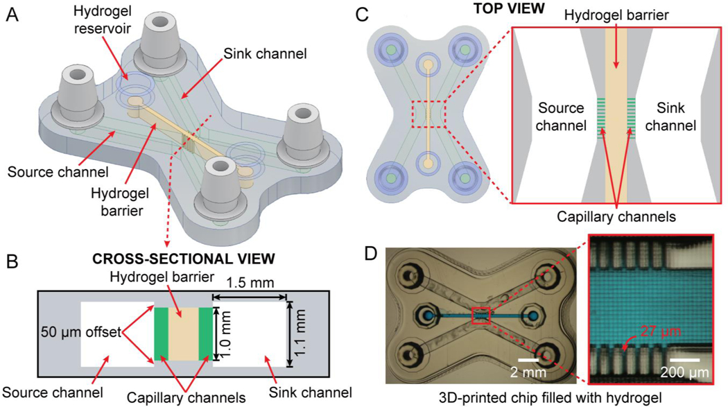
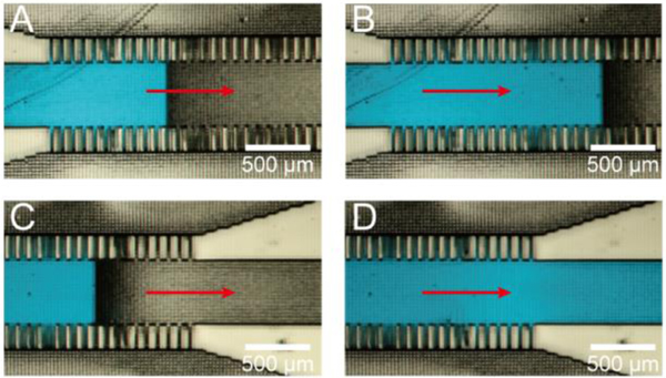
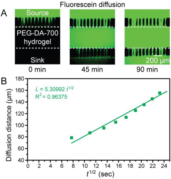
Hydrogels allow for controlling the diffusion rate and amount of solute according to the hydrogel network and thus have found many applications in drug delivery, biomaterials, toxicology, and tissue engineering. This paper describes a 3D-printed microfluidic chip for the straightforward partitioning of hydrogel barriers between microchannels. We use a previously-reported 3-channel architecture whereby the middle channel is filled with a hydrogel - acting like a porous barrier for diffusive transport - and the two side channels act as sink and source; the middle channel communicates with the side channels via orthogonal, small capillary channels that are also responsible for partitioning the hydrogel during filling. Our 3D-printed microfluidic chip is simple to fabricate by stereolithography (SL), inexpensive, reproducible, and convenient, so it is more adequate for transport studies than a microchip fabricated by photolithographic procedures. The chip was fabricated in a resin made of poly(ethylene glycol) diacrylate (PEG-DA) (MW = 258) (PEG-DA-258). The SL process allowed us to print high aspect ratio (37 : 1) capillary channels (27 μm-width and 1 mm-height) and enable the trapping of liquid-phase hydrogels in the hydrogel barrier middle channel. We studied the permeability of hydrogel barriers made of PEG-DA (MW = 700) (PEG-DA-700, 10% polymer content by wt. in water) - as a model of photopolymerizable barriers - and agarose (MW = 120 000, 2% polymer content by wt. in water) - as a model of thermally-gelled barriers. We measured the diffusion of fluorescein, 10k-dextran-Alexa 680 and BSA-Texas Red through these barriers. Fluorescein diffusion was observed through both 10% PEG-DA-700 and 2% agarose barriers while 10k-dextran-Alexa 680 and BSA-Texas Red diffused appreciably only through the 2% agarose hydrogel barrier. Our microfluidic chip facilitates the tuning of such barriers simply by altering the hydrogel materials. The straightforward trapping of selective barriers in 3D-printed microchannels should find wide applicability in drug delivery, tissue engineering, cell separation, and organ-on-a-chip platforms.
水凝胶可根据水凝胶网络控制溶质的扩散速率和数量,因此在药物输送、生物材料、毒理学和组织工程等领域有广泛的应用。本文描述了一种用于在微通道之间直接分隔水凝胶屏障的 3D 打印微流控芯片。我们使用了一种先前报道的 3 通道结构,其中中间通道充满水凝胶 - 充当扩散传输的多孔屏障 - 而两个侧通道充当源和汇;中间通道通过正交的小毛细管通道与侧通道连通,这些小毛细管通道也负责在填充过程中分隔水凝胶。我们的 3D 打印微流控芯片通过立体光刻 (SL) 制造简单、成本低、可重复且方便,因此比通过光刻工艺制造的微芯片更适合用于传输研究。芯片是由聚乙二醇二丙烯酸酯 (PEG-DA)(MW = 258)制成的树脂(PEG-DA-258)制造的。SL 工艺允许我们打印高纵横比(37:1)的毛细管通道(27 μm 宽,1 mm 高),并能够在水凝胶屏障中间通道中捕获液相水凝胶。我们研究了由聚乙二醇二丙烯酸酯 (MW = 700)(PEG-DA-700,水中 10%聚合物含量,重量百分比)制成的水凝胶屏障的通透性 - 作为光聚合屏障的模型 - 和琼脂糖(MW = 120000,水中 2%聚合物含量,重量百分比) - 作为热凝胶屏障的模型。我们测量了荧光素、10k-葡聚糖-Alexa 680 和 BSA-Texas Red 通过这些屏障的扩散。荧光素通过 10% PEG-DA-700 和 2%琼脂糖屏障扩散,而 10k-葡聚糖-Alexa 680 和 BSA-Texas Red 仅通过 2%琼脂糖水凝胶屏障明显扩散。我们的微流控芯片通过简单地改变水凝胶材料来方便地调整这些屏障。在 3D 打印微通道中直接捕获选择性屏障应在药物输送、组织工程、细胞分离和器官芯片平台中得到广泛应用。