• 文献检索
  • 文档翻译
  • 深度研究
  • 学术资讯
  • Suppr Zotero 插件Zotero 插件
  • 邀请有礼
  • 套餐&价格
  • 历史记录
应用&插件
Suppr Zotero 插件Zotero 插件浏览器插件Mac 客户端Windows 客户端微信小程序
定价
高级版会员购买积分包购买API积分包
服务
文献检索文档翻译深度研究API 文档MCP 服务
关于我们
关于 Suppr公司介绍联系我们用户协议隐私条款
关注我们

Suppr 超能文献

核心技术专利:CN118964589B侵权必究
粤ICP备2023148730 号-1Suppr @ 2026

文献检索

告别复杂PubMed语法,用中文像聊天一样搜索,搜遍4000万医学文献。AI智能推荐,让科研检索更轻松。

立即免费搜索

文件翻译

保留排版,准确专业,支持PDF/Word/PPT等文件格式,支持 12+语言互译。

免费翻译文档

深度研究

AI帮你快速写综述,25分钟生成高质量综述,智能提取关键信息,辅助科研写作。

立即免费体验

脱氧血红蛋白密度的轴向变化是血氧水平依赖信号低频时滞结构的来源。

Axial variation of deoxyhemoglobin density as a source of the low-frequency time lag structure in blood oxygenation level-dependent signals.

机构信息

Department of Psychiatry, Kyoto University Graduate School of Medicine, Kyoto, Japan.

Laboratory for Brain Connectomics Imaging, RIKEN Center for Biosystems Dynamics Research, Kobe, Japan.

出版信息

PLoS One. 2019 Sep 23;14(9):e0222787. doi: 10.1371/journal.pone.0222787. eCollection 2019.

DOI:10.1371/journal.pone.0222787
PMID:31545839
原文链接:https://pmc.ncbi.nlm.nih.gov/articles/PMC6756514/
Abstract

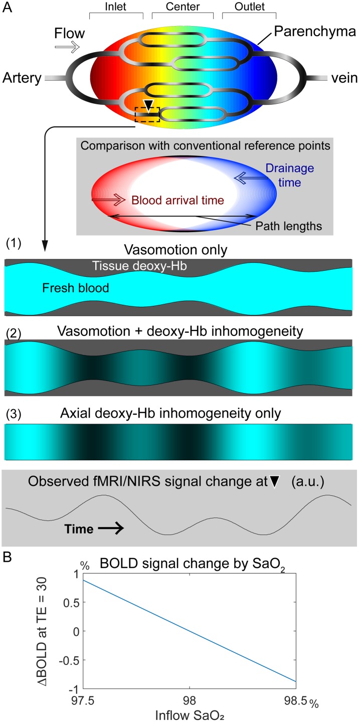
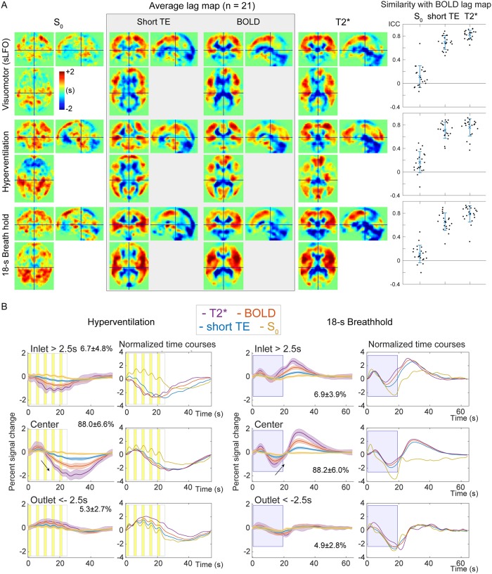
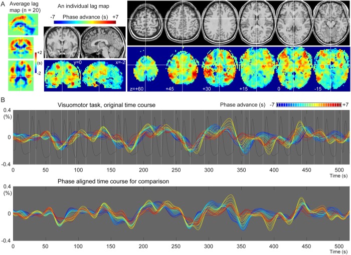
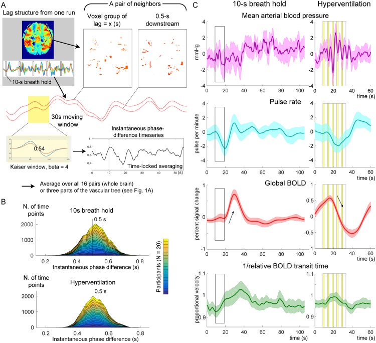
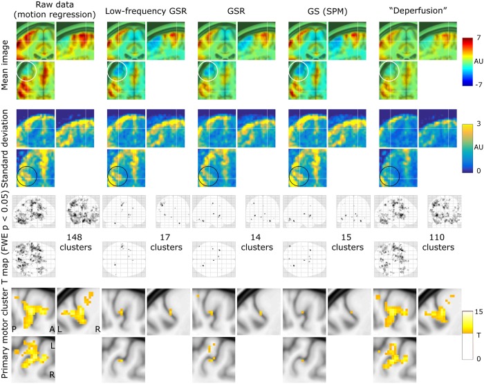
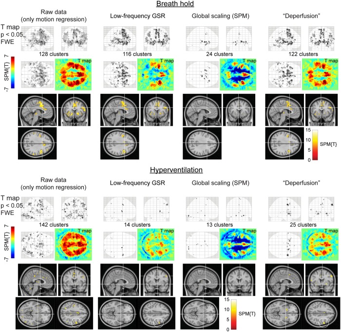
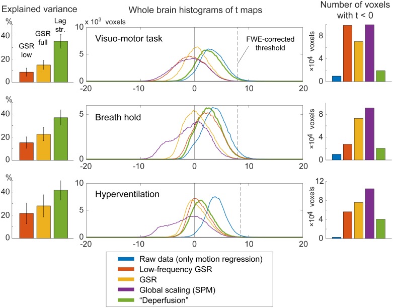
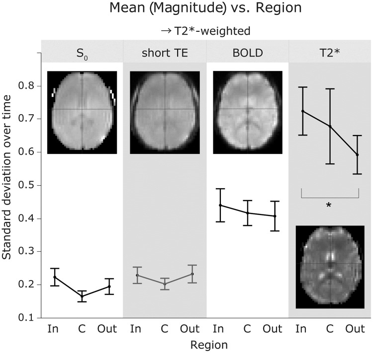
Perfusion-related information is reportedly embedded in the low-frequency component of a blood oxygen level-dependent (BOLD) functional magnetic resonance imaging (fMRI) signal. The blood-propagation pattern through the cerebral vascular tree is detected as an interregional lag variation of spontaneous low-frequency oscillations (sLFOs). Mapping of this lag, or phase, has been implicitly treated as a projection of the vascular tree structure onto real space. While accumulating evidence supports the biological significance of this signal component, the physiological basis of the "perfusion lag structure," a requirement for an integrative resting-state fMRI-signal model, is lacking. In this study, we conducted analyses furthering the hypothesis that the sLFO is not only largely of systemic origin, but also essentially intrinsic to blood, and hence behaves as a virtual tracer. By summing the small fluctuations of instantaneous phase differences between adjacent vascular regions, a velocity response to respiratory challenges was detected. Regarding the relationship to neurovascular coupling, the removal of the whole lag structure, which can be considered as an optimized global-signal regression, resulted in a reduction of inter-individual variance while preserving the fMRI response. Examination of the T2* and S0, or non-BOLD, components of the fMRI signal revealed that the lag structure is deoxyhemoglobin dependent, while paradoxically presenting a signal-magnitude reduction in the venous side of the cerebral vasculature. These findings provide insight into the origin of BOLD sLFOs, suggesting that they are highly intrinsic to the circulating blood.

摘要

据报道,灌注相关信息嵌入在血氧水平依赖(BOLD)功能磁共振成像(fMRI)信号的低频成分中。通过脑血管树的血液传播模式被检测为自发低频振荡(sLFOs)的区域间滞后变化。这种滞后或相位的映射一直被隐含地视为血管树结构在实空间上的投影。虽然越来越多的证据支持该信号成分的生物学意义,但作为整合静息态 fMRI 信号模型的要求,“灌注滞后结构”的生理基础仍然缺乏。在这项研究中,我们进行了分析,进一步假设 sLFO 不仅主要源于系统性,而且本质上是血液内在的,因此表现为虚拟示踪剂。通过对相邻血管区域之间瞬时相位差的小波动进行求和,检测到对呼吸挑战的速度响应。关于与神经血管耦合的关系,去除整个滞后结构(可以看作是优化的全局信号回归),在保留 fMRI 反应的同时,减少了个体间的方差。检查 fMRI 信号的 T2*和 S0,或非 BOLD 成分,发现滞后结构依赖于去氧血红蛋白,而在脑血管的静脉侧却表现出信号幅度减小的矛盾现象。这些发现提供了对 BOLD sLFO 起源的深入了解,表明它们与循环血液高度内在相关。

https://cdn.ncbi.nlm.nih.gov/pmc/blobs/8623/6756514/89fff9125018/pone.0222787.g009.jpg
https://cdn.ncbi.nlm.nih.gov/pmc/blobs/8623/6756514/1c7a964e3df4/pone.0222787.g001.jpg
https://cdn.ncbi.nlm.nih.gov/pmc/blobs/8623/6756514/6b83b5c2d0e5/pone.0222787.g002.jpg
https://cdn.ncbi.nlm.nih.gov/pmc/blobs/8623/6756514/7a1a0ad5f5e4/pone.0222787.g003.jpg
https://cdn.ncbi.nlm.nih.gov/pmc/blobs/8623/6756514/4b073c68dd78/pone.0222787.g004.jpg
https://cdn.ncbi.nlm.nih.gov/pmc/blobs/8623/6756514/abc34a4adb72/pone.0222787.g005.jpg
https://cdn.ncbi.nlm.nih.gov/pmc/blobs/8623/6756514/058e54132c0f/pone.0222787.g006.jpg
https://cdn.ncbi.nlm.nih.gov/pmc/blobs/8623/6756514/25c0930261dc/pone.0222787.g007.jpg
https://cdn.ncbi.nlm.nih.gov/pmc/blobs/8623/6756514/dad764541f42/pone.0222787.g008.jpg
https://cdn.ncbi.nlm.nih.gov/pmc/blobs/8623/6756514/89fff9125018/pone.0222787.g009.jpg

相似文献

1
Axial variation of deoxyhemoglobin density as a source of the low-frequency time lag structure in blood oxygenation level-dependent signals.脱氧血红蛋白密度的轴向变化是血氧水平依赖信号低频时滞结构的来源。
PLoS One. 2019 Sep 23;14(9):e0222787. doi: 10.1371/journal.pone.0222787. eCollection 2019.
2
The resting-state fMRI arterial signal predicts differential blood transit time through the brain.静息态 fMRI 动脉信号可预测大脑中血液通过时间的差异。
J Cereb Blood Flow Metab. 2019 Jun;39(6):1148-1160. doi: 10.1177/0271678X17753329. Epub 2018 Jan 15.
3
A Resilient, Non-neuronal Source of the Spatiotemporal Lag Structure Detected by BOLD Signal-Based Blood Flow Tracking.通过基于BOLD信号的血流追踪检测到的时空滞后结构的一个弹性非神经元来源。
Front Neurosci. 2017 May 11;11:256. doi: 10.3389/fnins.2017.00256. eCollection 2017.
4
Perfusion information extracted from resting state functional magnetic resonance imaging.从静息态功能磁共振成像中提取的灌注信息。
J Cereb Blood Flow Metab. 2017 Feb;37(2):564-576. doi: 10.1177/0271678X16631755. Epub 2016 Jul 20.
5
Vascular effects of caffeine found in BOLD fMRI.在 BOLD fMRI 中发现的咖啡因的血管效应。
J Neurosci Res. 2019 Apr;97(4):456-466. doi: 10.1002/jnr.24360. Epub 2018 Nov 29.
6
Physiological origin of low-frequency drift in blood oxygen level dependent (BOLD) functional magnetic resonance imaging (fMRI).血氧水平依赖(BOLD)功能磁共振成像(fMRI)中低频漂移的生理起源。
Magn Reson Med. 2009 Apr;61(4):819-27. doi: 10.1002/mrm.21902.
7
Estimating and mitigating the effects of systemic low frequency oscillations (sLFO) on resting state networks in awake non-human primates using time lag dependent methodology.使用时间滞后相关方法评估和减轻全身低频振荡(sLFO)对清醒非人类灵长类动物静息态网络的影响。
Front Neuroimaging. 2023 Jan 19;1:1031991. doi: 10.3389/fnimg.2022.1031991. eCollection 2022.
8
Detecting resting-state brain activity by spontaneous cerebral blood volume fluctuations using whole brain vascular space occupancy imaging.利用全脑血管空间占有率成像技术检测自发脑血容量波动的静息态脑活动。
Neuroimage. 2014 Jan 1;84:575-84. doi: 10.1016/j.neuroimage.2013.09.019. Epub 2013 Sep 20.
9
Regional variations in vascular density correlate with resting-state and task-evoked blood oxygen level-dependent signal amplitude.血管密度的区域差异与静息状态和任务诱发的血氧水平依赖信号幅度相关。
Hum Brain Mapp. 2014 May;35(5):1906-20. doi: 10.1002/hbm.22301. Epub 2013 Jul 11.
10
Quantitative mapping of cerebrovascular reactivity using resting-state BOLD fMRI: Validation in healthy adults.使用静息态BOLD功能磁共振成像对脑血管反应性进行定量映射:在健康成年人中的验证
Neuroimage. 2016 Sep;138:147-163. doi: 10.1016/j.neuroimage.2016.05.025. Epub 2016 May 11.

引用本文的文献

1
Systemic physiological "noise" in fMRI has clinical relevance.功能磁共振成像中的系统性生理“噪声”具有临床相关性。
medRxiv. 2025 Aug 25:2025.08.19.25333215. doi: 10.1101/2025.08.19.25333215.
2
Brain-wide functional connectivity artifactually inflates throughout functional magnetic resonance imaging scans.全脑功能连接在功能磁共振成像扫描中人为地膨胀。
Nat Hum Behav. 2024 Aug;8(8):1568-1580. doi: 10.1038/s41562-024-01908-6. Epub 2024 Jun 19.
3
Hemodynamic timing in resting-state and breathing-task BOLD fMRI.静息态和呼吸任务 BOLD fMRI 的血流动力学时程。

本文引用的文献

1
Non-invasive monitoring of longitudinal changes in cerebral hemodynamics in acute ischemic stroke using BOLD signal delay.使用 BOLD 信号延迟无创监测急性缺血性脑卒中患者大脑血液动力学的纵向变化。
J Cereb Blood Flow Metab. 2020 Jan;40(1):23-34. doi: 10.1177/0271678X18803951. Epub 2018 Oct 18.
2
Resting-state Functional Magnetic Resonance Imaging Identifies Cerebrovascular Reactivity Impairment in Patients With Arterial Occlusive Diseases: A Pilot Study.静息态功能磁共振成像识别动脉闭塞性疾病患者的脑血管反应性损伤:一项初步研究。
Neurosurgery. 2019 Nov 1;85(5):680-688. doi: 10.1093/neuros/nyy434.
3
Using temporal ICA to selectively remove global noise while preserving global signal in functional MRI data.
Neuroimage. 2023 Jul 1;274:120120. doi: 10.1016/j.neuroimage.2023.120120. Epub 2023 Apr 16.
4
Reproducibility of cerebral perfusion measurements using BOLD delay.使用 BOLD 延迟测量脑灌注的可重复性。
Hum Brain Mapp. 2023 May;44(7):2778-2789. doi: 10.1002/hbm.26244. Epub 2023 Feb 25.
5
Human CSF movement influenced by vascular low frequency oscillations and respiration.人类脑脊液的流动受血管低频振荡和呼吸的影响。
Front Physiol. 2022 Aug 19;13:940140. doi: 10.3389/fphys.2022.940140. eCollection 2022.
6
The relationship between attention and avoidance coping in anorexia nervosa: functional magnetic resonance imaging study.神经性厌食症中注意力与回避应对之间的关系:功能磁共振成像研究
BJPsych Open. 2021 Jul 13;7(4):e130. doi: 10.1192/bjo.2021.963.
7
The Role of Cerebrovascular-Reactivity Mapping in Functional MRI: Calibrated fMRI and Resting-State fMRI.脑血管反应性图谱在功能磁共振成像中的作用:校准功能磁共振成像和静息态功能磁共振成像
Front Physiol. 2021 Mar 25;12:657362. doi: 10.3389/fphys.2021.657362. eCollection 2021.
8
Timescales of spontaneous fMRI fluctuations relate to structural connectivity in the brain.功能磁共振成像自发波动的时间尺度与大脑中的结构连通性相关。
Netw Neurosci. 2020 Sep 1;4(3):788-806. doi: 10.1162/netn_a_00151. eCollection 2020.
9
Rude mechanicals in brain haemodynamics: non-neural actors that influence blood flow.脑血流动力学中的“粗笨技工”:影响血流的非神经因素
Philos Trans R Soc Lond B Biol Sci. 2021 Jan 4;376(1815):20190635. doi: 10.1098/rstb.2019.0635. Epub 2020 Nov 16.
10
The Effect of Scan Length on the Assessment of BOLD Delay in Ischemic Stroke.扫描长度对缺血性卒中脑血流动力学延迟评估的影响
Front Neurol. 2020 May 5;11:381. doi: 10.3389/fneur.2020.00381. eCollection 2020.
使用时间 ICA 选择性地去除功能磁共振成像数据中的全局噪声,同时保留全局信号。
Neuroimage. 2018 Nov 1;181:692-717. doi: 10.1016/j.neuroimage.2018.04.076. Epub 2018 Aug 2.
4
The resting-state fMRI arterial signal predicts differential blood transit time through the brain.静息态 fMRI 动脉信号可预测大脑中血液通过时间的差异。
J Cereb Blood Flow Metab. 2019 Jun;39(6):1148-1160. doi: 10.1177/0271678X17753329. Epub 2018 Jan 15.
5
Identifying and characterizing systematic temporally-lagged BOLD artifacts.识别和描述系统的时间滞后 BOLD 伪影。
Neuroimage. 2018 May 1;171:376-392. doi: 10.1016/j.neuroimage.2017.12.082. Epub 2017 Dec 26.
6
Alteration of Venous Drainage Route in Idiopathic Normal Pressure Hydrocephalus and Normal Aging.特发性正常压力脑积水和正常衰老中静脉引流途径的改变
Front Aging Neurosci. 2017 Nov 23;9:387. doi: 10.3389/fnagi.2017.00387. eCollection 2017.
7
Weak correlations between hemodynamic signals and ongoing neural activity during the resting state.静息状态下血流动力学信号与持续神经活动之间的弱相关性。
Nat Neurosci. 2017 Dec;20(12):1761-1769. doi: 10.1038/s41593-017-0007-y. Epub 2017 Nov 6.
8
A Resilient, Non-neuronal Source of the Spatiotemporal Lag Structure Detected by BOLD Signal-Based Blood Flow Tracking.通过基于BOLD信号的血流追踪检测到的时空滞后结构的一个弹性非神经元来源。
Front Neurosci. 2017 May 11;11:256. doi: 10.3389/fnins.2017.00256. eCollection 2017.
9
Investigating the spatiotemporal characteristics of the deoxyhemoglobin-related and deoxyhemoglobin-unrelated functional hemodynamic response across cortical layers in awake marmosets.在清醒的食蟹猴中研究各皮层层间脱氧血红蛋白相关和脱氧血红蛋白不相关功能血流动力学反应的时空特征。
Neuroimage. 2018 Jan 1;164:121-130. doi: 10.1016/j.neuroimage.2017.03.005. Epub 2017 Mar 6.
10
The global signal in fMRI: Nuisance or Information?功能磁共振成像中的全局信号:干扰因素还是信息?
Neuroimage. 2017 Apr 15;150:213-229. doi: 10.1016/j.neuroimage.2017.02.036. Epub 2017 Feb 16.