Cerebral Microcirculation Section, Laboratory of Functional and Molecular Imaging, National Institute of Neurological Disorders and Stroke, National Institutes of Health, Bethesda, MD 20892, USA.
Cerebral Microcirculation Section, Laboratory of Functional and Molecular Imaging, National Institute of Neurological Disorders and Stroke, National Institutes of Health, Bethesda, MD 20892, USA.
Neuroimage. 2018 Jan 1;164:121-130. doi: 10.1016/j.neuroimage.2017.03.005. Epub 2017 Mar 6.
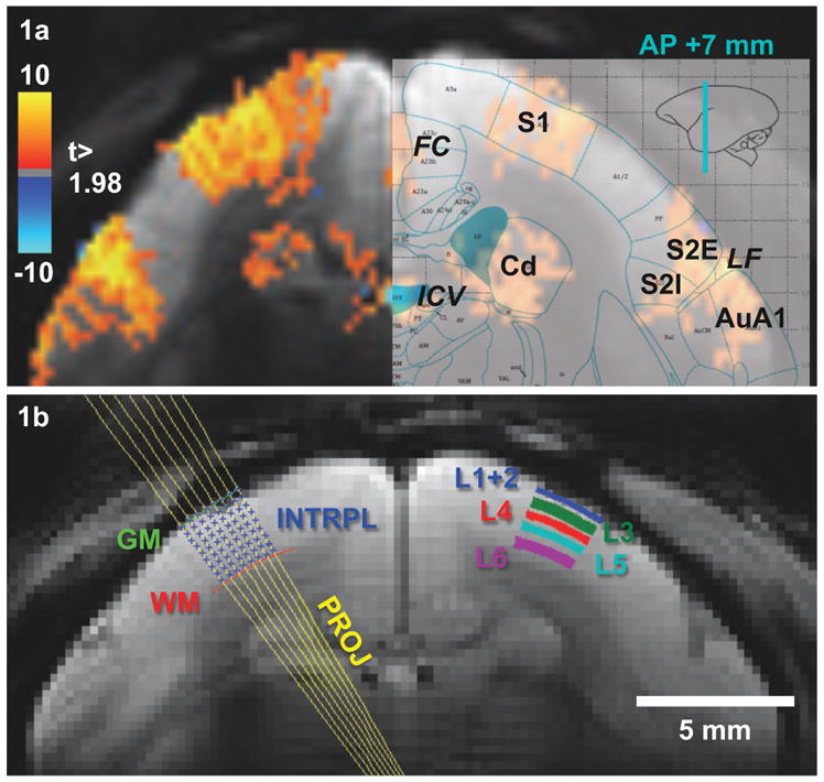
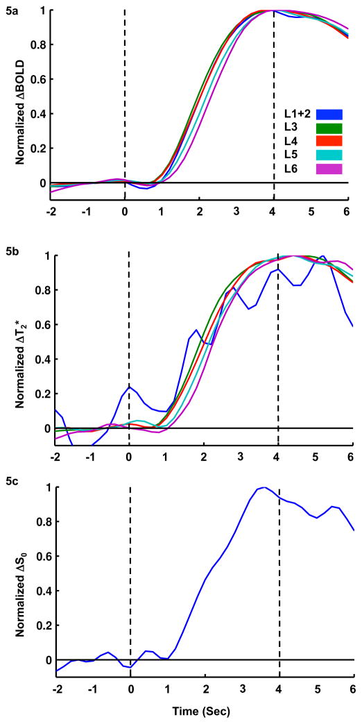
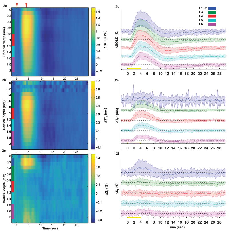
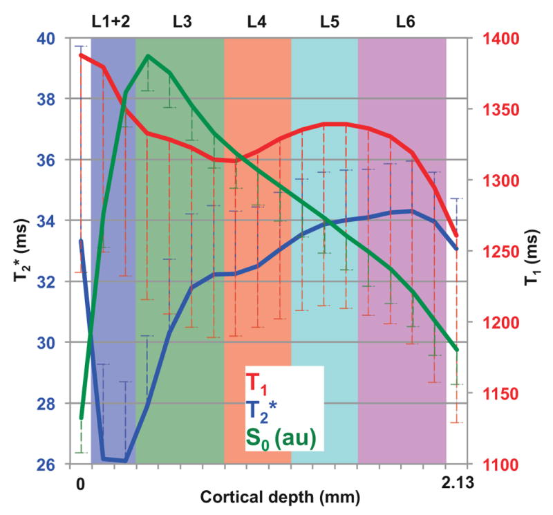
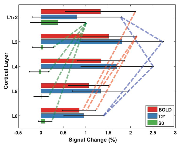
Blood oxygenation level dependent (BOLD) functional magnetic resonance imaging (fMRI) has become a major tool to map neural activity. However, the spatiotemporal characteristics of the BOLD functional hemodynamic response across the cortical layers remain poorly understood. While human fMRI studies suffer from low spatiotemporal resolution, the use of anesthesia in animal models introduces confounding factors. Additionally, inflow contributions to the fMRI signal become non-negligible when short repetition times (TRs) are used. In the present work, we mapped the BOLD fMRI response to somatosensory stimulation in awake marmosets. To address the above technical concerns, we used a dual-echo gradient-recalled echo planar imaging (GR-EPI) sequence to separate the deoxyhemoglobin-related response (absolute T* differences) from the deoxyhemoglobin-unrelated response (relative S changes). We employed a spatial saturation pulse to saturate incoming arterial spins and reduce inflow effects. Functional GR-EPI images were obtained from a single coronal slice with two different echo times (13.5 and 40.5ms) and TR=0.2s. BOLD, T*, and S images were calculated and their functional responses were detected in both hemispheres of primary somatosensory cortex, from which five laminar regions (L1+2, L3, L4, L5, and L6) were derived. The spatiotemporal distribution of the BOLD response across the cortical layers was heterogeneous, with the middle layers having the highest BOLD amplitudes and shortest onset times. ΔT* also showed a similar trend. However, functional S changes were detected only in L1+2, with a fast onset time. Because inflow effects were minimized, the source of S functional changes in L1+2 could be attributed to a reduction of cerebrospinal fluid volume fraction due to the functional increase in cerebral blood volume and to unmodeled T* changes in the extra- and intra-venous compartments. Caution should be exercised when interpreting laminar BOLD fMRI changes in superficial layers as surrogates of underlying neural activity.
血氧水平依赖(BOLD)功能磁共振成像(fMRI)已成为映射神经活动的主要工具。然而,皮质各层的 BOLD 功能血液动力学响应的时空特征仍知之甚少。虽然人类 fMRI 研究的空间和时间分辨率较低,但在动物模型中使用麻醉会引入混杂因素。此外,当使用短重复时间(TR)时,流入对 fMRI 信号的贡献变得不可忽略。在本工作中,我们在清醒的狨猴中绘制了体感刺激的 BOLD fMRI 响应。为了解决上述技术问题,我们使用双回波梯度回波平面成像(GR-EPI)序列来分离脱氧血红蛋白相关响应(绝对 T差异)和脱氧血红蛋白无关响应(相对 S 变化)。我们使用空间饱和脉冲来饱和进入的动脉自旋,减少流入效应。功能 GR-EPI 图像是从单个冠状切片获得的,具有两个不同的回波时间(13.5 和 40.5ms)和 TR=0.2s。计算了 BOLD、T和 S 图像,并在初级体感皮层的两个半球中检测了它们的功能响应,从中衍生出五个层区(L1+2、L3、L4、L5 和 L6)。皮质各层 BOLD 响应的时空分布是不均匀的,中层具有最高的 BOLD 幅度和最短的起始时间。ΔT也显示出类似的趋势。然而,只有在 L1+2 中检测到功能 S 变化,其起始时间较快。由于流入效应最小,L1+2 中 S 功能变化的来源可能归因于由于脑血流量增加而导致脑脊液体积分数减少,以及静脉外和静脉内腔室中未建模的 T变化。在解释浅层的层 BOLD fMRI 变化作为潜在神经活动的替代物时应谨慎。