Nancy E. and Peter C. Meinig School of Biomedical Engineering, Cornell University, Ithaca, NY, 14853, USA.
Nancy E. and Peter C. Meinig School of Biomedical Engineering, Cornell University, Ithaca, NY, 14853, USA; Department of Diagnostic Medicine/Pathobiology, Kansas State University College of Veterinary Medicine, Manhattan, KS, 66506, USA.
Biomaterials. 2019 Dec;224:119489. doi: 10.1016/j.biomaterials.2019.119489. Epub 2019 Sep 11.
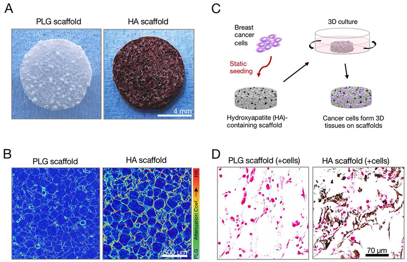
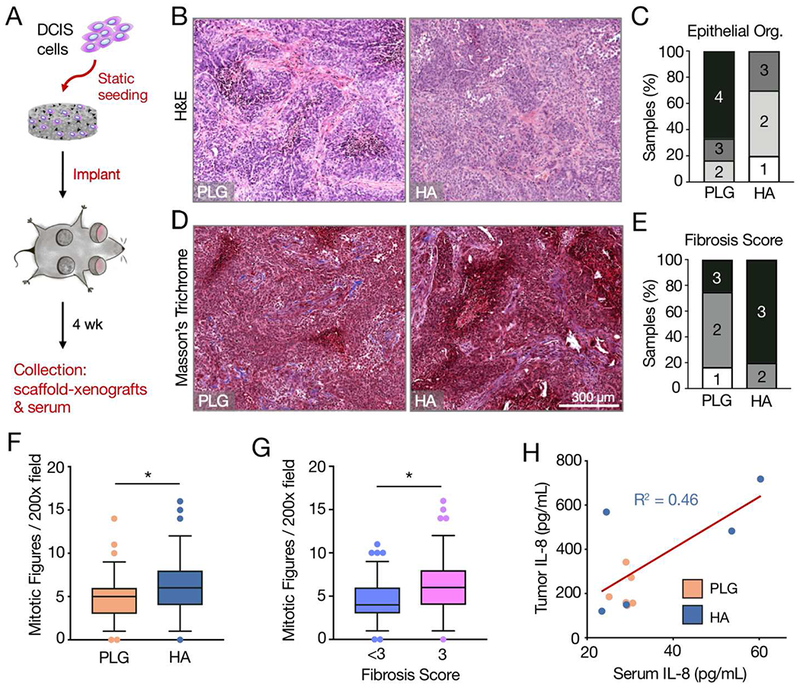
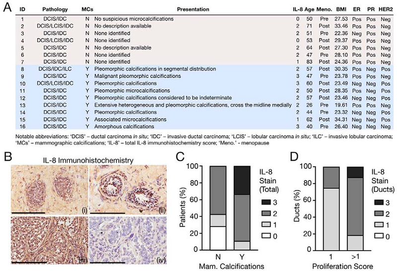
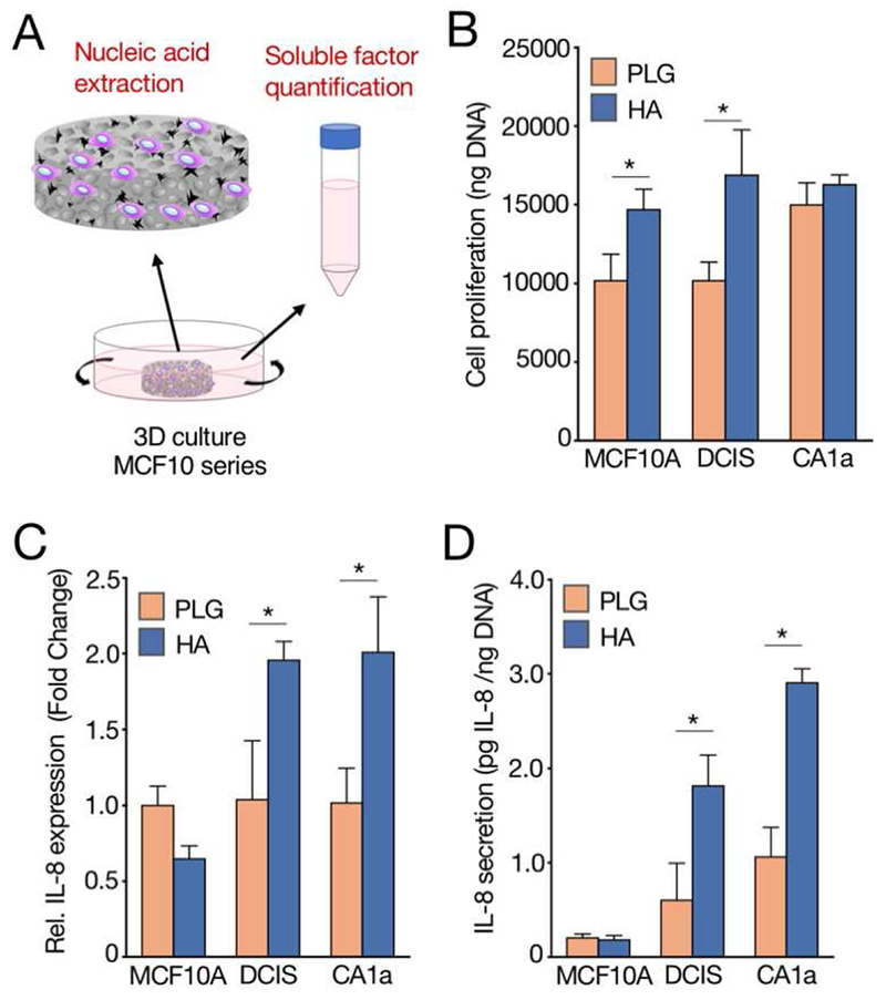
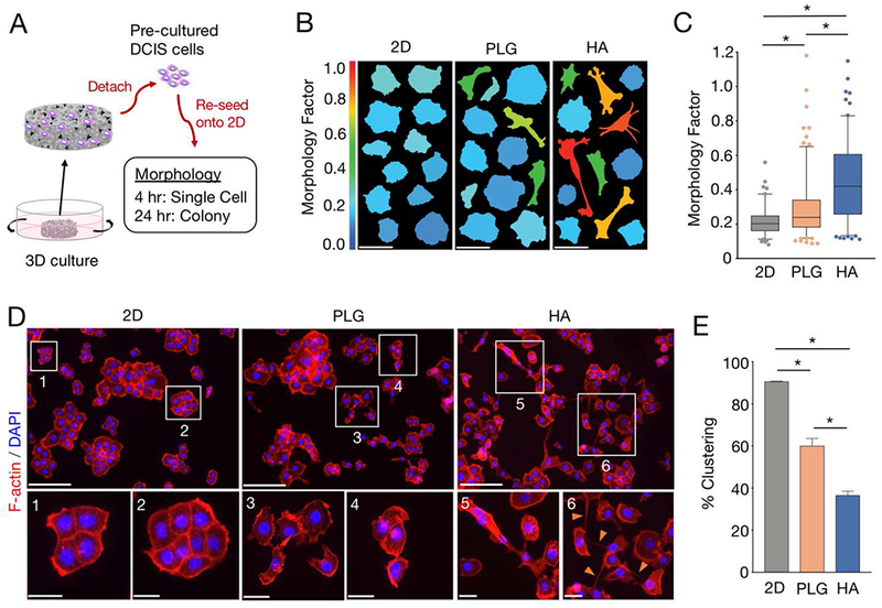
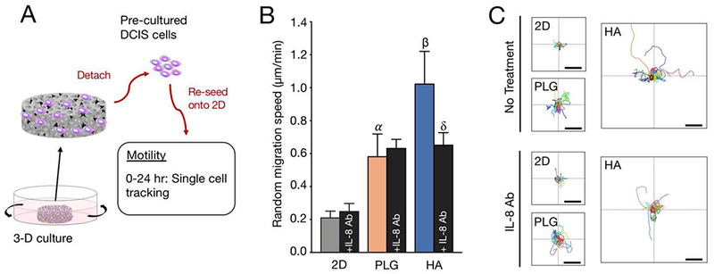
While ductal carcinoma in situ (DCIS) is known as a precursor lesion to most invasive breast carcinomas, the mechanisms underlying this transition remain enigmatic. DCIS is typically diagnosed by the mammographic detection of microcalcifications (MC). MCs consisting of non-stoichiometric hydroxyapatite (HA) mineral are frequently associated with malignant disease, yet it is unclear whether HA can actively promote malignancy. To investigate this outstanding question, we compared phenotypic outcomes of breast cancer cells cultured in control or HA-containing poly(lactide-co-glycolide) (PLG) scaffolds. Exposure to HA mineral in scaffolds increased the expression of pro-tumorigenic interleukin-8 (IL-8) among transformed but not benign cells. Notably, MCF10DCIS.com cells cultured in HA scaffolds adopted morphological changes associated with increased invasiveness and exhibited increased motility that were dependent on IL-8 signaling. Moreover, MCF10DCIS.com xenografts in HA scaffolds displayed evidence of enhanced malignant progression relative to xenografts in control scaffolds. These experimental findings were supported by a pathological analysis of clinical DCIS specimens, which correlated the presence of MCs with increased IL-8 staining and ductal proliferation. Collectively, our work suggests that HA mineral may stimulate malignancy in preinvasive DCIS cells and validate PLG scaffolds as useful tools to study cell-mineral interactions.
虽然导管原位癌 (DCIS) 被认为是大多数浸润性乳腺癌的前体病变,但这种转变的机制仍然是个谜。DCIS 通常通过乳腺 X 光检查发现微钙化 (MC) 来诊断。由非化学计量羟基磷灰石 (HA) 组成的 MC 常与恶性疾病相关,但尚不清楚 HA 是否能主动促进恶性转化。为了研究这一悬而未决的问题,我们比较了在对照或含有 HA 的聚 (乳酸-共-乙醇酸) (PLG) 支架中培养的乳腺癌细胞的表型结果。暴露于支架中的 HA 矿物增加了转化但不是良性细胞中促肿瘤生成的白细胞介素-8 (IL-8) 的表达。值得注意的是,在 HA 支架中培养的 MCF10DCIS.com 细胞采用了与侵袭性增加相关的形态变化,并表现出依赖于 IL-8 信号的运动性增加。此外,在 HA 支架中的 MCF10DCIS.com 异种移植物显示出与在对照支架中的异种移植物相比,恶性进展增强的证据。这些实验结果得到了对临床 DCIS 标本的病理分析的支持,该分析将 MC 的存在与 IL-8 染色和导管增殖增加相关联。总的来说,我们的工作表明,HA 矿物可能刺激前体 DCIS 细胞的恶性转化,并验证了 PLG 支架作为研究细胞-矿物相互作用的有用工具。