Department of Radiology, Seoul National University Hospital and Seoul National University College of Medicine, 101 Daehak-ro, Jongno-gu, Seoul, 03080, Republic of Korea.
Department of Biomedical Sciences, Seoul National University College of Medicine, 103 Daehak-ro, Jongno-gu, Seoul, 03080, Republic of Korea.
J Exp Clin Cancer Res. 2018 Aug 22;37(1):200. doi: 10.1186/s13046-018-0867-3.
The function of preadipocytes in the progression of early stage breast cancer has not been fully elucidated at the molecular level. To delineate the role of preadipocytes in breast cancer progression, we investigated the cross-talk between human breast ductal carcinoma in situ (DCIS) cells and preadipocytes with both an in vitro culture and xenograft tumor model.
GFP or RFP was transduced into human DCIS cell line MCF10DCIS.com cells or preadipocytes using lentivirus. Cell sorter was used to separate pure, viable populations of GFP- or RFP-transduced cells. Cell viability and proliferation was assessed by crystal violet assays and cell migration and invasion capability was assayed by the transwell strategy. Gene and protein levels were measured by western blot, RT-PCR and immunostaining. Adipokines and cytokines were quantified using ELISA. Human tumor xenografts in a nude mice model were used. Ultrasound imaging of tumors was performed to evaluate the therapeutic potential of a IL-6 neutralizing antibody.
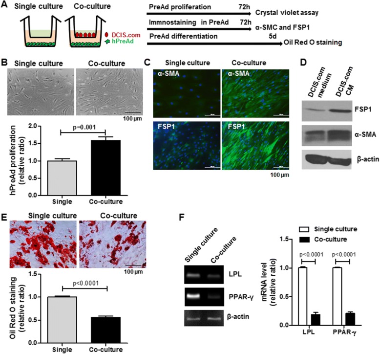
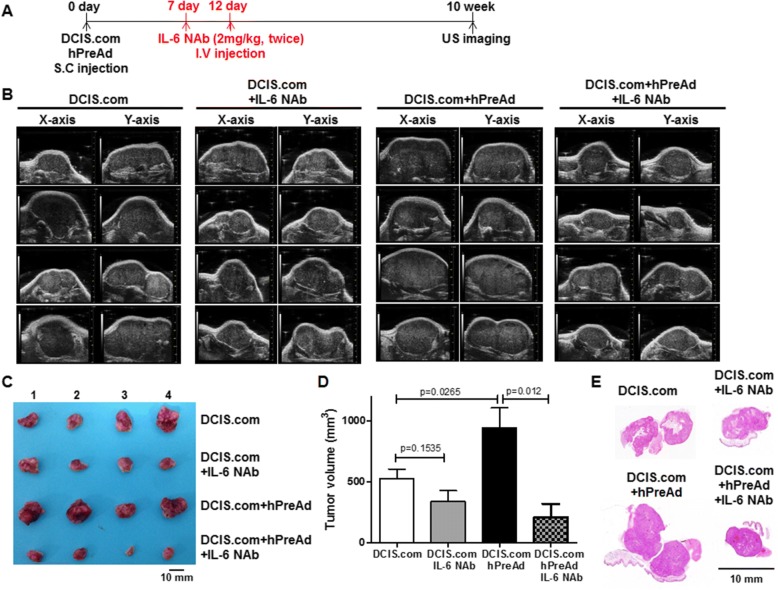
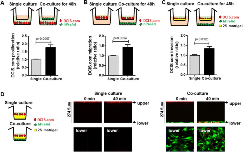
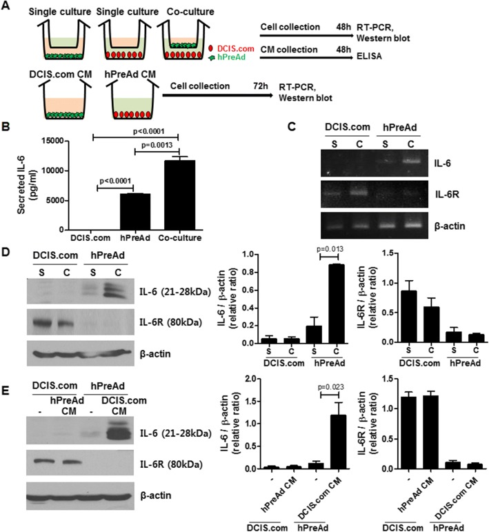
In the co-culture system with the MCF10DCIS.com and preadipocytes, MCF10DCIS.com proliferation, migration and invasion were enhanced by preadipocytes. Preadipocytes exhibited in an increased IL-6 secretion and cancer-associated fibroblast markers expression, FSP1 and α-SMC in co-culture with MCF10DCIS.com or in MCF10DCIS.com conditioned media, whereas the adipocyte differentiation capacity was suppressed by co-culture with MCF10DCIS.com. A neutralizing antibody of IL-6 or IL-6R suppressed the promotion of MCF10DCIS.com proliferation and migration by co-culture with preadipocytes. In the xenograft tumor model, the tumor growth of MCF10DCIS.com was enhanced by the co-injection of preadipocytes, and the administration of IL-6 neutralizing antibodies resulted in potent effects on tumor inhibition.
Our findings suggest that IL-6-mediated cross-talk between preadipocytes and breast DCIS cells can promote the progression of early stage breast cancer. Therefore, blocking IL-6 signaling might be a potential therapeutic strategy for breast DCIS characterized by pathological IL-6 overproduction.
前脂肪细胞在早期乳腺癌进展中的功能在分子水平上尚未完全阐明。为了阐明前脂肪细胞在乳腺癌进展中的作用,我们通过体外培养和异种移植肿瘤模型研究了人乳腺导管原位癌(DCIS)细胞与前脂肪细胞之间的串扰。
使用慢病毒将 GFP 或 RFP 转导到人 DCIS 细胞系 MCF10DCIS.com 细胞或前脂肪细胞中。细胞分选器用于分离纯的、活的 GFP-或 RFP 转导细胞群体。通过结晶紫测定法评估细胞活力和增殖,通过 Transwell 策略测定细胞迁移和侵袭能力。通过 Western blot、RT-PCR 和免疫染色测量基因和蛋白水平。使用 ELISA 定量测定脂肪因子和细胞因子。使用裸鼠模型中的人肿瘤异种移植物。进行肿瘤超声成像以评估 IL-6 中和抗体的治疗潜力。
在 MCF10DCIS.com 和前脂肪细胞的共培养系统中,前脂肪细胞增强了 MCF10DCIS.com 的增殖、迁移和侵袭。与 MCF10DCIS.com 共培养或在 MCF10DCIS.com 条件培养基中,前脂肪细胞表现出 IL-6 分泌和癌症相关成纤维细胞标志物表达增加,FSP1 和 α-SMC,而与 MCF10DCIS.com 共培养则抑制了脂肪细胞分化能力。IL-6 或 IL-6R 的中和抗体抑制了与前脂肪细胞共培养对 MCF10DCIS.com 增殖和迁移的促进作用。在异种移植肿瘤模型中,前脂肪细胞的共注射增强了 MCF10DCIS.com 的肿瘤生长,而 IL-6 中和抗体的给药对肿瘤抑制具有强大的作用。
我们的研究结果表明,IL-6 介导的前脂肪细胞与乳腺 DCIS 细胞之间的串扰可以促进早期乳腺癌的进展。因此,阻断 IL-6 信号可能是一种有潜力的治疗策略,适用于病理上 IL-6 过度产生的乳腺 DCIS。