• 文献检索
  • 文档翻译
  • 深度研究
  • 学术资讯
  • Suppr Zotero 插件Zotero 插件
  • 邀请有礼
  • 套餐&价格
  • 历史记录
应用&插件
Suppr Zotero 插件Zotero 插件浏览器插件Mac 客户端Windows 客户端微信小程序
定价
高级版会员购买积分包购买API积分包
服务
文献检索文档翻译深度研究API 文档MCP 服务
关于我们
关于 Suppr公司介绍联系我们用户协议隐私条款
关注我们

Suppr 超能文献

核心技术专利:CN118964589B侵权必究
粤ICP备2023148730 号-1Suppr @ 2026

文献检索

告别复杂PubMed语法,用中文像聊天一样搜索,搜遍4000万医学文献。AI智能推荐,让科研检索更轻松。

立即免费搜索

文件翻译

保留排版,准确专业,支持PDF/Word/PPT等文件格式,支持 12+语言互译。

免费翻译文档

深度研究

AI帮你快速写综述,25分钟生成高质量综述,智能提取关键信息,辅助科研写作。

立即免费体验

头部CT数据处理建议。

Recommendations for Processing Head CT Data.

作者信息

Muschelli John

机构信息

Department of Biostatistics, Johns Hopkins Bloomberg School of Public Health, Baltimore, MD, United States.

出版信息

Front Neuroinform. 2019 Sep 4;13:61. doi: 10.3389/fninf.2019.00061. eCollection 2019.

DOI:10.3389/fninf.2019.00061
PMID:31551745
原文链接:https://pmc.ncbi.nlm.nih.gov/articles/PMC6738271/
Abstract

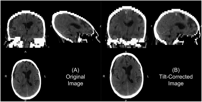
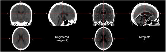
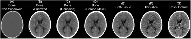
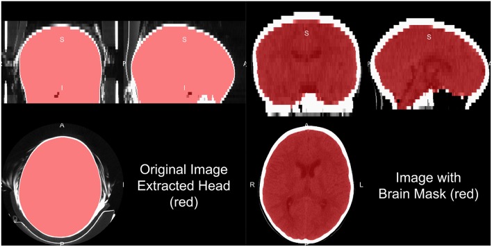
Many research applications of neuroimaging use magnetic resonance imaging (MRI). As such, recommendations for image analysis and standardized imaging pipelines exist. Clinical imaging, however, relies heavily on X-ray computed tomography (CT) scans for diagnosis and prognosis. Currently, there is only one image processing pipeline for head CT, which focuses mainly on head CT data with lesions. We present tools and a complete pipeline for processing CT data, focusing on open-source solutions, that focus on head CT but are applicable to most CT analyses. We describe going from raw DICOM data to a spatially normalized brain within CT presenting a full example with code. Overall, we recommend anonymizing data with Clinical Trials Processor, converting DICOM data to NIfTI using dcm2niix, using BET for brain extraction, and registration using a publicly-available CT template for analysis.

摘要

神经成像的许多研究应用都使用磁共振成像(MRI)。因此,存在图像分析和标准化成像流程的相关建议。然而,临床成像在很大程度上依赖于X射线计算机断层扫描(CT)进行诊断和预后评估。目前,只有一个用于头部CT的图像处理流程,其主要关注有病变的头部CT数据。我们展示了用于处理CT数据的工具和完整流程,重点是开源解决方案,这些工具和流程主要针对头部CT,但也适用于大多数CT分析。我们描述了从原始DICOM数据到CT内空间归一化大脑的过程,并给出了带有代码的完整示例。总体而言,我们建议使用临床试验处理器对数据进行匿名化处理,使用dcm2niix将DICOM数据转换为NIfTI格式,使用BET进行脑提取,并使用公开可用的CT模板进行配准以进行分析。

https://cdn.ncbi.nlm.nih.gov/pmc/blobs/1cf3/6738271/f555c9a98859/fninf-13-00061-g0004.jpg
https://cdn.ncbi.nlm.nih.gov/pmc/blobs/1cf3/6738271/7805414ed6e7/fninf-13-00061-g0001.jpg
https://cdn.ncbi.nlm.nih.gov/pmc/blobs/1cf3/6738271/c4b70e38a0e5/fninf-13-00061-g0002.jpg
https://cdn.ncbi.nlm.nih.gov/pmc/blobs/1cf3/6738271/f0641f124479/fninf-13-00061-g0003.jpg
https://cdn.ncbi.nlm.nih.gov/pmc/blobs/1cf3/6738271/f555c9a98859/fninf-13-00061-g0004.jpg

相似文献

1
Recommendations for Processing Head CT Data.头部CT数据处理建议。
Front Neuroinform. 2019 Sep 4;13:61. doi: 10.3389/fninf.2019.00061. eCollection 2019.
2
DICOM for quantitative imaging biomarker development: a standards based approach to sharing clinical data and structured PET/CT analysis results in head and neck cancer research.用于定量成像生物标志物开发的DICOM:一种基于标准的方法,用于在头颈癌研究中共享临床数据和结构化PET/CT分析结果。
PeerJ. 2016 May 24;4:e2057. doi: 10.7717/peerj.2057. eCollection 2016.
3
Validated automatic brain extraction of head CT images.头部CT图像的经验证的自动脑提取
Neuroimage. 2015 Jul 1;114:379-85. doi: 10.1016/j.neuroimage.2015.03.074. Epub 2015 Apr 7.
4
Radtools: R utilities for convenient extraction of medical image metadata.Radtools:用于便捷提取医学图像元数据的R实用工具。
F1000Res. 2018 Dec 24;7. doi: 10.12688/f1000research.17139.3. eCollection 2018.
5
NIfTI-MRS: A standard data format for magnetic resonance spectroscopy.NIfTI-MRS:磁共振波谱的标准数据格式。
Magn Reson Med. 2022 Dec;88(6):2358-2370. doi: 10.1002/mrm.29418. Epub 2022 Sep 11.
6
The first step for neuroimaging data analysis: DICOM to NIfTI conversion.神经影像数据分析的第一步:从DICOM格式转换为NIfTI格式。
J Neurosci Methods. 2016 May 1;264:47-56. doi: 10.1016/j.jneumeth.2016.03.001. Epub 2016 Mar 2.
7
A Neonatal Bimodal MR-CT Head Template.一种新生儿头部双模态磁共振成像-计算机断层扫描模板。
PLoS One. 2017 Jan 27;12(1):e0166112. doi: 10.1371/journal.pone.0166112. eCollection 2017.
8
Optimized partial-coverage functional analysis pipeline (OPFAP): a semi-automated pipeline for skull stripping and co-registration of partial-coverage, ultra-high-field functional images.优化的部分覆盖功能分析管道(OPFAP):一种用于部分覆盖、超高场功能图像的颅骨剥离和配准的半自动管道。
MAGMA. 2018 Oct;31(5):621-632. doi: 10.1007/s10334-018-0690-z. Epub 2018 May 29.
9
Magnetic Resonance Imaging Application in the Area of Mild and Acute Traumatic Brain Injury: Implications for Diagnostic Markers?磁共振成像在轻度和急性创伤性脑损伤领域的应用:对诊断标志物有何启示?
10
The Stroke Neuro-Imaging Phenotype Repository: An Open Data Science Platform for Stroke Research.中风神经影像表型库:一个用于中风研究的开放数据科学平台。
Front Neuroinform. 2021 Jun 24;15:597708. doi: 10.3389/fninf.2021.597708. eCollection 2021.

引用本文的文献

1
Brain and intracranial volumes are both enlarged and serve as potential risk factors in normal pressure hydrocephalus.脑容量和颅内容量均增大,是正常压力脑积水的潜在危险因素。
Sci Rep. 2025 Mar 8;15(1):8131. doi: 10.1038/s41598-025-92721-2.
2
A Probabilistic Method to Predict Classifier Accuracy on Larger Datasets given Small Pilot Data.一种在给定小样本先导数据的情况下预测较大数据集上分类器准确率的概率方法。
Proc Mach Learn Res. 2023 Dec;225:129-144.
3
Automated Detection of Normal Pressure Hydrocephalus Using CT Imaging for Calculating the Ventricle-to-Subarachnoid Volume Ratio.

本文引用的文献

1
Radtools: R utilities for convenient extraction of medical image metadata.Radtools:用于便捷提取医学图像元数据的R实用工具。
F1000Res. 2018 Dec 24;7. doi: 10.12688/f1000research.17139.3. eCollection 2018.
2
Deep learning algorithms for detection of critical findings in head CT scans: a retrospective study.深度学习算法在头部 CT 扫描中关键发现检测的应用:一项回顾性研究。
Lancet. 2018 Dec 1;392(10162):2388-2396. doi: 10.1016/S0140-6736(18)31645-3. Epub 2018 Oct 11.
3
Application of Machine Learning to Automated Analysis of Cerebral Edema in Large Cohorts of Ischemic Stroke Patients.
使用CT成像自动检测正常压力脑积水以计算脑室与蛛网膜下腔体积比
AJNR Am J Neuroradiol. 2025 Jan 8;46(1):141-146. doi: 10.3174/ajnr.A8451.
4
Deep learning biomarker of chronometric and biological ischemic stroke lesion age from unenhanced CT.基于平扫CT的计时性和生物性缺血性脑卒中病变年龄的深度学习生物标志物
NPJ Digit Med. 2024 Dec 6;7(1):338. doi: 10.1038/s41746-024-01325-z.
5
Deep learning-based defacing tool for CT angiography: CTA-DEFACE.基于深度学习的 CT 血管造影图像篡改工具:CTA-DEFACE。
Eur Radiol Exp. 2024 Oct 9;8(1):111. doi: 10.1186/s41747-024-00510-9.
6
Automated segmentation of ventricular volumes and subarachnoid hemorrhage from computed tomography images: Evaluation of a rule-based pipeline approach.基于计算机断层扫描图像的心室容积和蛛网膜下腔出血自动分割:基于规则的流水线方法评估
Neuroradiol J. 2025 Feb;38(1):30-43. doi: 10.1177/19714009241260791. Epub 2024 Jun 13.
7
A comparative analysis of different augmentations for brain images.不同脑图像增强方法的比较分析。
Med Biol Eng Comput. 2024 Oct;62(10):3123-3150. doi: 10.1007/s11517-024-03127-7. Epub 2024 May 24.
8
Anonymization and validation of three-dimensional volumetric renderings of computed tomography data using commercially available T1-weighted magnetic resonance imaging-based algorithms.使用基于商业可用的T1加权磁共振成像的算法对计算机断层扫描数据的三维容积渲染进行匿名化处理和验证。
J Med Imaging (Bellingham). 2023 Nov;10(6):066501. doi: 10.1117/1.JMI.10.6.066501. Epub 2023 Nov 22.
9
Simple approach to quantify hypoxic-ischemic brain injury severity from computed tomography imaging files after cardiac arrest.一种从心脏骤停后 CT 成像文件中量化缺氧缺血性脑损伤严重程度的简单方法。
Resuscitation. 2024 Feb;195:110050. doi: 10.1016/j.resuscitation.2023.110050. Epub 2023 Nov 15.
10
Segmentation stability of human head and neck cancer medical images for radiotherapy applications under de-identification conditions: Benchmarking data sharing and artificial intelligence use-cases.去识别化条件下用于放射治疗的人类头颈癌医学图像的分割稳定性:基准数据共享和人工智能用例
Front Oncol. 2023 Feb 28;13:1120392. doi: 10.3389/fonc.2023.1120392. eCollection 2023.
机器学习在大量缺血性中风患者脑水肿自动分析中的应用。
Front Neurol. 2018 Aug 21;9:687. doi: 10.3389/fneur.2018.00687. eCollection 2018.
4
TCIApathfinder: An R Client for the Cancer Imaging Archive REST API.TCIApathfinder:用于癌症成像档案 REST API 的 R 客户端。
Cancer Res. 2018 Aug 1;78(15):4424-4426. doi: 10.1158/0008-5472.CAN-18-0678. Epub 2018 Jun 5.
5
Neuroconductor: an R platform for medical imaging analysis.Neuroconductor:一个用于医学影像分析的 R 平台。
Biostatistics. 2019 Apr 1;20(2):218-239. doi: 10.1093/biostatistics/kxx068.
6
Automated Segmentation of Head Computed Tomography Images Using FSL.使用FSL对头计算机断层扫描图像进行自动分割
J Comput Assist Tomogr. 2018 Jan/Feb;42(1):104-110. doi: 10.1097/RCT.0000000000000660.
7
The brain imaging data structure, a format for organizing and describing outputs of neuroimaging experiments.脑影像数据结构,一种组织和描述神经影像实验结果的格式。
Sci Data. 2016 Jun 21;3:160044. doi: 10.1038/sdata.2016.44.
8
The first step for neuroimaging data analysis: DICOM to NIfTI conversion.神经影像数据分析的第一步:从DICOM格式转换为NIfTI格式。
J Neurosci Methods. 2016 May 1;264:47-56. doi: 10.1016/j.jneumeth.2016.03.001. Epub 2016 Mar 2.
9
Free DICOM de-identification tools in clinical research: functioning and safety of patient privacy.临床研究中的免费DICOM去识别工具:患者隐私的功能与安全性
Eur Radiol. 2015 Dec;25(12):3685-95. doi: 10.1007/s00330-015-3794-0. Epub 2015 Jun 3.
10
Validated automatic brain extraction of head CT images.头部CT图像的经验证的自动脑提取
Neuroimage. 2015 Jul 1;114:379-85. doi: 10.1016/j.neuroimage.2015.03.074. Epub 2015 Apr 7.