• 文献检索
  • 文档翻译
  • 深度研究
  • 学术资讯
  • Suppr Zotero 插件Zotero 插件
  • 邀请有礼
  • 套餐&价格
  • 历史记录
应用&插件
Suppr Zotero 插件Zotero 插件浏览器插件Mac 客户端Windows 客户端微信小程序
定价
高级版会员购买积分包购买API积分包
服务
文献检索文档翻译深度研究API 文档MCP 服务
关于我们
关于 Suppr公司介绍联系我们用户协议隐私条款
关注我们

Suppr 超能文献

核心技术专利:CN118964589B侵权必究
粤ICP备2023148730 号-1Suppr @ 2026

文献检索

告别复杂PubMed语法,用中文像聊天一样搜索,搜遍4000万医学文献。AI智能推荐,让科研检索更轻松。

立即免费搜索

文件翻译

保留排版,准确专业,支持PDF/Word/PPT等文件格式,支持 12+语言互译。

免费翻译文档

深度研究

AI帮你快速写综述,25分钟生成高质量综述,智能提取关键信息,辅助科研写作。

立即免费体验

基于深度学习的 CT 血管造影图像篡改工具:CTA-DEFACE。

Deep learning-based defacing tool for CT angiography: CTA-DEFACE.

机构信息

Department of Neuroradiology, Heidelberg University Hospital, Heidelberg, Germany.

Division for Computational Neuroimaging, Heidelberg University Hospital, Heidelberg, Germany.

出版信息

Eur Radiol Exp. 2024 Oct 9;8(1):111. doi: 10.1186/s41747-024-00510-9.

DOI:10.1186/s41747-024-00510-9
PMID:39382818
原文链接:https://pmc.ncbi.nlm.nih.gov/articles/PMC11465008/
Abstract

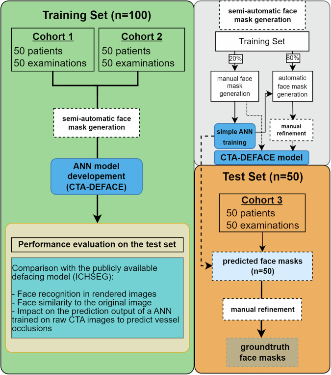
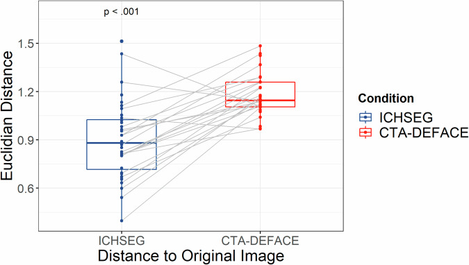
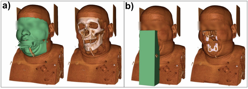
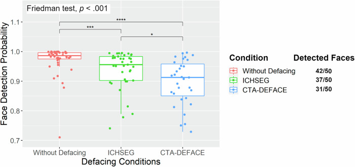
The growing use of artificial neural network (ANN) tools for computed tomography angiography (CTA) data analysis underscores the necessity for elevated data protection measures. We aimed to establish an automated defacing pipeline for CTA data. In this retrospective study, CTA data from multi-institutional cohorts were utilized to annotate facemasks (n = 100) and train an ANN model, subsequently tested on an external institution's dataset (n = 50) and compared to a publicly available defacing algorithm. Face detection (MTCNN) and verification (FaceNet) networks were applied to measure the similarity between the original and defaced CTA images. Dice similarity coefficient (DSC), face detection probability, and face similarity measures were calculated to evaluate model performance. The CTA-DEFACE model effectively segmented soft face tissue in CTA data achieving a DSC of 0.94 ± 0.02 (mean ± standard deviation) on the test set. Our model was benchmarked against a publicly available defacing algorithm. After applying face detection and verification networks, our model showed substantially reduced face detection probability (p < 0.001) and similarity to the original CTA image (p < 0.001). The CTA-DEFACE model enabled robust and precise defacing of CTA data. The trained network is publicly accessible at www.github.com/neuroAI-HD/CTA-DEFACE . RELEVANCE STATEMENT: The ANN model CTA-DEFACE, developed for automatic defacing of CT angiography images, achieves significantly lower face detection probabilities and greater dissimilarity from the original images compared to a publicly available model. The algorithm has been externally validated and is publicly accessible. KEY POINTS: The developed ANN model (CTA-DEFACE) automatically generates facemasks for CT angiography images. CTA-DEFACE offers superior deidentification capabilities compared to a publicly available model. By means of graphics processing unit optimization, our model ensures rapid processing of medical images. Our model underwent external validation, underscoring its reliability for real-world application.

摘要

人工神经网络(ANN)工具在计算机断层血管造影(CTA)数据分析中的应用日益广泛,这凸显了提高数据保护措施的必要性。我们旨在建立一个用于 CTA 数据的自动掩蔽流水线。在这项回顾性研究中,我们使用了来自多机构队列的 CTA 数据来注释面罩(n=100)并训练 ANN 模型,然后在外部机构的数据集(n=50)上进行测试,并与公开可用的掩蔽算法进行比较。应用人脸检测(MTCNN)和验证(FaceNet)网络来衡量原始和掩蔽后的 CTA 图像之间的相似性。计算 Dice 相似系数(DSC)、面部检测概率和面部相似性度量来评估模型性能。CTA-DEFACE 模型有效地分割了 CTA 数据中的软组织,在测试集上的 DSC 为 0.94±0.02(平均值±标准差)。我们的模型与公开可用的掩蔽算法进行了基准比较。在应用人脸检测和验证网络后,我们的模型显示出显著降低的人脸检测概率(p<0.001)和与原始 CTA 图像的相似性(p<0.001)。CTA-DEFACE 模型能够稳健而精确地掩蔽 CTA 数据。训练有素的网络可在 www.github.com/neuroAI-HD/CTA-DEFACE 上公开获取。相关性声明:开发的 ANN 模型 CTA-DEFACE 用于自动掩蔽 CT 血管造影图像,与公开可用的模型相比,实现了显著更低的人脸检测概率和更大的与原始图像的不相似性。该算法已通过外部验证并可公开获取。要点:开发的 ANN 模型(CTA-DEFACE)自动生成 CT 血管造影图像的面罩。CTA-DEFACE 提供了比公开可用的模型更优越的去识别能力。通过图形处理单元优化,我们的模型确保了医疗图像的快速处理。我们的模型经过了外部验证,强调了其在实际应用中的可靠性。

https://cdn.ncbi.nlm.nih.gov/pmc/blobs/8533/11465008/9954dd41b6d0/41747_2024_510_Fig4_HTML.jpg
https://cdn.ncbi.nlm.nih.gov/pmc/blobs/8533/11465008/78d77077213c/41747_2024_510_Fig1_HTML.jpg
https://cdn.ncbi.nlm.nih.gov/pmc/blobs/8533/11465008/79b5e52a068c/41747_2024_510_Fig2_HTML.jpg
https://cdn.ncbi.nlm.nih.gov/pmc/blobs/8533/11465008/c164499d359d/41747_2024_510_Fig3_HTML.jpg
https://cdn.ncbi.nlm.nih.gov/pmc/blobs/8533/11465008/9954dd41b6d0/41747_2024_510_Fig4_HTML.jpg

相似文献

1
Deep learning-based defacing tool for CT angiography: CTA-DEFACE.基于深度学习的 CT 血管造影图像篡改工具:CTA-DEFACE。
Eur Radiol Exp. 2024 Oct 9;8(1):111. doi: 10.1186/s41747-024-00510-9.
2
Segmentation stability of human head and neck cancer medical images for radiotherapy applications under de-identification conditions: Benchmarking data sharing and artificial intelligence use-cases.去识别化条件下用于放射治疗的人类头颈癌医学图像的分割稳定性:基准数据共享和人工智能用例
Front Oncol. 2023 Feb 28;13:1120392. doi: 10.3389/fonc.2023.1120392. eCollection 2023.
3
Reproducibility evaluation of the effects of MRI defacing on brain segmentation.MRI去标识对脑部分割影响的可重复性评估
J Med Imaging (Bellingham). 2023 Nov;10(6):064001. doi: 10.1117/1.JMI.10.6.064001. Epub 2023 Nov 8.
4
A reproducibility evaluation of the effects of MRI defacing on brain segmentation.MRI去标识化对脑部分割影响的可重复性评估
medRxiv. 2023 May 21:2023.05.15.23289995. doi: 10.1101/2023.05.15.23289995.
5
Application of a convolutional neural network to the quality control of MRI defacing.卷积神经网络在 MRI 伪影质量控制中的应用。
Comput Biol Med. 2022 Dec;151(Pt A):106211. doi: 10.1016/j.compbiomed.2022.106211. Epub 2022 Oct 18.
6
Assessing the Impact of Defacing Algorithms on Brain Volumetry Accuracy in MRI Analyses.评估毁损算法对MRI分析中脑容量测量准确性的影响。
Dement Neurocogn Disord. 2024 Jul;23(3):127-135. doi: 10.12779/dnd.2024.23.3.127. Epub 2024 May 8.
7
A Deep Learning Approach for Automated Bone Removal from Computed Tomography Angiography of the Brain.深度学习在脑 CT 血管造影去骨中的应用
J Digit Imaging. 2023 Jun;36(3):964-972. doi: 10.1007/s10278-023-00788-y. Epub 2023 Feb 13.
8
Impact of defacing on automated brain atrophy estimation.毁损对自动脑萎缩估计的影响。
Insights Imaging. 2022 Mar 26;13(1):54. doi: 10.1186/s13244-022-01195-7.
9
Evaluating a 3D deep learning pipeline for cerebral vessel and intracranial aneurysm segmentation from computed tomography angiography-digital subtraction angiography image pairs.评估用于从 CT 血管造影-数字减影血管造影图像对中分割脑血管和颅内动脉瘤的 3D 深度学习管道。
Neurosurg Focus. 2023 Jun;54(6):E13. doi: 10.3171/2023.3.FOCUS2374.
10
Automated vessel segmentation in lung CT and CTA images via deep neural networks.通过深度神经网络实现肺部CT和CTA图像中的血管自动分割。
J Xray Sci Technol. 2021;29(6):1123-1137. doi: 10.3233/XST-210955.

本文引用的文献

1
TotalSegmentator: Robust Segmentation of 104 Anatomic Structures in CT Images.全段分割器:CT图像中104种解剖结构的稳健分割
Radiol Artif Intell. 2023 Jul 5;5(5):e230024. doi: 10.1148/ryai.230024. eCollection 2023 Sep.
2
Deep-learning based detection of vessel occlusions on CT-angiography in patients with suspected acute ischemic stroke.基于深度学习的 CT 血管造影疑似急性缺血性脑卒中患者血管闭塞检测。
Nat Commun. 2023 Aug 15;14(1):4938. doi: 10.1038/s41467-023-40564-8.
3
Facial Anonymization and Privacy Concerns in Total-Body PET/CT.
全身正电子发射断层扫描/计算机断层扫描中的面部匿名化与隐私问题。
J Nucl Med. 2023 Aug;64(8):1304-1309. doi: 10.2967/jnumed.122.265280. Epub 2023 Jun 2.
4
Radiomics Signatures of Carotid Plaque on Computed Tomography Angiography : An Approach to Identify Symptomatic Plaques.基于 CT 血管造影的颈动脉斑块影像组学特征:识别易损斑块的一种方法。
Clin Neuroradiol. 2023 Dec;33(4):931-941. doi: 10.1007/s00062-023-01289-9. Epub 2023 May 17.
5
Deep Learning-based Assessment of Internal Carotid Artery Anatomy to Predict Difficult Intracranial Access in Endovascular Recanalization of Acute Ischemic Stroke.基于深度学习的颈内动脉解剖结构评估对急性缺血性脑卒中血管内再通中预测困难颅内通路的价值
Clin Neuroradiol. 2023 Sep;33(3):783-792. doi: 10.1007/s00062-023-01276-0. Epub 2023 Mar 16.
6
Face recognition from research brain PET: An unexpected PET problem.从研究大脑正电子发射断层扫描图像中进行人脸识别:一个意想不到的正电子发射断层扫描问题。
Neuroimage. 2022 Sep;258:119357. doi: 10.1016/j.neuroimage.2022.119357. Epub 2022 Jun 3.
7
Utilization of Artificial Intelligence-based Intracranial Hemorrhage Detection on Emergent Noncontrast CT Images in Clinical Workflow.基于人工智能的颅内出血检测在急诊非增强CT图像临床工作流程中的应用。
Radiol Artif Intell. 2022 Feb 9;4(2):e210168. doi: 10.1148/ryai.210168. eCollection 2022 Mar.
8
Automated detection and segmentation of intracranial hemorrhage suspect hyperdensities in non-contrast-enhanced CT scans of acute stroke patients.自动化检测和分割急性脑卒中患者非增强 CT 扫描中的疑似颅内出血高密度影。
Eur Radiol. 2022 Apr;32(4):2246-2254. doi: 10.1007/s00330-021-08352-4. Epub 2021 Nov 13.
9
Multisite Comparison of MRI Defacing Software Across Multiple Cohorts.多队列中MRI去标识软件的多中心比较
Front Psychiatry. 2021 Feb 24;12:617997. doi: 10.3389/fpsyt.2021.617997. eCollection 2021.
10
Changing the face of neuroimaging research: Comparing a new MRI de-facing technique with popular alternatives.改变神经影像学研究的面貌:比较一种新的 MRI 去面技术与流行的替代方法。
Neuroimage. 2021 May 1;231:117845. doi: 10.1016/j.neuroimage.2021.117845. Epub 2021 Feb 11.