Gomez Hernandez Maria Paula, Bates Amber M, Starman Emily E, Lanzel Emily A, Comnick Carissa, Xie Xian Jin, Brogden Kim A
Iowa Institute for Oral Health Research, College of Dentistry, University of Iowa, Iowa City, IA 52242, USA.
Department of Human Oncology, University of Wisconsin School of Medicine and Public Health, University of Wisconsin-Madison, Madison, WI 53705, USA.
Antibiotics (Basel). 2019 Sep 24;8(4):161. doi: 10.3390/antibiotics8040161.
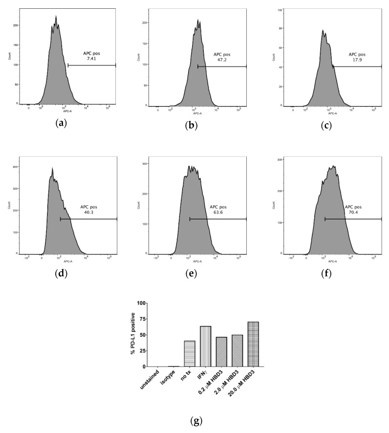
Human β-defensin 3 (HBD3) is an antimicrobial peptide up-regulated in the oral tissues of individuals with head and neck squamous cell carcinomas (HNSCC) and oral squamous cell carcinomas (SCC) and present in high concentrations in their saliva. In this study, we determined if HBD3 contributes to HNSCC pathogenesis by inducing programmed death-ligand 1 (PD-L1) expression on HNSCC cell lines. For this, SCC cell lines SCC4, SCC15, SCC19, SCC25, and SCC99 (5.0 × 10 viable cells) were used. Cells were incubated with IFNγ (0.6 µM) and HBD3 (0.2, 2.0, or 20.0 µM) for 24 h. Cells alone served as controls. Cells were then treated with anti-human APC-CD274 (PD-L1) and Live/Dead Fixable Green Dead Cell Stain. Cells treated with an isotype antibody and cells alone served as controls. All cell suspensions were analyzed in a LSR II Violet Flow Cytometer. Cytometric data was analyzed using FlowJo software. Treatment with IFNγ (0.6 µM) increased the number of cells expressing PD-L1 ( < 0.05) with respect to controls. Treatment with HBD3 (20.0 µM) also increased the number of cells expressing PD-L1 ( < 0.05) with respect to controls. However, treatment with IFNγ (0.6 µM) was not significantly different from treatment with HBD3 (20.0 µM) and the numbers of cells expressing PD-L1 were similar ( = 1). Thus, HBD3 increases the number of cells expressing PD-L1. This is a novel concept, but the role HBD3 contributes to HNSCC pathogenesis by inducing PD-L1 expression in tumors will have to be determined.
人β-防御素3(HBD3)是一种抗菌肽,在头颈部鳞状细胞癌(HNSCC)和口腔鳞状细胞癌(SCC)患者的口腔组织中上调,并在其唾液中高浓度存在。在本研究中,我们确定HBD3是否通过诱导HNSCC细胞系上的程序性死亡配体1(PD-L1)表达来促进HNSCC发病机制。为此,使用了SCC细胞系SCC4、SCC15、SCC19、SCC25和SCC99(5.0×10个活细胞)。将细胞与IFNγ(0.6μM)和HBD3(0.2、2.0或20.0μM)孵育24小时。仅细胞用作对照。然后用抗人APC-CD274(PD-L1)和活/死可固定绿色死细胞染色剂处理细胞。用同型抗体处理的细胞和仅细胞用作对照。所有细胞悬液在LSR II紫罗兰色流式细胞仪中进行分析。使用FlowJo软件分析细胞计数数据。与对照相比,用IFNγ(0.6μM)处理增加了表达PD-L1的细胞数量(<0.05)。与对照相比,用HBD3(20.0μM)处理也增加了表达PD-L1的细胞数量(<0.05)。然而,用IFNγ(0.