Charité - Universitätsmedizin Berlin, corporate member of Freie Universität Berlin, Humboldt-Universität zu Berlin, and Berlin Institute of Health, Charitéplatz 1, 10117, Berlin, Germany.
Department of Veterinary Medicine, Institute of Animal Welfare, Animal Behavior and Laboratory Animal Science, Freie Universität Berlin, Königsweg 67, Building 21, 14163, Berlin, Germany.
Sci Rep. 2019 Sep 25;9(1):13827. doi: 10.1038/s41598-019-50100-8.
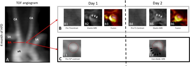
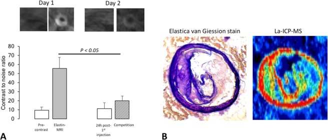
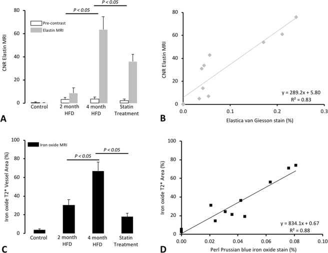
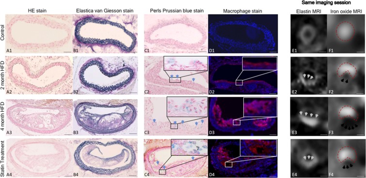
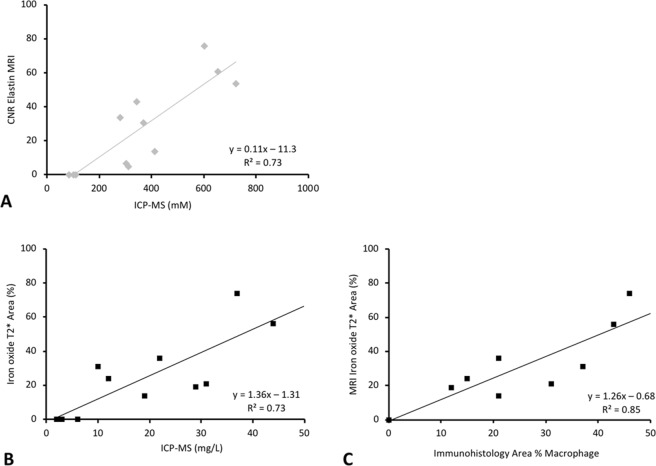
Molecular MRI is a promising in-vivo modality to detect and quantify morphological and molecular vessel-wall changes in atherosclerosis. The combination of different molecular biomarkers may improve the risk stratification of patients. This study aimed to investigate the feasibility of simultaneous visualization and quantification of plaque-burden and inflammatory activity by dual-probe molecular MRI in a mouse-model of progressive atherosclerosis and in response-to-therapy. Homozygous apolipoprotein E knockout mice (ApoE) were fed a high-fat-diet (HFD) for up to four-months prior to MRI of the brachiocephalic-artery. To assess response-to-therapy, a statin was administered for the same duration. MR imaging was performed before and after administration of an elastin-specific gadolinium-based and a macrophage-specific iron-oxide-based probe. Following in-vivo MRI, samples were analyzed using histology, immunohistochemistry, inductively-coupled-mass-spectrometry and laser-inductively-coupled-mass-spectrometry. In atherosclerotic-plaques, intraplaque expression of elastic-fibers and inflammatory activity were not directly linked. While the elastin-specific probe demonstrated the highest accumulation in advanced atherosclerotic-plaques after four-months of HFD, the iron-oxide-based probe showed highest accumulation in early atherosclerotic-plaques after two-months of HFD. In-vivo measurements for the elastin and iron-oxide-probe were in good agreement with ex-vivo histopathology (Elastica-van-Giesson stain: y = 298.2 + 5.8, R = 0.83, p < 0.05; Perls' Prussian-blue-stain: y = 834.1 + 0.67, R = 0.88, p < 0.05). Contrast-to-noise-ratio (CNR) measurements of the elastin probe were in good agreement with ICP-MS (y = 0.11x-11.3, R² = 0.73, p < 0.05). Late stage atherosclerotic-plaques displayed the strongest increase in both CNR and gadolinium concentration (p < 0.05). The gadolinium probe did not affect the visualization of the iron-oxide-probe and vice versa. This study demonstrates the feasibility of simultaneous assessment of plaque-burden and inflammatory activity by dual-probe molecular MRI of progressive atherosclerosis. The in-vivo detection and quantification of different MR biomarkers in a single scan could be useful to improve characterization of atherosclerotic-lesions.
分子磁共振成像是一种有前途的体内模态,可以检测和量化动脉粥样硬化中形态和分子血管壁的变化。不同分子生物标志物的结合可能会改善患者的风险分层。本研究旨在探讨在进展性动脉粥样硬化的小鼠模型中以及在治疗反应中,通过双探针分子磁共振成像同时可视化和定量斑块负担和炎症活性的可行性。载脂蛋白 E 基因敲除(ApoE)纯合子小鼠(ApoE)在进行磁共振成像之前接受高脂肪饮食(HFD)喂养长达四个月。为了评估治疗反应,给予他汀类药物治疗相同的时间。在给予弹性蛋白特异性钆基探针和巨噬细胞特异性氧化铁探针之前和之后进行磁共振成像。在体内 MRI 后,使用组织学、免疫组织化学、电感耦合质谱和激光诱导耦合质谱分析样本。在动脉粥样硬化斑块中,斑块内弹性纤维的表达与炎症活性没有直接联系。虽然在 HFD 喂养四个月后,弹性蛋白特异性探针在进展性动脉粥样硬化斑块中显示出最高的积累,但在 HFD 喂养两个月后,氧化铁探针在早期动脉粥样硬化斑块中显示出最高的积累。体内测量的弹性蛋白和氧化铁探针与体外组织病理学非常吻合(弹性纤维van-Giesson 染色:y=298.2+5.8,R=0.83,p<0.05;普鲁士蓝染色:y=834.1+0.67,R=0.88,p<0.05)。弹性蛋白探针的对比噪声比(CNR)测量与 ICP-MS 非常吻合(y=0.11x-11.3,R²=0.73,p<0.05)。晚期动脉粥样硬化斑块在 CNR 和钆浓度方面显示出最强的增加(p<0.05)。钆探针不会影响氧化铁探针的可视化,反之亦然。本研究证明了通过进展性动脉粥样硬化的双探针分子磁共振成像同时评估斑块负担和炎症活性的可行性。在单次扫描中检测和定量不同的磁共振生物标志物可能有助于改善对动脉粥样硬化病变的特征描述。