Institute of Medical Biology, UiT The Arctic University of Norway, Tromsø, Norway.
Department of Clinical Pathology, University Hospital of North Norway, Tromsø, Norway.
J Transl Med. 2019 Oct 3;17(1):334. doi: 10.1186/s12967-019-2086-x.
MicroRNAs (miRNAs) are promising biomarkers due to their structural stability and distinct expression profile in various cancers. We wanted to explore the miRNA expression in benign breast tissue and breast cancer subgroups in the Norwegian Women and Cancer study.
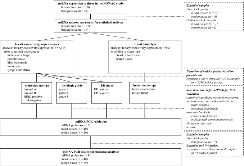
Specimens and histopathological data from study participants in Northern Norway diagnosed with breast cancer, and benign tissue from breast reduction surgery were collected. Main molecular subtypes were based on surrogate markers; luminal A (ER+ and/or PR+, HER2- and Ki67 ≤ 30%), luminal B (ER+ and/or PR+, HER2- and Ki67 > 30% or ER+ and/or PR+ and HER2+), HER2 positive (ER- and PR- and HER2+) and triple-negative (ER-, PR- and HER2-). RNA was extracted from formalin-fixed paraffin-embedded (FFPE) tissue, and miRNAs were successfully analyzed in 102 cancers and 36 benign controls using the 7th generation miRCURY LNA microarray containing probes targeting all human miRNAs as annotated in miRBASE version 19.0. Validation with RT-qPCR was performed.
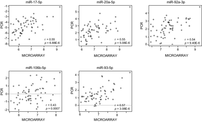
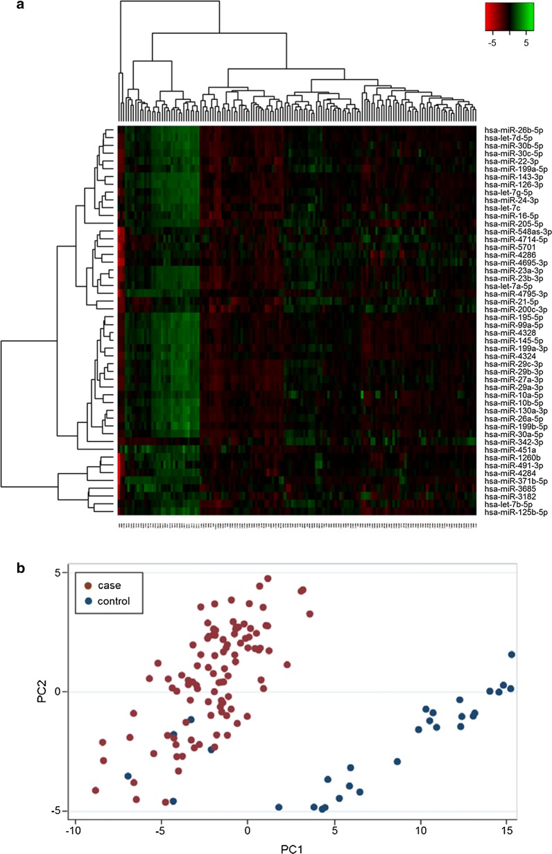
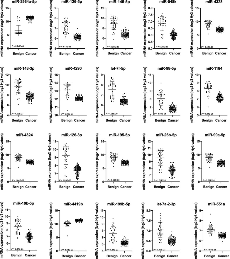
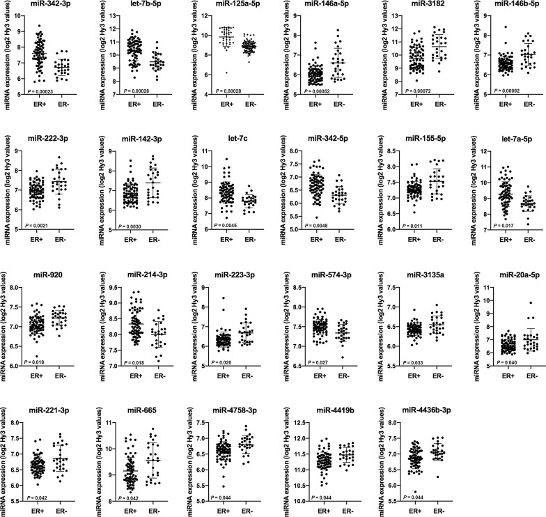
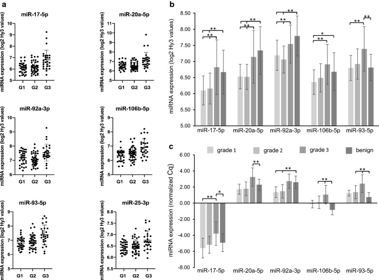
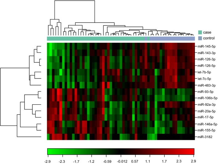
On average, 450 miRNAs were detected in each sample, and 304 miRNAs were significantly different between malignant and benign tissue. Subgroup analyses of cancer cases revealed 23 miRNAs significantly different between ER+ and ER- tumors, and 47 miRNAs different between tumors stratified according to grade. Significantly higher levels were found in high grade tumors for miR-17-5p (p = 0.006), miR-20a-5p (p = 0.007), miR-106b-5p (p = 0.007), miR-93-5p (p = 0.007) and miR-25-3p (p = 0.015) from the paralogous clusters miR-17-92 and miR-106b-25. Expression of miR-17-5p (p = 0.0029), miR-20a-5p (p = 0.0021), miR-92a-3p (p = 0.011) and miR-106b-5p (p = 0.021) was significantly higher in triple-negative tumors compared to the rest, and miR-17-5p and miR-20a-5p were significantly lower in luminal A tumors.
miRNA expression profiles were significantly different between malignant and benign tissue and between cancer subgroups according to ER- status, grade and molecular subtype. miRNAs in the miR-17-92 cluster and miR-17 family were overexpressed in high grade and triple-negative tumors associated with aggressive behavior. The expression and functional role of these miRNAs should be further studied in breast cancer to explore their potential as biomarkers in diagnostic pathology and clinical oncology.
微小 RNA(miRNAs)是很有前途的生物标志物,因为它们在各种癌症中具有结构稳定性和独特的表达谱。我们希望在挪威妇女与癌症研究中探索良性乳腺组织和乳腺癌亚组中的 miRNA 表达。
收集了来自挪威北部患有乳腺癌的研究参与者的标本和组织病理学数据,以及乳房缩小手术的良性组织。主要分子亚型基于替代标志物;腔 A(ER+ 和/或 PR+,HER2-和 Ki67≤30%),腔 B(ER+ 和/或 PR+,HER2-和 Ki67>30%或 ER+ 和/或 PR+和 HER2+),HER2 阳性(ER-和 PR-和 HER2+)和三阴性(ER-,PR-和 HER2-)。使用包含 miRBASE 版本 19.0 中注释的所有人类 miRNA 探针的第 7 代 miRCURY LNA 微阵列从福尔马林固定石蜡包埋(FFPE)组织中提取 RNA,并成功分析了 102 例癌症和 36 例良性对照中的 miRNA。使用 RT-qPCR 进行了验证。
平均每个样本检测到 450 个 miRNA,恶性和良性组织之间有 304 个 miRNA 存在显著差异。对癌症病例的亚组分析显示,ER+和 ER-肿瘤之间有 23 个 miRNA 存在显著差异,根据分级分层的肿瘤之间有 47 个 miRNA 存在差异。高级别肿瘤中 miR-17-5p(p=0.006)、miR-20a-5p(p=0.007)、miR-106b-5p(p=0.007)、miR-93-5p(p=0.007)和 miR-25-3p(p=0.015)的表达水平更高,这些 miRNA 来自 miRNA-17-92 和 miRNA-106b-25 的基因簇。miR-17-5p(p=0.0029)、miR-20a-5p(p=0.0021)、miR-92a-3p(p=0.011)和 miR-106b-5p(p=0.021)在三阴性肿瘤中的表达明显高于其他肿瘤,而 miR-17-5p 和 miR-20a-5p 在腔 A 肿瘤中的表达明显较低。
恶性和良性组织之间以及根据 ER-状态、分级和分子亚型的癌症亚组之间的 miRNA 表达谱存在显著差异。miR-17-92 簇和 miR-17 家族中的 miRNA 在高级别和三阴性肿瘤中过度表达,与侵袭性行为相关。这些 miRNA 的表达和功能作用应在乳腺癌中进一步研究,以探索其作为诊断病理学和临床肿瘤学中生物标志物的潜力。