Jiangsu key Laboratory of Oral Diseases, Department of Conservative Dentistry and Endodontics, Stomatological Hospital, Nanjing Medical University, Nanjing, China.
The Affiliated Suzhou Science and Technology Town Hospital of Nanjing Medical University, Suzhou, China.
BMC Oral Health. 2019 Oct 7;19(1):216. doi: 10.1186/s12903-019-0899-x.
The resin bond strength of sclerotic dentine is significantly lower than that of the normal dentine, which paused a challenge for bonding procedures clinically. The aim of this study was to compare the effects of different surface pretreatments on the micro-tensile bond strength and microstructure between sclerotic dentine and normal dentine.
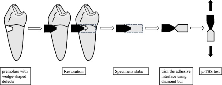
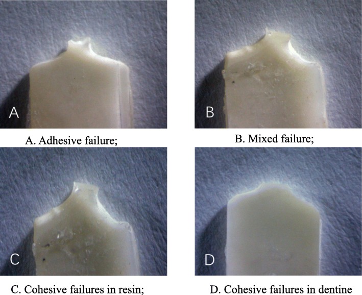
Eighty teeth that were collected, forty premolars with typical wedge-shaped defects visually graded as class III were assigned as the sclerotic dentine group (SD), the other forty normal premolars with artificial wedge-shaped defects were assigned as the normal dentine group (ND). Each group was randomly subdivided into eight subgroups according to the solution used: 35% phosphoric acid, 15% EDTA, 5% or 10% NaClO. Then the dentine surface was examined using a scanning electron microscope (SEM). The lesions were restored using self-etching adhesive and the subsequent resin composite. The teeth were sectioned into sticks for the micro-tensile bond strength analysis, and the data were analysed using the SPSS17.0 software package (α = 0.05).
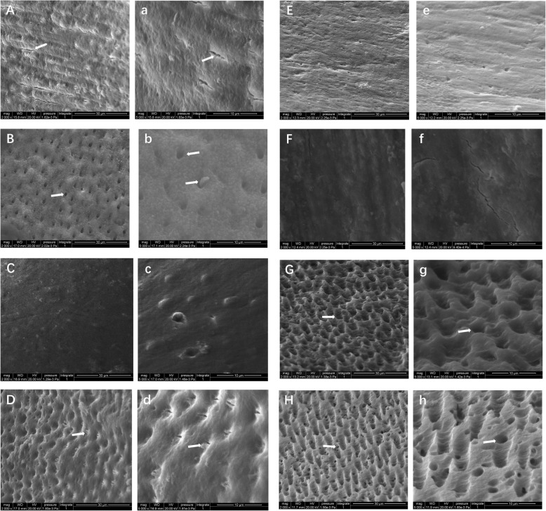
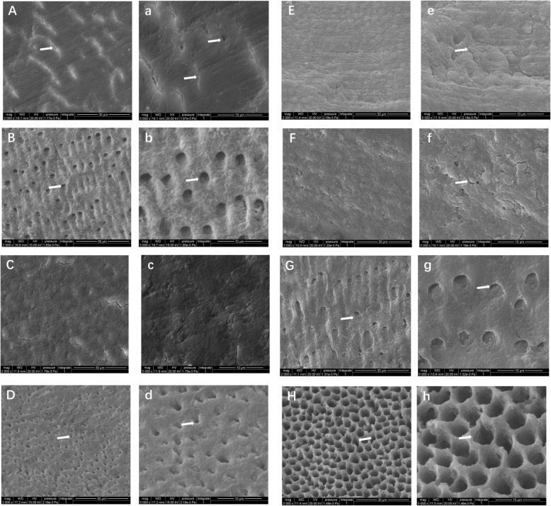
First, for the ND groups, after pretreatment using 35% phosphoric acid, and 35% phosphoric acid + 5% or 10% sodium hypochlorite, the bonding strengths of the normal dentine were higher than that of the other groups (P < 0.05). Second, for the SD groups, after pretreatment using 35% phosphoric acid, 15% EDTA, and 35% phosphoric acid + 5% or 10% sodium hypochlorite, the bonding strengths of the sclerotic dentine were higher than that of the other groups (P < 0.05). Third, the bond strengths of the sclerotic dentine were lower than that of the normal dentine without any pretreatment (P < 0.05). After pretreatment using 35% phosphoric acid + 5% or 10% sodium hypochlorite, the bonding strengths of the sclerotic dentine were higher than that of the normal dentine (P < 0.05). SEM observation showed that the appearances of dentine surface were changed after pretreatment using the above solutions, with the reduced smear layer, opened small groove and increased dentinal tubules.
Pretreatment of dentine using 35% phosphoric acid+ 5% or + 10% sodium hypochlorite changed the microstructure of the sclerotic dentine surface and subsequently increased the micro-tensile bond strength.
硬化牙本质的树脂粘结强度明显低于正常牙本质,这给临床粘结程序带来了挑战。本研究的目的是比较不同表面预处理方法对硬化牙本质和正常牙本质之间微拉伸粘结强度和微观结构的影响。
收集 80 颗牙齿,40 颗前磨牙有典型的楔形缺损,肉眼评估为 III 级,分为硬化牙本质组(SD),另 40 颗具有人工楔形缺损的正常前磨牙分为正常牙本质组(ND)。每组根据所用溶液随机分为 8 个亚组:35%磷酸、15% EDTA、5%或 10%次氯酸钠。然后使用扫描电子显微镜(SEM)检查牙本质表面。使用自酸蚀粘结剂和随后的树脂复合材料修复病变。将牙齿切成棒状进行微拉伸粘结强度分析,使用 SPSS17.0 软件包(α=0.05)进行数据分析。
首先,对于 ND 组,在用 35%磷酸和 35%磷酸+5%或 10%次氯酸钠预处理后,正常牙本质的粘结强度高于其他组(P<0.05)。其次,对于 SD 组,在用 35%磷酸、15% EDTA 和 35%磷酸+5%或 10%次氯酸钠预处理后,硬化牙本质的粘结强度高于其他组(P<0.05)。第三,未经预处理的硬化牙本质粘结强度低于正常牙本质(P<0.05)。用 35%磷酸+5%或 10%次氯酸钠预处理后,硬化牙本质的粘结强度高于正常牙本质(P<0.05)。SEM 观察显示,用上述溶液预处理后牙本质表面外观发生变化,玷污层减少,小沟槽打开,牙本质小管增多。
用 35%磷酸+5%或 10%次氯酸钠预处理牙本质改变了硬化牙本质表面的微观结构,随后增加了微拉伸粘结强度。