Kim Gangmi, Kim Sun Il, Lee Kang Young
Department of Surgery, Severance Hospital, Yonsei University College of Medicine, Seoul, 03722, South Korea.
Department of Pathology, Severance Hospital, Yonsei University College of Medicine, Seoul, 03722, South Korea.
F1000Res. 2019 May 13;8:652. doi: 10.12688/f1000research.19110.1. eCollection 2019.
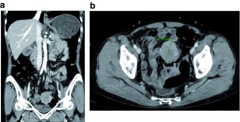
Schwannomas are tumors originating in Schwann cells of the peripheral nerve system and uncommonly develop in the gastrointestinal tract. Sigmoid colon schwannomas are very rare and only 28 cases have been reported. This study aims to report a case of a sigmoid colon schwannoma and present a literature review. We report a case of a 66-year-old female with asymptomatic sigmoid colon schwannoma. The patient underwent a screening colonoscopy and about 4cm sized submucosal tumor was identified at the sigmoid colon. A colonoscopic biopsy was performed and the microscopic exam revealed an ulcerated lesion with a proliferation of fibroblast-like spindle cells beneath ulcer, which was insufficient for diagnosis. Abdominopelvic computerized tomography (CT) scan showed a well-defined, well-enhancing, round shaped and slightly heterogenous mass at the sigmoid colon. No distant metastasis was identified in abdominopelvic CT and chest CT scans. Carcinoembryonic antigen level was within a normal range (1.33ng/mL). The patient underwent laparoscopic anterior resection. Immunohistochemical staining of the resected specimen showed positivity for S-100 protein in tumor cells and schwannoma was diagnosed post-surgically. Surgical resection margins were free from tumor and no regional lymph node metastasis was reported. Colon schwannomas are rare diseases. Most cases of colon schwannomas are accidentally identified during screening colonoscopy. The tumors usually present as submucosal masses and colonoscopic biopsies are mostly non-diagnostic. Surgical resection is required, and definitive diagnosis is made by confirming S-100 positive tumor cells in immunohistochemical analysis. Most cases are benign; a few cases have been reported to be malignant. Surgical resection with free negative margins is the treatment of choice.
施万细胞瘤起源于周围神经系统的施万细胞,在胃肠道中罕见发生。乙状结肠施万细胞瘤极为罕见,仅有28例报道。本研究旨在报告1例乙状结肠施万细胞瘤病例并进行文献综述。我们报告1例66岁无症状乙状结肠施万细胞瘤女性患者。患者接受结肠镜筛查,在乙状结肠发现一个约4cm大小的黏膜下肿瘤。进行了结肠镜活检,显微镜检查显示溃疡病变,溃疡下方有成纤维细胞样梭形细胞增生,这不足以确诊。腹盆腔计算机断层扫描(CT)显示乙状结肠有一个边界清晰、强化良好、圆形且稍不均匀的肿块。腹盆腔CT和胸部CT扫描未发现远处转移。癌胚抗原水平在正常范围内(1.33ng/mL)。患者接受了腹腔镜前切除术。切除标本的免疫组化染色显示肿瘤细胞S-100蛋白阳性,术后诊断为施万细胞瘤。手术切缘无肿瘤,未报告区域淋巴结转移。结肠施万细胞瘤是罕见疾病。大多数结肠施万细胞瘤病例是在结肠镜筛查时偶然发现的。肿瘤通常表现为黏膜下肿块,结肠镜活检大多无法确诊。需要手术切除,并通过免疫组化分析确认S-100阳性肿瘤细胞来做出明确诊断。大多数病例为良性;少数病例报告为恶性。手术切除切缘阴性是首选治疗方法。