Department of Mechanical Engineering, Pohang University of Science and Technology, 77 Cheongam-ro, Nam-gu, Pohang, Gyeoungbuk, 37673, Republic of Korea.
Division of Integrative Biosciences and Biotechnology, Pohang University of Science and Technology, 77 Cheongam-ro, Nam-gu, Pohang, Gyeoungbuk, 37673, Republic of Korea.
Sci Rep. 2019 Oct 29;9(1):15457. doi: 10.1038/s41598-019-51893-4.
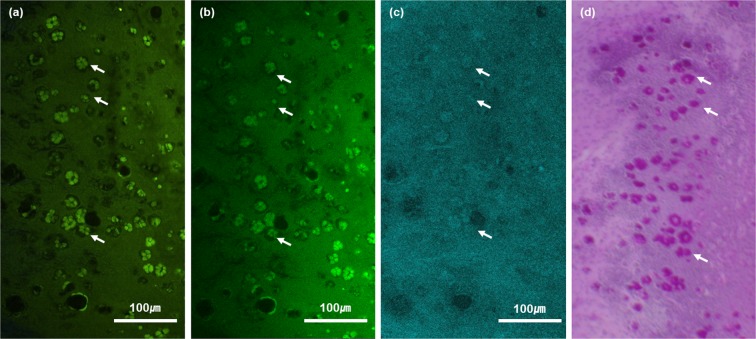
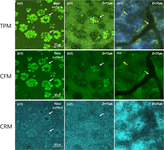
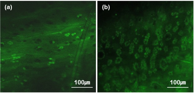
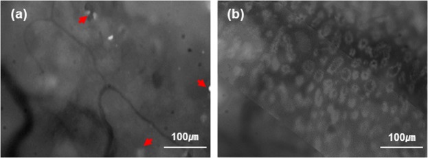
Conjunctival goblet cells (GCs) are specialized epithelial cells that secrete mucins onto the ocular surface to maintain the wet environment. Assessment of GCs is important because various ocular surface diseases are associated with their loss. Although there are GC assessment methods available, the current methods are either invasive or difficult to use. In this report, we developed a simple and non-invasive GC assessment method based on fluorescence imaging. Moxifloxacin ophthalmic solution was used to label GCs via topical administration, and then various fluorescence microscopies could image GCs in high contrasts. Fluorescence imaging of GCs in the mouse conjunctiva was confirmed by both confocal reflection microscopy and histology with Periodic acid-Schiff (PAS) labeling. Real-time in-vivo conjunctival GC imaging was demonstrated in a rat model by using both confocal fluorescence microscopy and simple wide-field fluorescence microscopy. Different GC densities were observed in the forniceal and bulbar conjunctivas of the rat eye. Moxifloxacin based fluorescence imaging provides high-contrast images of conjunctival GCs non-invasively and could be useful for the study or diagnosis of GC related ocular surface diseases.
结膜杯状细胞(GCs)是分泌粘蛋白到眼表面以维持湿润环境的特化上皮细胞。评估 GCs 很重要,因为各种眼表面疾病都与它们的丧失有关。尽管有可用的 GCs 评估方法,但目前的方法要么具有侵入性,要么难以使用。在本报告中,我们开发了一种基于荧光成像的简单、非侵入性的 GCs 评估方法。莫西沙星滴眼液通过局部给药标记 GCs,然后各种荧光显微镜可以高对比度地成像 GCs。共聚焦反射显微镜和过碘酸雪夫(PAS)标记的组织学证实了小鼠结膜 GCs 的荧光成像。通过共聚焦荧光显微镜和简单的宽场荧光显微镜,在大鼠模型中证明了实时活体结膜 GC 成像。在大鼠眼睛的穹窿和球结膜中观察到不同的 GC 密度。莫西沙星荧光成像提供了非侵入性的高对比度的结膜 GCs 图像,可用于研究或诊断与 GC 相关的眼表面疾病。