Marek Josef, Lubanda Jean-Claude, Cifkova Renata, Kuchynka Petr, Golan Lubor, Nemcek Eduard, Linhart Ales
2nd Department of Medicine - Department of Cardiovascular Medicine, First Faculty of Medicine, Charles University and General University Hospital in Prague, U Nemocnice 2, 128 08, Praha 2, Czech Republic.
Center for Cardiovascular Prevention, First Faculty of Medicine, Charles University in Prague and Thomayer Hospital, Prague, Czech Republic.
Cardiovasc Ultrasound. 2019 Oct 30;17(1):22. doi: 10.1186/s12947-019-0172-0.
Normative reference values for echocardiographic chamber quantification are of great importance; however, this can be challenging. Our aim was to derive these values including degrees of abnormality from a random Central European population sample with a homogeneous subset of healthy subjects.
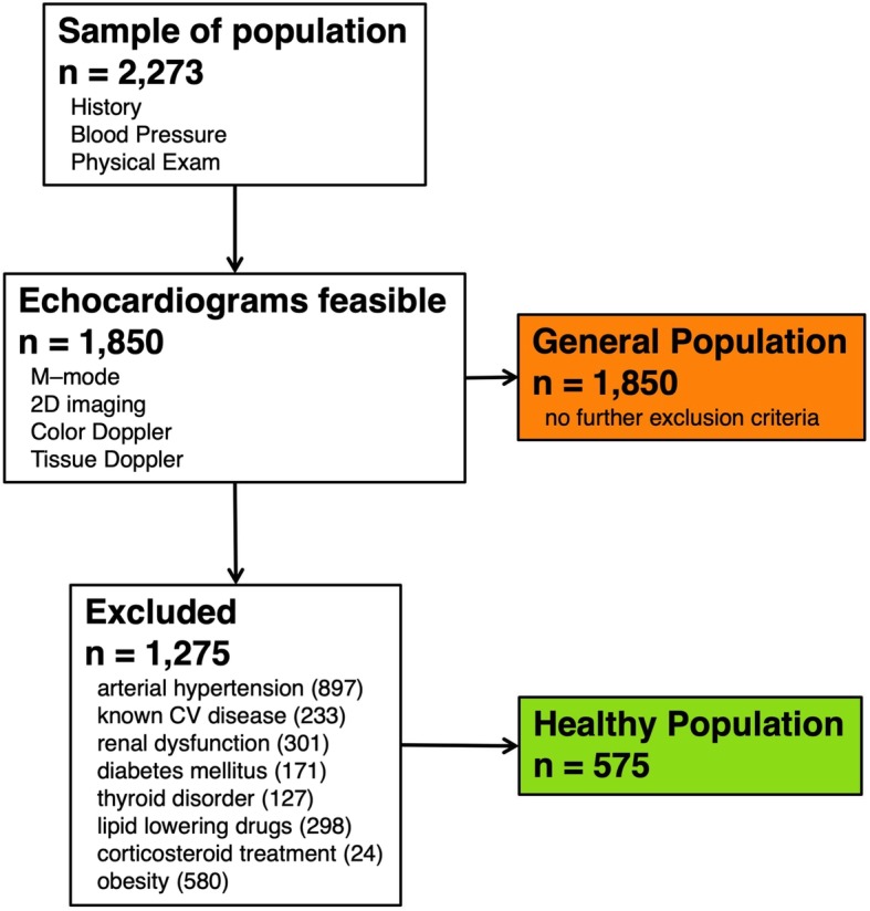
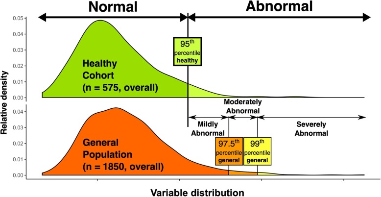
We analysed echocardiograms obtained in a randomly selected population sample during the Czech post-MONICA survey in 2007/2008. Overall, 1850 out of 2273 persons of the whole sample of three districts had adequate echocardiograms (81.4%). A healthy subgroup defined by the absence of known cardiovascular disease was used to define normal reference range limits (n = 575, median age 42 years [IQR 34-52], 57% females). The whole population sample with predefined percentile cut-offs was used to define degrees of abnormality.
Left ventricular (LV) size tended to decrease with age, while LV mass increased with age in both males and females and in both the healthy and general populations. LV dimensions were larger in males, except for body surface area-indexed LV diameter. M-mode derived LV measurements were larger and LV mass higher compared to 2D measurements. Right ventricle basal dimension was larger in males.
Our study provides reference ranges for echocardiographic measurements obtained in a healthy subgroup derived from an epidemiological study of a Central European population. Where feasible, degrees of abnormality are provided based on the whole population sample including patients with disease. Our data show that age, gender and measurement method significantly affect cardiac dimensions and function and should be always taken into account.
超声心动图腔室定量的正常参考值非常重要;然而,这可能具有挑战性。我们的目的是从具有健康受试者同质亚组的随机中欧人群样本中得出这些值,包括异常程度。
我们分析了在2007/2008年捷克莫尼卡调查期间从随机选择的人群样本中获得的超声心动图。总体而言,三个区的整个样本中的2273人中有1850人有足够的超声心动图(81.4%)。由无已知心血管疾病定义的健康亚组用于定义正常参考范围界限(n = 575,中位年龄42岁[四分位间距34 - 52],57%为女性)。具有预定义百分位数截断值的整个人群样本用于定义异常程度。
左心室(LV)大小倾向于随年龄减小,而左心室质量在男性和女性以及健康人群和总体人群中均随年龄增加。男性的左心室尺寸更大,但体表面积指数化的左心室直径除外。与二维测量相比,M型得出的左心室测量值更大,左心室质量更高。男性的右心室基底尺寸更大。
我们的研究提供了从对中欧人群的流行病学研究中得出的健康亚组的超声心动图测量参考范围。在可行的情况下,基于包括患病患者在内的整个人群样本提供异常程度。我们的数据表明,年龄、性别和测量方法会显著影响心脏尺寸和功能,应始终予以考虑。