• 文献检索
  • 文档翻译
  • 深度研究
  • 学术资讯
  • Suppr Zotero 插件Zotero 插件
  • 邀请有礼
  • 套餐&价格
  • 历史记录
应用&插件
Suppr Zotero 插件Zotero 插件浏览器插件Mac 客户端Windows 客户端微信小程序
定价
高级版会员购买积分包购买API积分包
服务
文献检索文档翻译深度研究API 文档MCP 服务
关于我们
关于 Suppr公司介绍联系我们用户协议隐私条款
关注我们

Suppr 超能文献

核心技术专利:CN118964589B侵权必究
粤ICP备2023148730 号-1Suppr @ 2026

文献检索

告别复杂PubMed语法,用中文像聊天一样搜索,搜遍4000万医学文献。AI智能推荐,让科研检索更轻松。

立即免费搜索

文件翻译

保留排版,准确专业,支持PDF/Word/PPT等文件格式,支持 12+语言互译。

免费翻译文档

深度研究

AI帮你快速写综述,25分钟生成高质量综述,智能提取关键信息,辅助科研写作。

立即免费体验

宫颈癌 T2 加权和弥散加权 MRI 影像组学特征:低体积肿瘤行子宫颈广泛切除术的预后价值。

Radiomic features of cervical cancer on T2-and diffusion-weighted MRI: Prognostic value in low-volume tumors suitable for trachelectomy.

机构信息

MRI Unit, Division of Radiotherapy and Imaging, The Institute of Cancer Research and the Royal Marsden NHS Foundation Trust, Sutton, UK.

Department of Gynaecological Oncology, The Royal Marsden NHS Foundation Trust, London, UK; St George's University of London, Tooting, London, UK.

出版信息

Gynecol Oncol. 2020 Jan;156(1):107-114. doi: 10.1016/j.ygyno.2019.10.010. Epub 2019 Nov 2.

DOI:10.1016/j.ygyno.2019.10.010
PMID:31685232
原文链接:https://pmc.ncbi.nlm.nih.gov/articles/PMC7001101/
Abstract

BACKGROUND

Textural features extracted from MRI potentially provide prognostic information additional to volume for influencing surgical management of cervical cancer.

PURPOSE

To identify textural features that differ between cervical tumors above and below the volume threshold of eligibility for trachelectomy and determine their value in predicting recurrence in patients with low-volume tumors.

METHODS

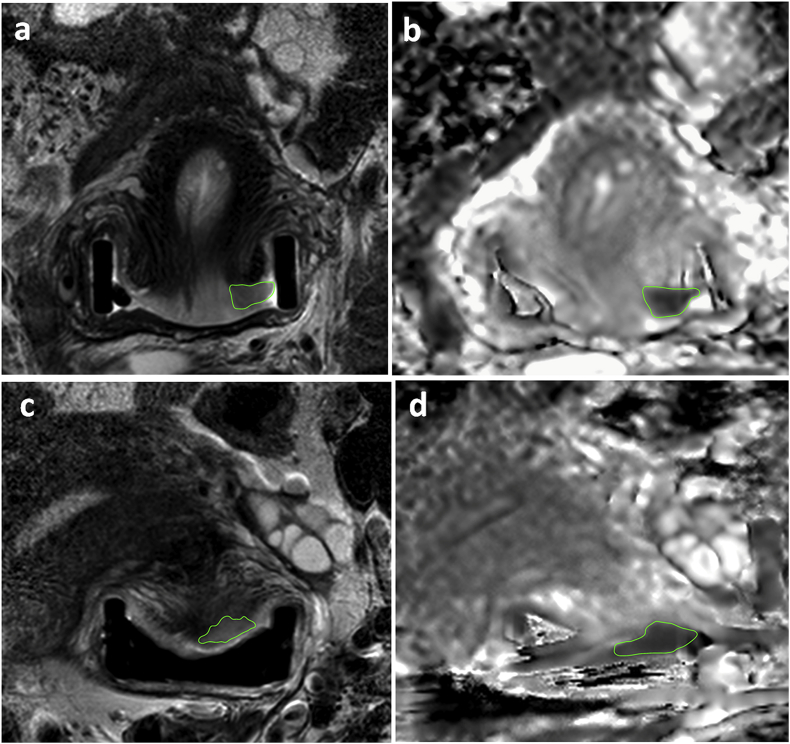
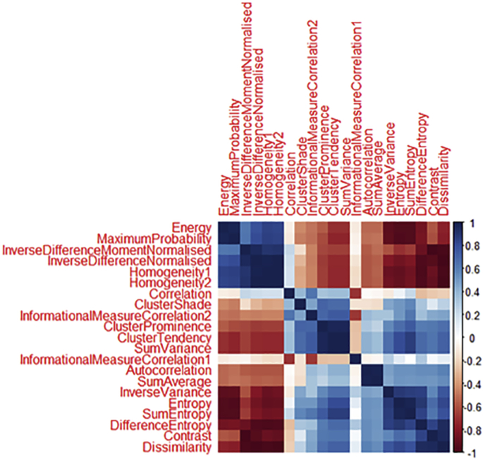
Of 378 patients with Stage1-2 cervical cancer imaged prospectively (3T, endovaginal coil), 125 had well-defined, histologically-confirmed squamous or adenocarcinomas with >100 voxels (>0.07 cm) suitable for radiomic analysis. Regions-of-interest outlined the whole tumor on T2-W images and apparent diffusion coefficient (ADC) maps. Textural features based on grey-level co-occurrence matrices were compared (Mann-Whitney test with Bonferroni correction) between tumors greater (n = 46) or less (n = 79) than 4.19 cm. Clustering eliminated correlated variables. Significantly different features were used to predict recurrence (regression modelling) in surgically-treated patients with low-volume tumors and compared with a model using clinico-pathological features.

RESULTS

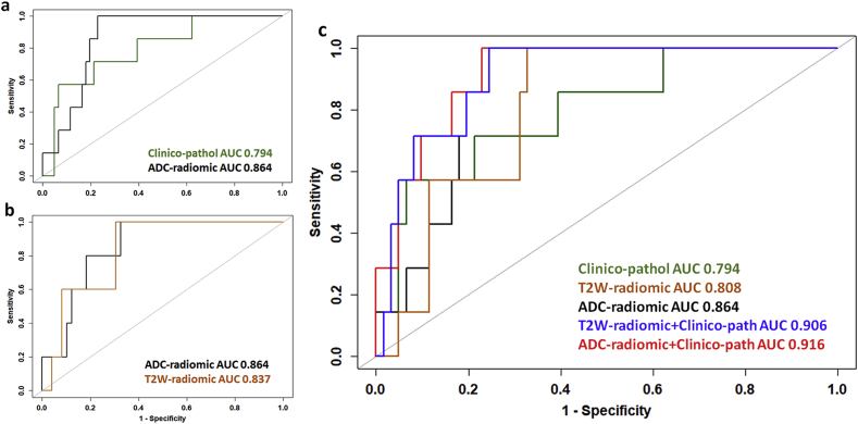
Textural features (Dissimilarity, Energy, ClusterProminence, ClusterShade, InverseVariance, Autocorrelation) in 6 of 10 clusters from T2-W and ADC data differed between high-volume (mean ± SD 15.3 ± 11.7 cm) and low-volume (mean ± SD 1.3 ± 1.2 cm) tumors. (p < 0.02). In low-volume tumors, predicting recurrence was indicated by: Dissimilarity, Energy (ADC-radiomics, AUC = 0.864); Dissimilarity, ClusterProminence, InverseVariance (T2-W-radiomics, AUC = 0.808); Volume, Depth of Invasion, LymphoVascular Space Invasion (clinico-pathological features, AUC = 0.794). Combining ADC-radiomic (but not T2-radiomic) and clinico-pathological features improved prediction of recurrence compared to the clinico-pathological model (AUC = 0.916, p = 0.006). Findings were supported by bootstrap re-sampling (n = 1000).

CONCLUSION

Textural features from ADC maps and T2-W images differ between high- and low-volume tumors and potentially predict recurrence in low-volume tumors.

摘要

背景

从 MRI 提取的纹理特征可能提供比体积更有预后意义的信息,从而影响宫颈癌的手术治疗。

目的

确定肿瘤体积是否超过行宫颈锥切术的阈值,识别肿瘤纹理特征,并确定其在预测低体积肿瘤患者复发中的价值。

方法

对 378 例经前瞻性 MRI 检查的Ⅰ-Ⅱ期宫颈癌患者(3T 场强,经阴道线圈)进行分析,其中 125 例为边界清晰、经组织学证实的鳞癌或腺癌,肿瘤体积>100 个体素(>0.07cm),适用于放射组学分析。在 T2-W 图像和表观扩散系数(ADC)图上勾画感兴趣区(ROI)以勾画整个肿瘤。比较(校正后的 Mann-Whitney 检验)肿瘤体积大于(n=46)或小于(n=79)4.19cm 的肿瘤的纹理特征(灰度共生矩阵)。聚类消除了相关变量。利用在接受手术治疗的低体积肿瘤患者中预测复发的显著不同的特征(回归模型),并与使用临床病理特征的模型进行比较。

结果

在 T2-W 和 ADC 数据的 10 个聚类中,有 6 个聚类的纹理特征(差异性、能量、聚类突出度、聚类阴影、逆方差、自相关)在高体积(平均值±标准差 15.3±11.7cm)和低体积(平均值±标准差 1.3±1.2cm)肿瘤之间存在差异(p<0.02)。在低体积肿瘤中,预测复发的指标包括:差异性、能量(ADC-放射组学,AUC=0.864);差异性、聚类突出度、逆方差(T2-W-放射组学,AUC=0.808);体积、浸润深度、脉管侵犯(临床病理特征,AUC=0.794)。与临床病理模型(AUC=0.916,p=0.006)相比,结合 ADC-放射组学(而非 T2-放射组学)和临床病理特征可提高预测复发的能力。Bootstrap 重采样(n=1000)支持该发现。

结论

高、低体积肿瘤之间的 ADC 图和 T2-W 图像的纹理特征不同,且可能预测低体积肿瘤的复发。

https://cdn.ncbi.nlm.nih.gov/pmc/blobs/08c0/7001101/966d6263d018/gr3.jpg
https://cdn.ncbi.nlm.nih.gov/pmc/blobs/08c0/7001101/26bbf38800da/gr1.jpg
https://cdn.ncbi.nlm.nih.gov/pmc/blobs/08c0/7001101/5075fe925bb4/gr2.jpg
https://cdn.ncbi.nlm.nih.gov/pmc/blobs/08c0/7001101/966d6263d018/gr3.jpg
https://cdn.ncbi.nlm.nih.gov/pmc/blobs/08c0/7001101/26bbf38800da/gr1.jpg
https://cdn.ncbi.nlm.nih.gov/pmc/blobs/08c0/7001101/5075fe925bb4/gr2.jpg
https://cdn.ncbi.nlm.nih.gov/pmc/blobs/08c0/7001101/966d6263d018/gr3.jpg

相似文献

1
Radiomic features of cervical cancer on T2-and diffusion-weighted MRI: Prognostic value in low-volume tumors suitable for trachelectomy.宫颈癌 T2 加权和弥散加权 MRI 影像组学特征:低体积肿瘤行子宫颈广泛切除术的预后价值。
Gynecol Oncol. 2020 Jan;156(1):107-114. doi: 10.1016/j.ygyno.2019.10.010. Epub 2019 Nov 2.
2
Radiomics analysis of apparent diffusion coefficient in cervical cancer: A preliminary study on histological grade evaluation.宫颈癌表观扩散系数的放射组学分析:组织学分级评估的初步研究。
J Magn Reson Imaging. 2019 Jan;49(1):280-290. doi: 10.1002/jmri.26192. Epub 2018 May 14.
3
Preoperative imaging in patients undergoing trachelectomy for cervical cancer: validation of a combined T2- and diffusion-weighted endovaginal MRI technique at 3.0 T.宫颈癌行根治性宫颈切除术患者的术前影像学检查:3.0T 下 T2 与弥散加权联合的经阴道内 MRI 技术的验证。
Gynecol Oncol. 2014 May;133(2):326-32. doi: 10.1016/j.ygyno.2014.02.026. Epub 2014 Feb 26.
4
Texture Analysis as Imaging Biomarker for recurrence in advanced cervical cancer treated with CCRT.纹理分析作为影像学生物标志物预测接受 CCRT 治疗的晚期宫颈癌复发
Sci Rep. 2018 Jul 30;8(1):11399. doi: 10.1038/s41598-018-29838-0.
5
MRI-based radiomic signatures for pretreatment prognostication in cervical cancer.基于 MRI 的放射组学特征用于宫颈癌的术前预后预测。
Cancer Med. 2023 Oct;12(20):20251-20265. doi: 10.1002/cam4.6526. Epub 2023 Oct 16.
6
Deep learning for fully automated tumor segmentation and extraction of magnetic resonance radiomics features in cervical cancer.深度学习在宫颈癌全自动肿瘤分割和磁共振影像组学特征提取中的应用。
Eur Radiol. 2020 Mar;30(3):1297-1305. doi: 10.1007/s00330-019-06467-3. Epub 2019 Nov 11.
7
Relationship between imaging biomarkers of stage I cervical cancer and poor-prognosis histologic features: quantitative histogram analysis of diffusion-weighted MR images.I 期宫颈癌影像学生物标志物与预后不良组织学特征的关系:扩散加权磁共振图像的定量直方图分析。
AJR Am J Roentgenol. 2013 Feb;200(2):314-20. doi: 10.2214/AJR.12.9545.
8
Diffusion-weighted imaging in cervical cancer with an endovaginal technique: potential value for improving tumor detection in stage Ia and Ib1 disease.采用经阴道技术的宫颈癌扩散加权成像:对提高Ia期和Ib1期疾病肿瘤检测的潜在价值。
Radiology. 2008 Nov;249(2):541-50. doi: 10.1148/radiol.2491072165.
9
Radiomics Analysis of Multiparametric MRI Evaluates the Pathological Features of Cervical Squamous Cell Carcinoma.多参数 MRI 放射组学分析评估宫颈鳞状细胞癌的病理特征。
J Magn Reson Imaging. 2019 Apr;49(4):1141-1148. doi: 10.1002/jmri.26301. Epub 2018 Sep 19.
10
Novel T2 Mapping for Evaluating Cervical Cancer Features by Providing Quantitative T2 Maps and Synthetic Morphologic Images: A Preliminary Study.通过提供定量T2图谱和合成形态学图像评估宫颈癌特征的新型T2图谱:一项初步研究
J Magn Reson Imaging. 2020 Dec;52(6):1859-1869. doi: 10.1002/jmri.27297. Epub 2020 Aug 14.

引用本文的文献

1
Multi-parametric MRI-based radiomics nomogram for predicting lymphovascular space invasion in early-stage cervical adenocarcinoma.基于多参数磁共振成像的影像组学列线图预测早期宫颈腺癌的淋巴血管间隙浸润
Front Oncol. 2025 Aug 21;15:1612691. doi: 10.3389/fonc.2025.1612691. eCollection 2025.
2
A 20-feature radiomic signature of triple-negative breast cancer identifies patients at high risk of death.三阴性乳腺癌的一种20特征放射组学特征可识别出死亡风险高的患者。
NPJ Breast Cancer. 2025 Jul 26;11(1):79. doi: 10.1038/s41523-025-00790-3.
3
A T2 weighted imaging-based radiomics nomogram for the classification of hepatic blood-rich lesions: hepatocellular carcinoma and benign liver lesions.

本文引用的文献

1
Radiomics Nomogram Building From Multiparametric MRI to Predict Grade in Patients With Glioma: A Cohort Study.基于多参数 MRI 的影像组学列线图构建预测胶质瘤患者分级的模型:一项队列研究。
J Magn Reson Imaging. 2019 Mar;49(3):825-833. doi: 10.1002/jmri.26265. Epub 2018 Sep 8.
2
Early Cervical Cancer: Predictive Relevance of Preoperative 3-Tesla Multiparametric Magnetic Resonance Imaging.早期宫颈癌:术前3特斯拉多参数磁共振成像的预测相关性
Int J Surg Oncol. 2018 Aug 1;2018:9120753. doi: 10.1155/2018/9120753. eCollection 2018.
3
Radiomic analysis in T2W and SPAIR T2W MRI: predict treatment response to chemoradiotherapy in esophageal squamous cell carcinoma.
基于T2加权成像的放射组学列线图用于肝血供丰富病变的分类:肝细胞癌和良性肝病变
Discov Oncol. 2025 Jul 1;16(1):1223. doi: 10.1007/s12672-025-02868-7.
4
Integrating MRI-based radiomics and clinicopathological features for preoperative prognostication of early-stage cervical adenocarcinoma patients: in comparison to deep learning approach.基于 MRI 的放射组学与临床病理特征相结合对早期宫颈腺癌患者进行术前预后预测:与深度学习方法的比较。
Cancer Imaging. 2024 Aug 1;24(1):101. doi: 10.1186/s40644-024-00747-y.
5
Endovaginal coil for pelvic high-resolution magnetic resonance imaging of cervical cancer: a preliminary parameter optimization study.用于宫颈癌盆腔高分辨率磁共振成像的阴道内线圈:一项初步参数优化研究。
Quant Imaging Med Surg. 2024 Jun 1;14(6):3851-3862. doi: 10.21037/qims-23-1718. Epub 2024 May 24.
6
Radiomic profiles improve prognostication and reveal targets for therapy in cervical cancer.放射组学特征可改善宫颈癌的预后,并揭示治疗靶点。
Sci Rep. 2024 May 17;14(1):11339. doi: 10.1038/s41598-024-61271-4.
7
An integrated machine learning-based model for joint diagnosis of ovarian cancer with multiple test indicators.基于集成机器学习的多检测指标联合诊断卵巢癌模型
J Ovarian Res. 2024 Feb 20;17(1):45. doi: 10.1186/s13048-024-01365-9.
8
Radiomics analysis based on multiparametric magnetic resonance imaging for differentiating early stage of cervical cancer.基于多参数磁共振成像的影像组学分析用于鉴别宫颈癌早期阶段
Front Med (Lausanne). 2024 Feb 2;11:1336640. doi: 10.3389/fmed.2024.1336640. eCollection 2024.
9
An Update on the Role of MRI in Treatment Stratification of Patients with Cervical Cancer.MRI在宫颈癌患者治疗分层中作用的最新进展
Cancers (Basel). 2023 Oct 23;15(20):5105. doi: 10.3390/cancers15205105.
10
Establishment and validation of a radiological-radiomics model for predicting high-grade patterns of lung adenocarcinoma less than or equal to 3 cm.用于预测直径小于或等于3cm的肺腺癌高级别模式的放射学-放射组学模型的建立与验证
Front Oncol. 2022 Sep 15;12:964322. doi: 10.3389/fonc.2022.964322. eCollection 2022.
T2加权和短TI反转恢复序列T2加权磁共振成像中的影像组学分析:预测食管鳞状细胞癌对放化疗的治疗反应
J Thorac Dis. 2018 Apr;10(4):2256-2267. doi: 10.21037/jtd.2018.03.123.
4
Radiomics analysis of apparent diffusion coefficient in cervical cancer: A preliminary study on histological grade evaluation.宫颈癌表观扩散系数的放射组学分析:组织学分级评估的初步研究。
J Magn Reson Imaging. 2019 Jan;49(1):280-290. doi: 10.1002/jmri.26192. Epub 2018 May 14.
5
The European Society of Gynaecological Oncology/European Society for Radiotherapy and Oncology/European Society of Pathology guidelines for the management of patients with cervical cancer.欧洲妇科肿瘤学会/欧洲放射肿瘤学会/欧洲病理学会宫颈癌管理患者指南。
Radiother Oncol. 2018 Jun;127(3):404-416. doi: 10.1016/j.radonc.2018.03.003. Epub 2018 May 1.
6
Shell feature: a new radiomics descriptor for predicting distant failure after radiotherapy in non-small cell lung cancer and cervix cancer.壳特征:一种新的放射组学描述符,用于预测非小细胞肺癌和宫颈癌放疗后的远处失败。
Phys Med Biol. 2018 May 2;63(9):095007. doi: 10.1088/1361-6560/aabb5e.
7
Obstetric Outcomes in Women With Early Bulky Cervical Cancer Downstaged by Neoadjuvant Chemotherapy to Allow for Fertility-Sparing Surgery: A Meta-analysis and Metaregression.新辅助化疗降期后保留生育功能手术治疗早期大块宫颈癌的产科结局:Meta 分析和 Meta 回归。
Int J Gynecol Cancer. 2018 May;28(4):794-801. doi: 10.1097/IGC.0000000000001232.
8
Prediction of outcome using pretreatment F-FDG PET/CT and MRI radiomics in locally advanced cervical cancer treated with chemoradiotherapy.使用预处理 F-FDG PET/CT 和 MRI 放射组学预测接受放化疗的局部晚期宫颈癌的结局。
Eur J Nucl Med Mol Imaging. 2018 May;45(5):768-786. doi: 10.1007/s00259-017-3898-7. Epub 2017 Dec 9.
9
Apparent diffusion coefficient for prediction of parametrial invasion in cervical cancer: a critical evaluation based on stratification to a Likert scale using T2-weighted imaging.表观扩散系数预测宫颈癌宫旁浸润:基于T2加权成像按李克特量表分层的批判性评估
Radiol Med. 2018 Mar;123(3):209-216. doi: 10.1007/s11547-017-0823-x. Epub 2017 Oct 20.
10
Value of whole-lesion apparent diffusion coefficient (ADC) first-order statistics and texture features in clinical staging of cervical cancers.全病灶表观扩散系数(ADC)一阶统计量及纹理特征在宫颈癌临床分期中的价值
Clin Radiol. 2017 Nov;72(11):951-958. doi: 10.1016/j.crad.2017.06.115. Epub 2017 Jul 18.