• 文献检索
  • 文档翻译
  • 深度研究
  • 学术资讯
  • Suppr Zotero 插件Zotero 插件
  • 邀请有礼
  • 套餐&价格
  • 历史记录
应用&插件
Suppr Zotero 插件Zotero 插件浏览器插件Mac 客户端Windows 客户端微信小程序
定价
高级版会员购买积分包购买API积分包
服务
文献检索文档翻译深度研究API 文档MCP 服务
关于我们
关于 Suppr公司介绍联系我们用户协议隐私条款
关注我们

Suppr 超能文献

核心技术专利:CN118964589B侵权必究
粤ICP备2023148730 号-1Suppr @ 2026

文献检索

告别复杂PubMed语法,用中文像聊天一样搜索,搜遍4000万医学文献。AI智能推荐,让科研检索更轻松。

立即免费搜索

文件翻译

保留排版,准确专业,支持PDF/Word/PPT等文件格式,支持 12+语言互译。

免费翻译文档

深度研究

AI帮你快速写综述,25分钟生成高质量综述,智能提取关键信息,辅助科研写作。

立即免费体验

在坐骨软组织上多层注射羟基磷灰石填充剂,以恢复慢性坐压疮的前期状态。

Multilayered injection of calcium hydroxylapatite filler on ischial soft tissue to rejuvenate the previous phase of chronic sitting pressure sore.

作者信息

Kim JongSeo

机构信息

Kim-Jongseo Plastic Surgery Clinic, Seoul, Republic of Korea.

出版信息

Clin Cosmet Investig Dermatol. 2019 Oct 15;12:771-784. doi: 10.2147/CCID.S212599. eCollection 2019.

DOI:10.2147/CCID.S212599
PMID:31686889
原文链接:https://pmc.ncbi.nlm.nih.gov/articles/PMC6800547/
Abstract

INTRODUCTION

During a sitting position, the pressure distribution is located below the ischial tuberosity. Many women have skin atrophy on the ischial area. To treat atrophic changes on the skin above the ischium, volumization and improving skin texture are acquired simultaneously. Two methods of automatic and manual injections using a hard filler with a stitmulating effect were administered respectively to both the dermis and subdermis layers. A biopsy study using various straining evaluated histological tissue reactions after the filler injections.

METHODS

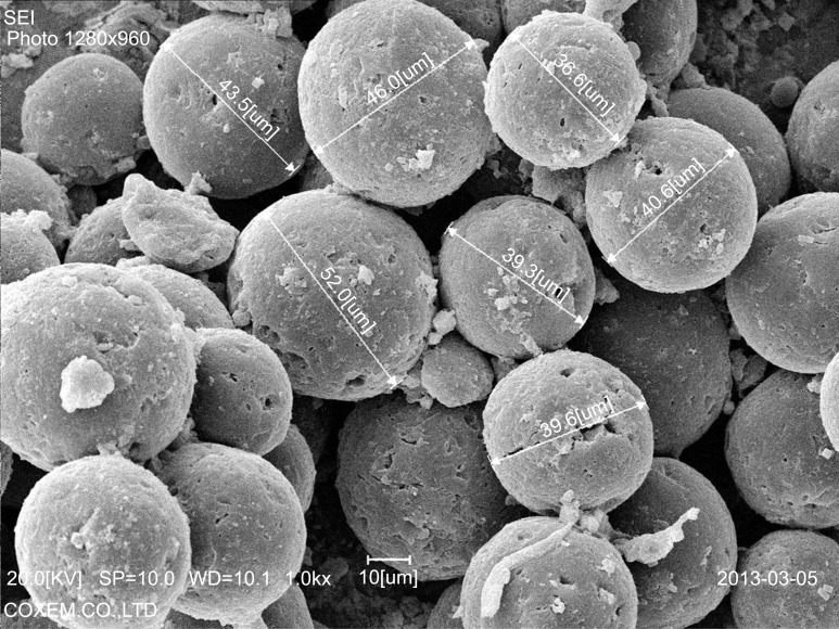
This study focused on rejuvenating soft tissue on the atrophic ischeal areas, as described by the author as the previous phase of chronic sitting pressure sore, by using the multi-layered injection of calcium-hydroxylapatite (CaHA) filler. Sixteen women (mean, 38.5 years) were treated from 2012 January to 2019 April. Prior to the injection, 1.5cc of Radiesse (calcium hydroxylapatite filler; Merz, Germany) was diluted with 1cc of normal saline and 0.5cc of lidocaine, and 3cc of filler mixture (1:1 dilution) was made. All subjects received the intradermal injection and multi layered subdermal injection with 2.5cc of diluted CaHA filler. A second session for booster treatment was performed at 6 months using the same method. Photography was taken by a camera and a dermascope observation before and 7 months after. Before and 7 months after the first injection, soft tissue depression, skin discoloration, and roughness were assessed. Standard deviations and coefficients of variation were also calculated for changes in depression, discoloration and roughness after the treatment. Biopsy specimens (3×5 mm) were taken from three patients 7 months after the first session. The specimens were analyzed using various stainins.

RESULTS

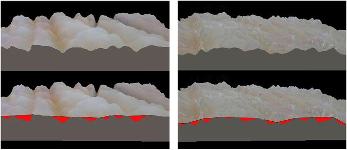
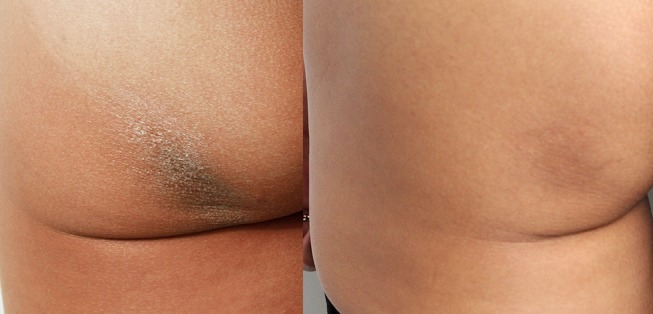
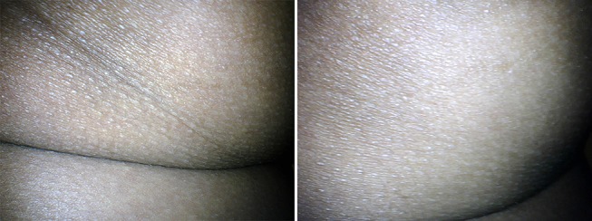
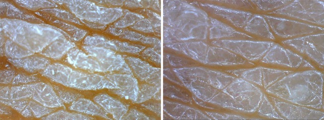
The improvements of skin quality, skin fold, and roughness were visible at physical examination, medical photography and also at high-resolution dermascope examination in all patients. Post-treatment the depressed amounts on the ischial areas reduced with increased volume.

CONCLUSION

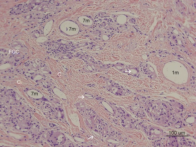
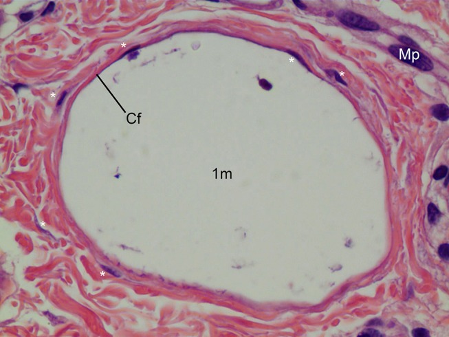
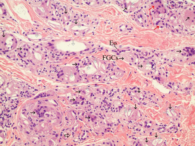
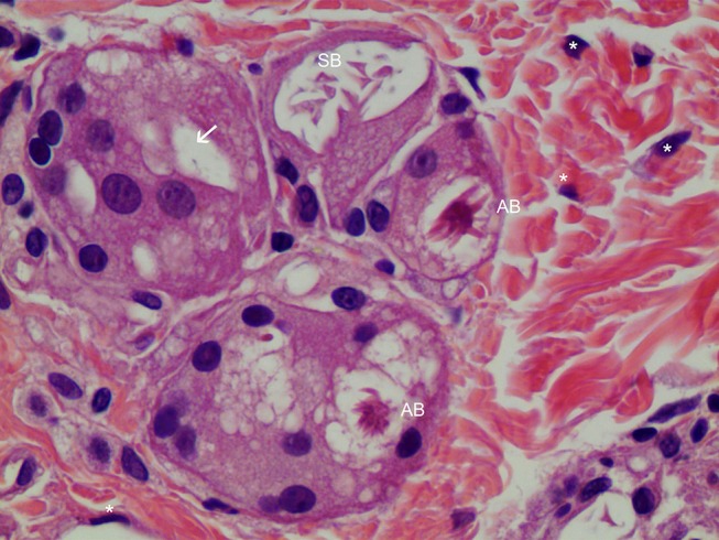
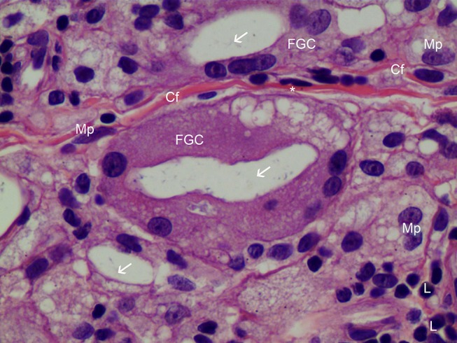
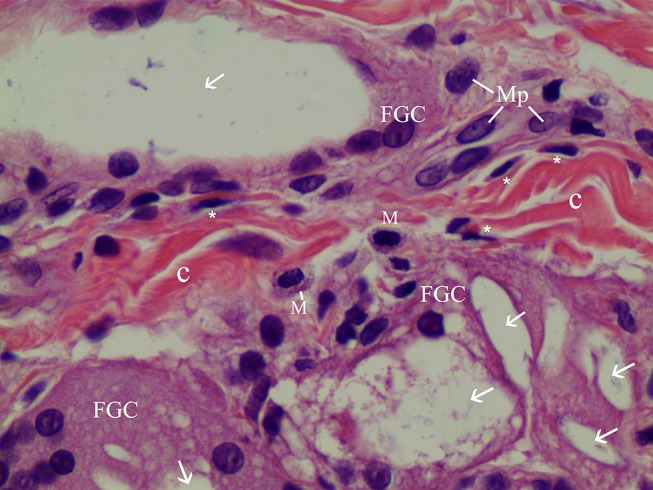
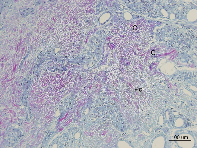
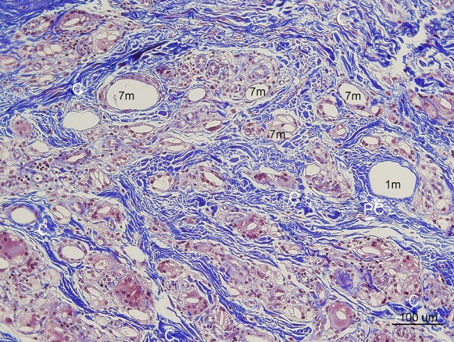
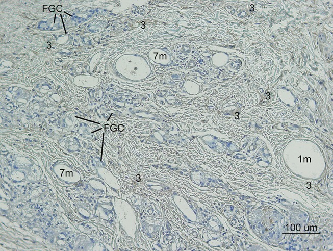
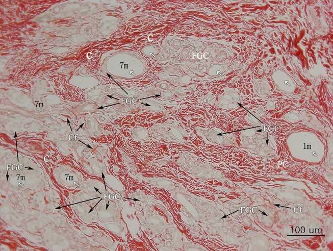
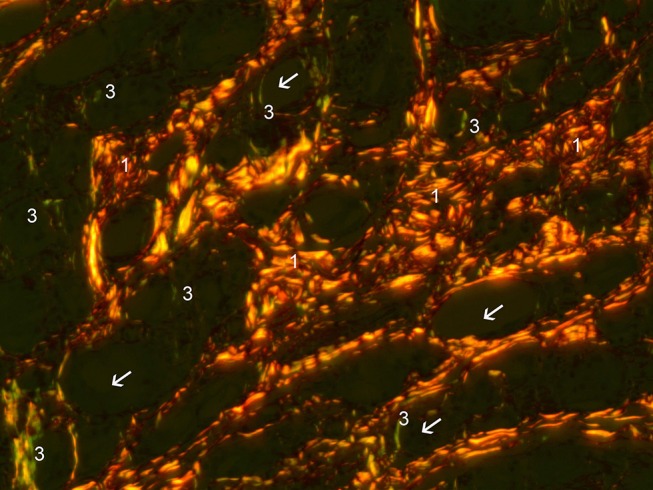
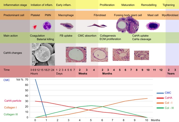
Depressed soft tissue and skin folds on ischial areas were significantly improved by volumization of subdermal filler injection. The skin quality, roughness, and pigmentation on ischial areas improved, and these improvements may be caused by intradermal micro-droplet injections of CaHA filler which may be influenced by neocollagenesis by numerous fibroblasts and increased micro-blood circulation (neovascularization). This is the first article to show the scientific evidence of neocollagenesis and tissue reaction after an injection of CaHA filler in the dermis, especially using various histological staining and to show various stages of inflammation and foreign body reaction around CaHA particles. Numerous fibroblasts were present around CaHA particles, but plasma cells were not found. Interestingly a few eosinophils were found around CaHA filler. After a significant period of time, multi-layered injections of diluted CaHA tightened and remodeled atrophic ischial skin. The multi layered injection approach was safe and effectively treated ischial soft tissue atrophy without significant side effects, such as infection or delayed swelling or lumps.

https://cdn.ncbi.nlm.nih.gov/pmc/blobs/4661/6800547/5aeff68b4d1c/CCID-12-771-g0018.jpg
https://cdn.ncbi.nlm.nih.gov/pmc/blobs/4661/6800547/703ce9529a85/CCID-12-771-g0001.jpg
https://cdn.ncbi.nlm.nih.gov/pmc/blobs/4661/6800547/746792171fb3/CCID-12-771-g0002.jpg
https://cdn.ncbi.nlm.nih.gov/pmc/blobs/4661/6800547/8490b3df8ec0/CCID-12-771-g0003.jpg
https://cdn.ncbi.nlm.nih.gov/pmc/blobs/4661/6800547/92128d867266/CCID-12-771-g0004.jpg
https://cdn.ncbi.nlm.nih.gov/pmc/blobs/4661/6800547/b772c0a570f5/CCID-12-771-g0005.jpg
https://cdn.ncbi.nlm.nih.gov/pmc/blobs/4661/6800547/faebdc2bff02/CCID-12-771-g0006.jpg
https://cdn.ncbi.nlm.nih.gov/pmc/blobs/4661/6800547/b53b51803065/CCID-12-771-g0007.jpg
https://cdn.ncbi.nlm.nih.gov/pmc/blobs/4661/6800547/d26849355725/CCID-12-771-g0008.jpg
https://cdn.ncbi.nlm.nih.gov/pmc/blobs/4661/6800547/2102ae501f56/CCID-12-771-g0009.jpg
https://cdn.ncbi.nlm.nih.gov/pmc/blobs/4661/6800547/9b7eeeb0e1b7/CCID-12-771-g0010.jpg
https://cdn.ncbi.nlm.nih.gov/pmc/blobs/4661/6800547/73e183860d17/CCID-12-771-g0011.jpg
https://cdn.ncbi.nlm.nih.gov/pmc/blobs/4661/6800547/b674253f8b5d/CCID-12-771-g0012.jpg
https://cdn.ncbi.nlm.nih.gov/pmc/blobs/4661/6800547/6db508812179/CCID-12-771-g0013.jpg
https://cdn.ncbi.nlm.nih.gov/pmc/blobs/4661/6800547/f9f67b973d0c/CCID-12-771-g0014.jpg
https://cdn.ncbi.nlm.nih.gov/pmc/blobs/4661/6800547/3055443bc303/CCID-12-771-g0015.jpg
https://cdn.ncbi.nlm.nih.gov/pmc/blobs/4661/6800547/d42d5232b108/CCID-12-771-g0016.jpg
https://cdn.ncbi.nlm.nih.gov/pmc/blobs/4661/6800547/98f3316f5501/CCID-12-771-g0017.jpg
https://cdn.ncbi.nlm.nih.gov/pmc/blobs/4661/6800547/5aeff68b4d1c/CCID-12-771-g0018.jpg
摘要

引言

坐姿时,压力分布位于坐骨结节下方。许多女性坐骨区域存在皮肤萎缩。为治疗坐骨上方皮肤的萎缩性改变,需同时实现容积增加和皮肤质地改善。分别对真皮层和皮下层采用自动注射和手动注射两种方法,使用具有刺激作用的硬填充剂。通过各种染色的活检研究评估填充剂注射后的组织学组织反应。

方法

本研究聚焦于通过多层注射羟基磷灰石钙(CaHA)填充剂来使萎缩性坐骨区域的软组织恢复活力,作者将其描述为慢性坐姿压疮的前期阶段。2012年1月至2019年4月对16名女性(平均年龄38.5岁)进行了治疗。注射前,将1.5cc瑞蓝(羟基磷灰石钙填充剂;德国默克公司)用1cc生理盐水和0.5cc利多卡因稀释,制成3cc填充剂混合物(1:1稀释)。所有受试者接受2.5cc稀释后CaHA填充剂的皮内注射和多层皮下注射。6个月时采用相同方法进行加强治疗。在注射前及注射后7个月用相机拍照并进行皮肤镜观察。在首次注射前及注射后7个月评估软组织凹陷、皮肤变色和粗糙度。还计算了治疗后凹陷、变色和粗糙度变化的标准差和变异系数。首次治疗7个月后从3名患者身上获取活检标本(3×5mm)。使用各种染色对标本进行分析。

结果

在体格检查、医学摄影以及所有患者的高分辨率皮肤镜检查中均可见皮肤质量、皮肤褶皱和粗糙度的改善。治疗后,坐骨区域的凹陷量随容积增加而减少。

结论

皮下填充剂注射增加容积显著改善了坐骨区域凹陷的软组织和皮肤褶皱。坐骨区域的皮肤质量、粗糙度和色素沉着得到改善,这些改善可能是由于CaHA填充剂的皮内微滴注射,这可能受到众多成纤维细胞的新胶原形成和微血液循环增加(新生血管形成)的影响。这是第一篇展示真皮内注射CaHA填充剂后新胶原形成和组织反应科学证据的文章,特别是使用各种组织学染色,并展示CaHA颗粒周围炎症和异物反应各个阶段的文章。CaHA颗粒周围存在众多成纤维细胞,但未发现浆细胞。有趣的是,在CaHA填充剂周围发现了一些嗜酸性粒细胞。经过较长一段时间后,多层注射稀释后的CaHA使萎缩的坐骨皮肤紧致并重塑。多层注射方法安全有效,可治疗坐骨软组织萎缩,且无明显副作用,如感染、延迟肿胀或肿块。

相似文献

1
Multilayered injection of calcium hydroxylapatite filler on ischial soft tissue to rejuvenate the previous phase of chronic sitting pressure sore.在坐骨软组织上多层注射羟基磷灰石填充剂,以恢复慢性坐压疮的前期状态。
Clin Cosmet Investig Dermatol. 2019 Oct 15;12:771-784. doi: 10.2147/CCID.S212599. eCollection 2019.
2
Improved Neocollagenesis and Skin Mechanical Properties After Injection of Diluted Calcium Hydroxylapatite in the Neck and Décolletage:A Pilot Study.颈部和胸部注射稀释羟基磷灰石后新胶原形成及皮肤力学性能改善的初步研究
J Drugs Dermatol. 2017 Jan 1;16(1):68-74.
3
Calcium hydroxylapatite treatment of human skin: evidence of collagen turnover through picrosirius red staining and circularly polarized microscopy.羟基磷灰石钙对人体皮肤的治疗:通过天狼星红染色和圆偏振显微镜观察胶原蛋白更新的证据
Clin Cosmet Investig Dermatol. 2018 Jan 15;11:29-35. doi: 10.2147/CCID.S143015. eCollection 2018.
4
Combining topical dermal infused exosomes with injected calcium hydroxylapatite for enhanced tissue biostimulation.将局部真皮注入的外泌体与注射用羟基磷灰石相结合以增强组织生物刺激作用。
J Cosmet Dermatol. 2023 Mar;22 Suppl 1:15-27. doi: 10.1111/jocd.15695.
5
Hand recontouring with calcium hydroxylapatite (Radiesse).使用羟基磷灰石钙(瑞蓝)进行手部轮廓重塑。
J Cosmet Dermatol. 2009 Mar;8(1):44-51. doi: 10.1111/j.1473-2165.2009.00423.x.
6
Hyperdilution of CaHA fillers for the improvement of age and hereditary volume deficits in East Asian patients.用于改善东亚患者年龄和遗传性容量不足的羟基磷灰石钙填充剂超稀释法
Clin Cosmet Investig Dermatol. 2018 Jul 16;11:357-363. doi: 10.2147/CCID.S159752. eCollection 2018.
7
Calcium hydroxylapatite microspheres activate fibroblasts through direct contact to stimulate neocollagenesis.羟基磷灰石微球通过直接接触激活成纤维细胞,刺激新胶原生成。
J Cosmet Dermatol. 2023 Feb;22(2):426-432. doi: 10.1111/jocd.15521. Epub 2022 Dec 27.
8
Open-Label, Post-Marketing Study to Evaluate the Performance and Safety of Calcium Hydroxylapatite With Integral Lidocaine to Correct Facial Volume Loss.评估含利多卡因的羟基磷灰石矫正面部容量缺失的性能和安全性的开放标签上市后研究。
J Drugs Dermatol. 2019 Jan 1;18(1):86-91.
9
A randomized, split-face, histomorphologic study comparing a volumetric calcium hydroxylapatite and a hyaluronic acid-based dermal filler.一项比较体积型羟基磷灰石钙和透明质酸基真皮填充剂的随机、半脸、组织形态学研究。
J Drugs Dermatol. 2014 Sep;13(9):1047-52.
10
Clinical, histologic and electron microscopic findings after injection of a calcium hydroxylapatite filler.注射羟基磷灰石钙填充剂后的临床、组织学和电子显微镜检查结果
J Cosmet Laser Ther. 2004 Dec;6(4):223-6. doi: 10.1080/147641704100003048.

引用本文的文献

1
The Immunologic Spectrum of Biostimulators and Its Clinical Importance.生物刺激剂的免疫谱及其临床重要性。
Plast Reconstr Surg Glob Open. 2025 Aug 5;13(8):e7001. doi: 10.1097/GOX.0000000000007001. eCollection 2025 Aug.
2
Dual Benefits of Calcium Hydroxyapatite Filler: A Prospective Study on Midface Volume Restoration and Skin Quality Enhancement.羟基磷灰石填充剂的双重益处:一项关于中面部容积恢复和皮肤质量改善的前瞻性研究。
J Cosmet Dermatol. 2025 Jun;24(6):e70265. doi: 10.1111/jocd.70265.
3
Arterioembolic Characteristics of Differentially Diluted CaHA-CMC Gels Within An Artificial Macrovascular Perfusion Model.

本文引用的文献

1
Changes in Dermal Thickness in Biopsy Study of Histologic Findings After a Single Injection of Polycaprolactone-Based Filler into the Dermis.聚己内酯类填充剂单次真皮内注射后组织学发现的活检中真皮厚度的变化。
Aesthet Surg J. 2019 Nov 13;39(12):NP484-NP494. doi: 10.1093/asj/sjz050.
2
Detailed Sonographic Anatomy of Dorsal Hand Augmentation With Hyaluronic Acid and Calcium Hydroxyapatite Fillers.手背增强中应用透明质酸和钙羟磷灰石填充剂的详细超声解剖学
Aesthet Surg J. 2019 Sep 13;39(10):1096-1106. doi: 10.1093/asj/sjy227.
3
Type III collagen affects dermal and vascular collagen fibrillogenesis and tissue integrity in a mutant Col3a1 transgenic mouse model.
人工大血管灌注模型中不同稀释度的羟基磷灰石-羧甲基纤维素凝胶的动脉栓塞特性
Aesthet Surg J. 2025 May 15;45(6):645-653. doi: 10.1093/asj/sjaf028.
4
Aesthetic Efficacy and Safety of Combined Microfocused Ultrasound With Visualization and Calcium Hydroxylapatite Treatment: A Systematic Review of Human Evidence.联合微聚焦超声与可视化及羟基磷灰石治疗的美学效果与安全性:对人类证据的系统评价
Aesthet Surg J. 2025 May 15;45(6):638-642. doi: 10.1093/asj/sjae239.
5
Polylactic Acid-Based Polymers Used for Facial Rejuvenation: A Narrative Review.用于面部年轻化的聚乳酸基聚合物:一项叙述性综述。
Aesthetic Plast Surg. 2024 Dec 2. doi: 10.1007/s00266-024-04543-8.
6
Comparative Rheology of Hyaluronic Acid Fillers, Poly-l-lactic Acid, and Varying Dilutions of Calcium Hydroxylapatite.透明质酸填充剂、聚左旋乳酸和不同稀释度的羟基磷灰石的比较流变学
Plast Reconstr Surg Glob Open. 2024 Aug 15;12(8):e6068. doi: 10.1097/GOX.0000000000006068. eCollection 2024 Aug.
7
Hyperdiluting Calcium Hydroxylapatite With Platelet-Rich Plasma and Hyaluronidase for Improving Neck Laxity and Wrinkle Severity.用富含血小板血浆和透明质酸酶超稀释羟基磷灰石以改善颈部松弛和皱纹严重程度。
Cureus. 2024 Jul 6;16(7):e63969. doi: 10.7759/cureus.63969. eCollection 2024 Jul.
8
Supraperiosteal technique protocol for forehead filling with a mixture of calcium hydroxyapatite and hyaluronic acid: Double-blind, randomized controlled clinical trial.羟基磷灰石与透明质酸混合物用于前额填充的骨膜上技术方案:双盲随机对照临床试验
J Cosmet Dermatol. 2024 Dec;23(12):4116-4122. doi: 10.1111/jocd.16488. Epub 2024 Aug 4.
9
A morphological analysis of calcium hydroxylapatite and poly-l-lactic acid biostimulator particles.钙羟基磷灰石和聚左旋乳酸生物刺激剂颗粒的形态分析。
Skin Res Technol. 2024 Jun;30(6):e13764. doi: 10.1111/srt.13764.
10
A Structured Approach for Treating Calcium Hydroxylapatite Focal Accumulations.治疗羟基磷灰石钙局灶性聚集的结构化方法。
Aesthet Surg J. 2024 Jul 15;44(8):869-879. doi: 10.1093/asj/sjae031.
III 型胶原影响突变 Col3a1 转基因小鼠模型的皮肤和血管胶原原纤维生成和组织完整性。
Matrix Biol. 2018 Sep;70:72-83. doi: 10.1016/j.matbio.2018.03.008. Epub 2018 Mar 15.
4
A randomized, split-face, histomorphologic study comparing a volumetric calcium hydroxylapatite and a hyaluronic acid-based dermal filler.一项比较体积型羟基磷灰石钙和透明质酸基真皮填充剂的随机、半脸、组织形态学研究。
J Drugs Dermatol. 2014 Sep;13(9):1047-52.
5
Ion channels regulating mast cell biology.调节肥大细胞生物学的离子通道。
Clin Exp Allergy. 2013 May;43(5):491-502. doi: 10.1111/cea.12043.
6
Quantitative multispectral imaging of Herovici's polychrome for the assessment of collagen content and tissue remodelling.赫罗维茨多彩染色定量多光谱成像评估胶原含量和组织重塑。
J Tissue Eng Regen Med. 2013 Feb;7(2):139-48. doi: 10.1002/term.508. Epub 2011 Nov 9.
7
Calcium hydroxylapatite filler for facial rejuvenation: a histologic and immunohistochemical analysis.用于面部年轻化的羟基磷灰石钙填充物:组织学和免疫组织化学分析
Dermatol Surg. 2008 Jun;34 Suppl 1:S64-7. doi: 10.1111/j.1524-4725.2008.34245.x.
8
Looking older: fibroblast collapse and therapeutic implications.显老:成纤维细胞衰退及其治疗意义
Arch Dermatol. 2008 May;144(5):666-72. doi: 10.1001/archderm.144.5.666.
9
Implementation and analysis of relief patterns of the surface of benign and malignant lesions of the skin by microtopography.通过微观形貌对皮肤良恶性病变表面的起伏模式进行实施与分析。
Phys Med Biol. 2005 Dec 7;50(23):5535-43. doi: 10.1088/0031-9155/50/23/008. Epub 2005 Nov 8.
10
Clinical, histologic and electron microscopic findings after injection of a calcium hydroxylapatite filler.注射羟基磷灰石钙填充剂后的临床、组织学和电子显微镜检查结果
J Cosmet Laser Ther. 2004 Dec;6(4):223-6. doi: 10.1080/147641704100003048.