Alzheimer Research Unit, Department of Neurology, Massachusetts General Hospital and Harvard Medical School, 114, 16th St, Charlestown, MA, 02129, USA.
Department of Physics, Chemistry and Biology, Linköping University, 581 83, Linköping, Sweden.
Acta Neuropathol Commun. 2019 Nov 8;7(1):171. doi: 10.1186/s40478-019-0832-1.
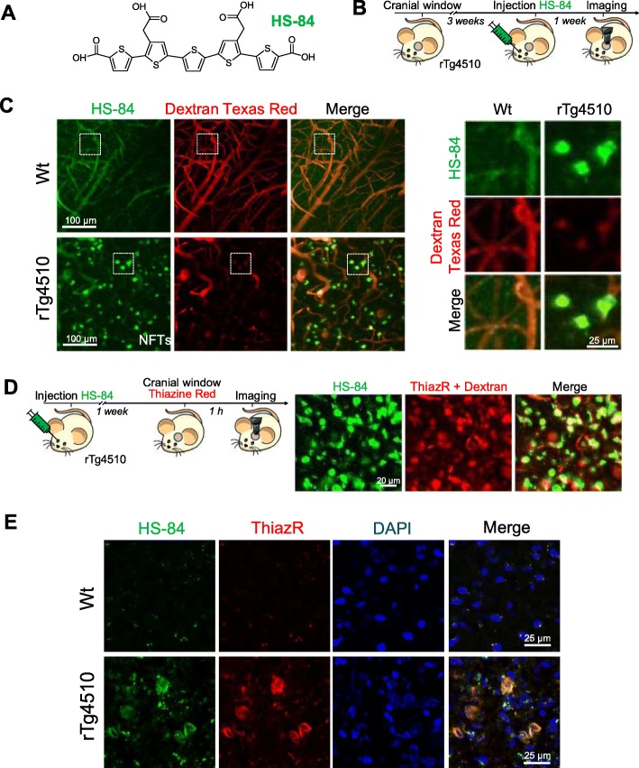
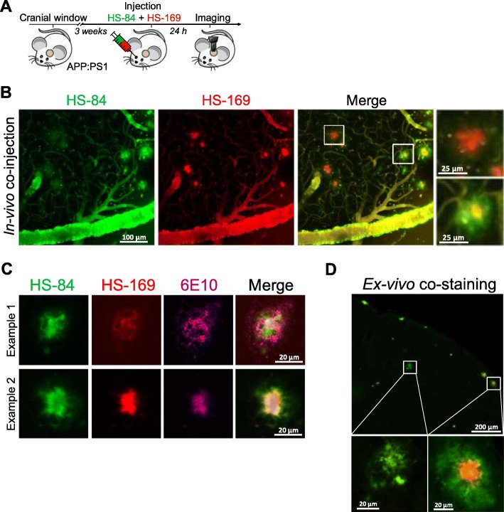
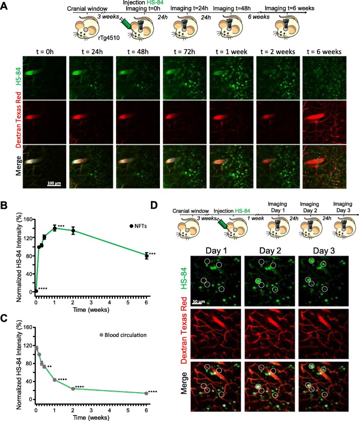
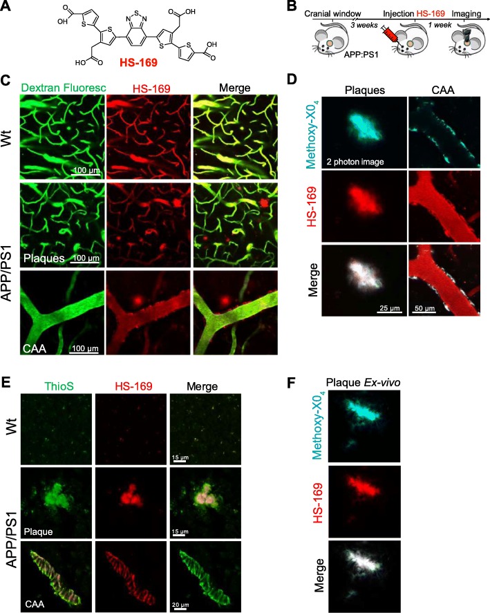
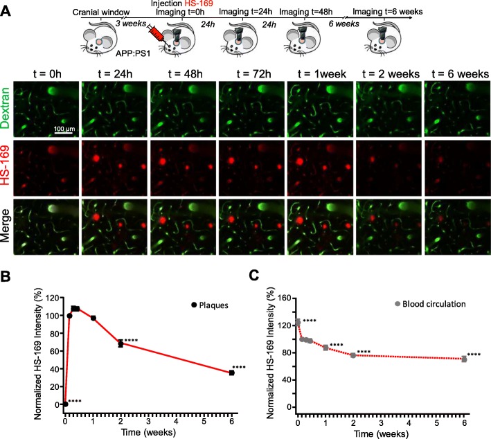
The detection of amyloid beta deposits and neurofibrillary tangles, both hallmarks of Alzheimer's disease (AD), is key to understanding the mechanisms underlying these pathologies. Luminescent conjugated oligothiophenes (LCOs) enable fluorescence imaging of these protein aggregates. Using LCOs and multiphoton microscopy, individual tangles and amyloid beta deposits were labeled in vivo and imaged longitudinally in a mouse model of tauopathy and cerebral amyloidosis, respectively. Importantly, LCO HS-84, whose emission falls in the green region of the spectrum, allowed for the first time longitudinal imaging of tangle dynamics following a single intravenous injection. In addition, LCO HS-169, whose emission falls in the red region of the spectrum, successfully labeled amyloid beta deposits, allowing multiplexing with other reporters whose emission falls in the green region of the spectrum. In conclusion, this method can provide a new approach for longitudinal in vivo imaging using multiphoton microscopy of AD pathologies as well as other neurodegenerative diseases associated with protein aggregation in mouse models.
淀粉样β沉积和神经原纤维缠结的检测是了解这些病变潜在机制的关键,淀粉样β沉积和神经原纤维缠结都是阿尔茨海默病(AD)的标志性特征。 发荧光的共轭寡聚噻吩(LCOs)可使这些蛋白聚集体实现荧光成像。 使用 LCO 和多光子显微镜,分别在神经tau 病变和脑淀粉样血管病的小鼠模型中,对单个缠结和淀粉样β沉积进行了体内标记和纵向成像。 重要的是,发射处于光谱绿光区域的 LCO HS-84 首次允许在单次静脉注射后对缠结动力学进行纵向成像。 此外,发射处于光谱红光区域的 LCO HS-169 成功标记了淀粉样β沉积,使其与发射处于光谱绿光区域的其他报告基因实现了多重标记。 总之,该方法可以为使用多光子显微镜对 AD 病变以及与小鼠模型中蛋白聚集相关的其他神经退行性疾病进行纵向体内成像提供一种新方法。