Wellcome / EPSRC Centre for Interventional and Surgical Sciences, University College London, London, UK; School of Biomedical Engineering and Imaging Sciences, King's College London, London, UK.
School of Mechanical and Electrical Engineering, University of Electronic Science and Technology of China, Chengdu, China; Wellcome / EPSRC Centre for Interventional and Surgical Sciences, University College London, London, UK; School of Biomedical Engineering and Imaging Sciences, King's College London, London, UK.
Neuroimage. 2020 Feb 1;206:116324. doi: 10.1016/j.neuroimage.2019.116324. Epub 2019 Nov 6.
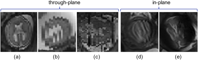
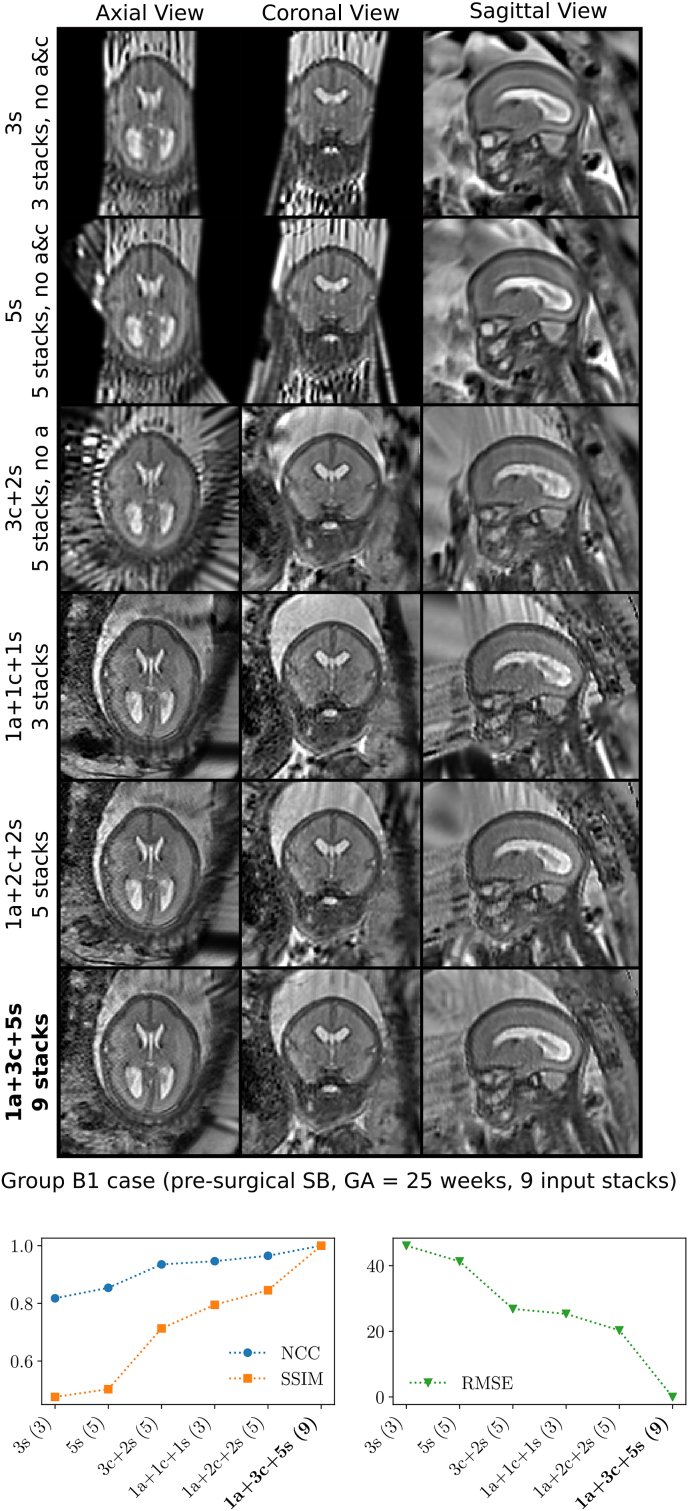
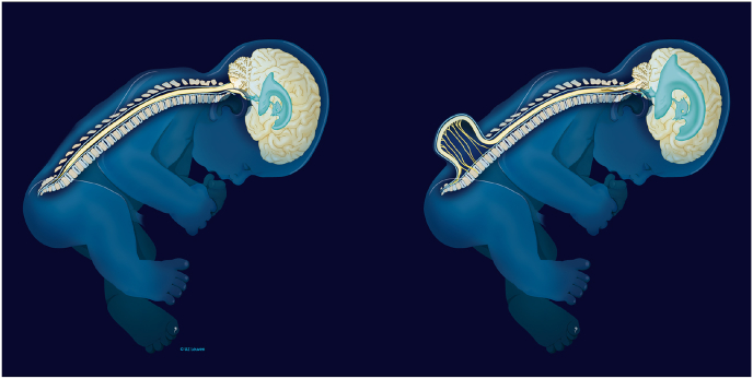
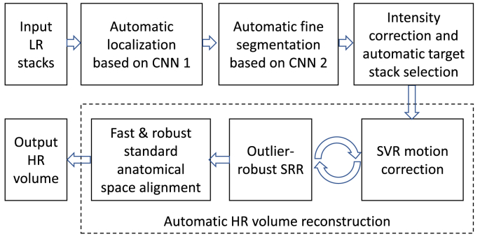
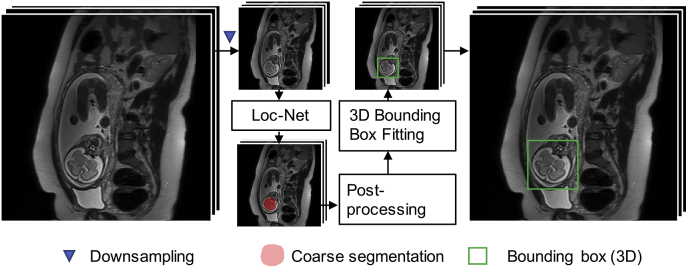
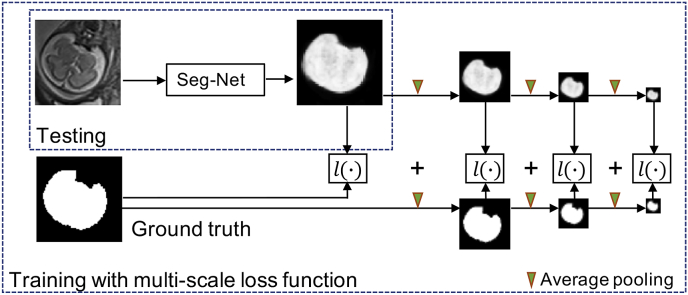
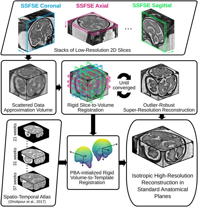
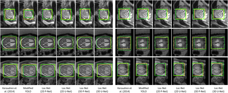
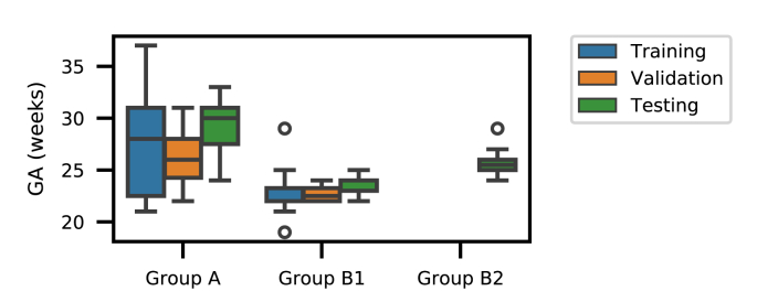
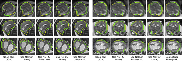
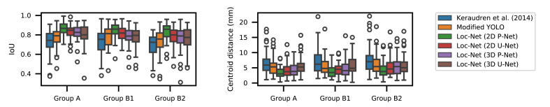
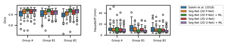
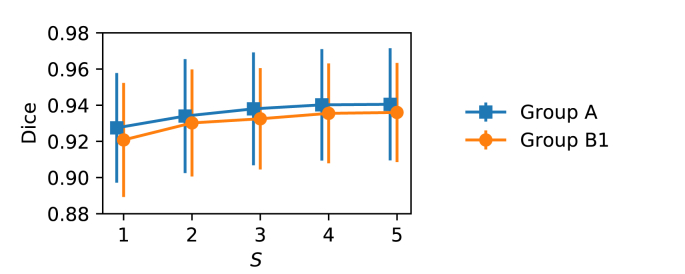
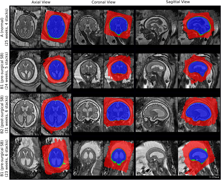
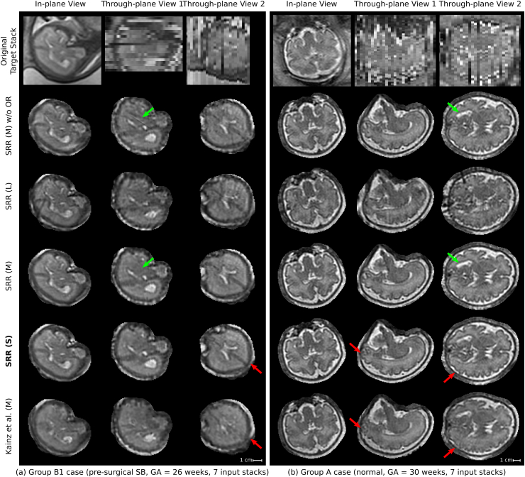
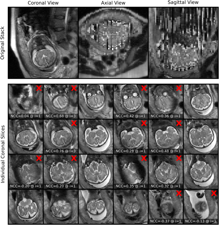
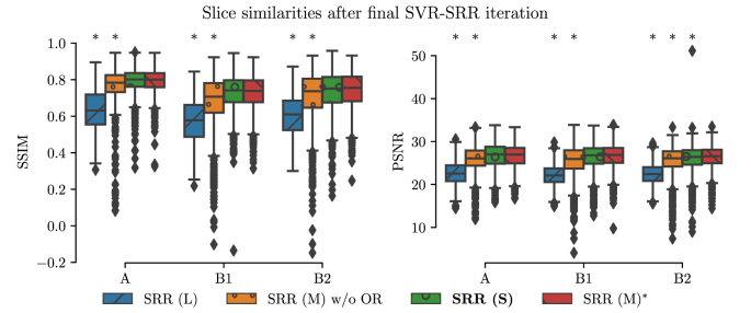
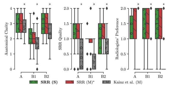
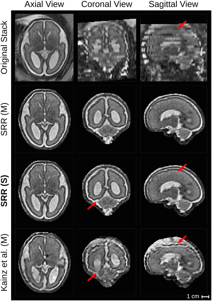
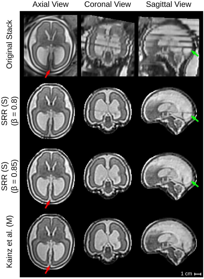
High-resolution volume reconstruction from multiple motion-corrupted stacks of 2D slices plays an increasing role for fetal brain Magnetic Resonance Imaging (MRI) studies. Currently existing reconstruction methods are time-consuming and often require user interactions to localize and extract the brain from several stacks of 2D slices. We propose a fully automatic framework for fetal brain reconstruction that consists of four stages: 1) fetal brain localization based on a coarse segmentation by a Convolutional Neural Network (CNN), 2) fine segmentation by another CNN trained with a multi-scale loss function, 3) novel, single-parameter outlier-robust super-resolution reconstruction, and 4) fast and automatic high-resolution visualization in standard anatomical space suitable for pathological brains. We validated our framework with images from fetuses with normal brains and with variable degrees of ventriculomegaly associated with open spina bifida, a congenital malformation affecting also the brain. Experiments show that each step of our proposed pipeline outperforms state-of-the-art methods in both segmentation and reconstruction comparisons including expert-reader quality assessments. The reconstruction results of our proposed method compare favorably with those obtained by manual, labor-intensive brain segmentation, which unlocks the potential use of automatic fetal brain reconstruction studies in clinical practice.
从多个运动伪影的二维切片堆栈中进行高分辨率体积重建,在胎儿磁共振成像(MRI)研究中发挥着越来越重要的作用。目前现有的重建方法既耗时又经常需要用户交互来定位和从几堆二维切片中提取大脑。我们提出了一种全自动的胎儿脑重建框架,该框架由四个阶段组成:1)基于卷积神经网络(CNN)的粗略分割的胎儿脑定位,2)使用多尺度损失函数训练的另一个 CNN 进行精细分割,3)新颖的、单参数抗离群点鲁棒超分辨率重建,4)在标准解剖空间中快速自动进行适用于病理脑的高分辨率可视化。我们使用来自正常大脑的胎儿图像和伴有开放性脊柱裂(一种也影响大脑的先天性畸形)的不同程度脑室扩大的胎儿图像来验证我们的框架。实验表明,我们提出的管道的每个步骤在分割和重建比较中都优于最先进的方法,包括专家读者质量评估。与手动、劳动强度大的脑分割相比,我们的方法的重建结果更有优势,这为临床实践中自动胎儿脑重建研究的应用提供了可能。