• 文献检索
  • 文档翻译
  • 深度研究
  • 学术资讯
  • Suppr Zotero 插件Zotero 插件
  • 邀请有礼
  • 套餐&价格
  • 历史记录
应用&插件
Suppr Zotero 插件Zotero 插件浏览器插件Mac 客户端Windows 客户端微信小程序
定价
高级版会员购买积分包购买API积分包
服务
文献检索文档翻译深度研究API 文档MCP 服务
关于我们
关于 Suppr公司介绍联系我们用户协议隐私条款
关注我们

Suppr 超能文献

核心技术专利:CN118964589B侵权必究
粤ICP备2023148730 号-1Suppr @ 2026

文献检索

告别复杂PubMed语法,用中文像聊天一样搜索,搜遍4000万医学文献。AI智能推荐,让科研检索更轻松。

立即免费搜索

文件翻译

保留排版,准确专业,支持PDF/Word/PPT等文件格式,支持 12+语言互译。

免费翻译文档

深度研究

AI帮你快速写综述,25分钟生成高质量综述,智能提取关键信息,辅助科研写作。

立即免费体验

心脏磁共振延迟钆增强成像检测左心室血栓后的长期栓塞结局:一项匹配队列研究。

Long-Term Embolic Outcomes After Detection of Left Ventricular Thrombus by Late Gadolinium Enhancement Cardiovascular Magnetic Resonance Imaging: A Matched Cohort Study.

机构信息

Cardiovascular Division, Department of Medicine (P.S.V., K.-H.A.C., F.K., P.S.N., O.O., C.S.), University of Minnesota Medical School, Minneapolis, MN.

Department of Medicine (C.C.), University of Minnesota Medical School, Minneapolis, MN.

出版信息

Circ Cardiovasc Imaging. 2019 Nov;12(11):e009723. doi: 10.1161/CIRCIMAGING.119.009723. Epub 2019 Nov 11.

DOI:10.1161/CIRCIMAGING.119.009723
PMID:31707810
原文链接:https://pmc.ncbi.nlm.nih.gov/articles/PMC6941143/
Abstract

BACKGROUND

Late gadolinium enhancement (LGE) cardiovascular magnetic resonance (CMR) imaging is more sensitive than echocardiography for the detection of intracardiac thrombus because of its unique ability to identify thrombus based on tissue characteristics related to avascularity. The long-term prognostic significance of left ventricular (LV) thrombus detected by LGE CMR is unknown.

METHODS

We performed a matched cohort study of consecutive adult patients with LV thrombus detected by LGE CMR who were matched on the date of CMR, age, and LV ejection fraction to up to 3 patients without LV thrombus. We investigated the long-term incidence of a composite of embolic events: stroke, transient ischemic attack, or extracranial systemic arterial embolism. We also compared outcomes among patients with LV thrombus detected by LGE CMR stratified by whether the LV thrombus was also detected by echocardiography or not.

RESULTS

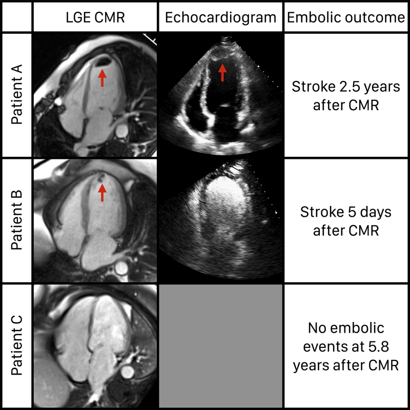
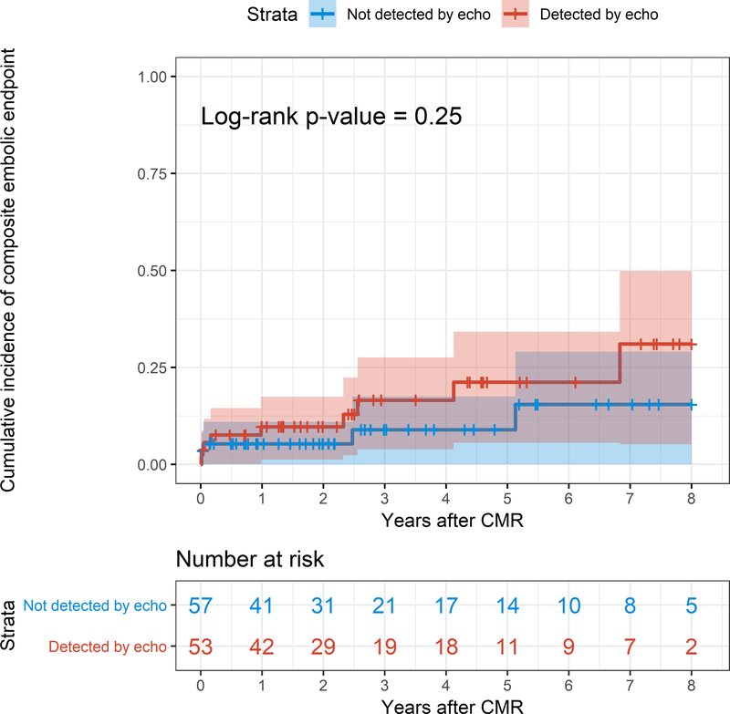
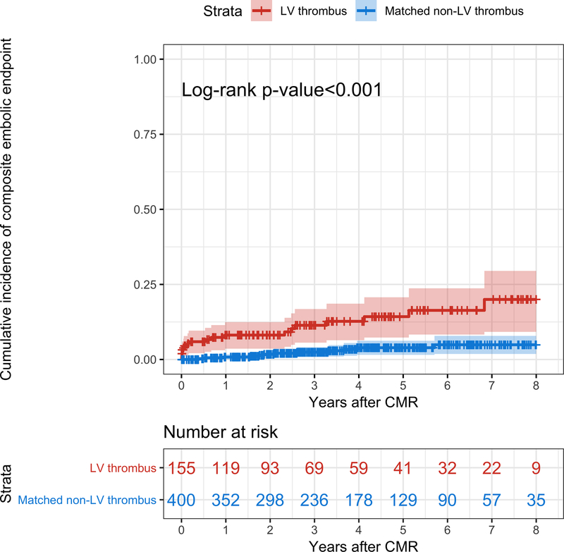
Of 157 LV thrombus patients, 155 were matched to 400 non-LV thrombus patients. During a median follow-up of 3.3 years, the cumulative incidence of embolism was significantly higher in LV thrombus patients compared with the matched non-LV thrombus patients (<0.001), with annualized rates of 3.7% and 0.8% for LV thrombus and matched non-LV thrombus patients, respectively. LV thrombus was the only independent predictor of the composite embolic end point (hazard ratio, 3.99 [95% CI, 1.54-10.35]; =0.004). The cumulative incidence of embolism was not different in patients with LV thrombus that was also detected by echocardiography versus patients with LV thrombus not detected by echocardiography (=0.25).

CONCLUSIONS

Despite contemporary antithrombotic treatment, LV thrombus detected by LGE CMR is associated with a 4-fold higher long-term incidence of embolism compared with matched non-LV thrombus patients. LV thrombus detected by LGE CMR but not by echocardiography is associated with a similar risk of embolism as that detected by both LGE CMR and echocardiography.

摘要

背景

相较于超声心动图,钆延迟增强(late gadolinium enhancement,LGE)心血管磁共振(cardiovascular magnetic resonance,CMR)成像在检测心腔内血栓方面更具优势,因为其具有基于与无血管组织特征相关的组织特征识别血栓的独特能力。LGE CMR 检测到的左心室(left ventricular,LV)血栓的长期预后意义尚不清楚。

方法

我们对连续接受 LGE CMR 检查且发现 LV 血栓的成年患者进行了匹配队列研究,按照 CMR 检查日期、年龄和左心室射血分数,将这些患者与最多 3 例未发现 LV 血栓的患者进行了匹配。我们调查了 LGE CMR 检测到的 LV 血栓患者发生复合栓塞事件(卒中和短暂性脑缺血发作或颅外全身动脉栓塞)的长期发生率。我们还比较了根据 LGE CMR 是否检测到 LV 血栓以及是否同时检测到超声心动图进行分层的 LV 血栓患者的结局。

结果

在 157 例 LV 血栓患者中,155 例与 400 例非 LV 血栓患者相匹配。在中位随访 3.3 年期间,LV 血栓患者的栓塞累积发生率明显高于匹配的非 LV 血栓患者(P<0.001),LV 血栓患者和匹配的非 LV 血栓患者的年化发生率分别为 3.7%和 0.8%。LV 血栓是复合栓塞终点的唯一独立预测因子(风险比,3.99[95%置信区间,1.54~10.35];P=0.004)。在同时通过超声心动图检测到 LV 血栓的患者与未通过超声心动图检测到 LV 血栓的患者中,栓塞的累积发生率无差异(P=0.25)。

结论

尽管接受了当代抗血栓治疗,LGE CMR 检测到的 LV 血栓与匹配的非 LV 血栓患者相比,其长期栓塞发生率仍高出 4 倍。LGE CMR 检测到但未通过超声心动图检测到的 LV 血栓与同时通过 LGE CMR 和超声心动图检测到的 LV 血栓的栓塞风险相似。

https://cdn.ncbi.nlm.nih.gov/pmc/blobs/e446/6941143/448aca577d20/nihms-1064628-f0003.jpg
https://cdn.ncbi.nlm.nih.gov/pmc/blobs/e446/6941143/30105c038bf1/nihms-1064628-f0001.jpg
https://cdn.ncbi.nlm.nih.gov/pmc/blobs/e446/6941143/105f870fb1eb/nihms-1064628-f0002.jpg
https://cdn.ncbi.nlm.nih.gov/pmc/blobs/e446/6941143/448aca577d20/nihms-1064628-f0003.jpg
https://cdn.ncbi.nlm.nih.gov/pmc/blobs/e446/6941143/30105c038bf1/nihms-1064628-f0001.jpg
https://cdn.ncbi.nlm.nih.gov/pmc/blobs/e446/6941143/105f870fb1eb/nihms-1064628-f0002.jpg
https://cdn.ncbi.nlm.nih.gov/pmc/blobs/e446/6941143/448aca577d20/nihms-1064628-f0003.jpg

相似文献

1
Long-Term Embolic Outcomes After Detection of Left Ventricular Thrombus by Late Gadolinium Enhancement Cardiovascular Magnetic Resonance Imaging: A Matched Cohort Study.心脏磁共振延迟钆增强成像检测左心室血栓后的长期栓塞结局:一项匹配队列研究。
Circ Cardiovasc Imaging. 2019 Nov;12(11):e009723. doi: 10.1161/CIRCIMAGING.119.009723. Epub 2019 Nov 11.
2
Prevalence and prognostic significance of left ventricular myocardial late gadolinium enhancement in severe aortic stenosis.重度主动脉瓣狭窄患者左心室心肌延迟钆增强的患病率及预后意义
Indian Heart J. 2017 Nov-Dec;69(6):742-750. doi: 10.1016/j.ihj.2017.05.027. Epub 2017 Jun 1.
3
Contrast-enhanced anatomic imaging as compared to contrast-enhanced tissue characterization for detection of left ventricular thrombus.与对比增强组织特征分析相比,对比增强解剖成像用于检测左心室血栓。
JACC Cardiovasc Imaging. 2009 Aug;2(8):969-79. doi: 10.1016/j.jcmg.2009.03.017.
4
Left Ventricular Thrombi: Insights from Cardiac Magnetic Resonance Imaging.左心室血栓:心脏磁共振成像的新视角。
Tomography. 2021 May 12;7(2):180-188. doi: 10.3390/tomography7020016.
5
Diagnostic utility and clinical implication of late gadolinium enhancement cardiac magnetic resonance for detection of catheter associated right atrial thrombus.晚期钆增强心脏磁共振对导管相关右房血栓的诊断价值及临床意义。
Clin Imaging. 2020 Jun;62:17-22. doi: 10.1016/j.clinimag.2020.01.021. Epub 2020 Jan 29.
6
Long-Term Prognostic Value of Cardiac Magnetic Resonance in Left Ventricle Noncompaction: A Prospective Multicenter Study.左心室心肌致密化不全的心脏磁共振长期预后价值:一项前瞻性多中心研究。
J Am Coll Cardiol. 2016 Nov 15;68(20):2166-2181. doi: 10.1016/j.jacc.2016.08.053.
7
Prognostic Implications of Left Ventricular Scar Determined by Late Gadolinium Enhanced Cardiac Magnetic Resonance in Patients With Atrial Fibrillation.钆延迟增强心脏磁共振成像测定左心室瘢痕对心房颤动患者的预后影响
Am J Cardiol. 2016 Oct 1;118(7):991-7. doi: 10.1016/j.amjcard.2016.06.054. Epub 2016 Jul 18.
8
Incidence and prognostic significance of myocardial late gadolinium enhancement in patients with sarcoidosis without cardiac manifestation.心肌磁共振钆延迟增强在无心脏表现的肉状瘤病患者中的发生率和预后意义。
Chest. 2014 Oct;146(4):1064-1072. doi: 10.1378/chest.14-0139.
9
Myocardial deformation imaging by two-dimensional speckle-tracking echocardiography for prediction of global and segmental functional changes after acute myocardial infarction: a comparison with late gadolinium enhancement cardiac magnetic resonance.二维斑点追踪超声心动图心肌形变成像预测急性心肌梗死后整体和节段性功能变化:与延迟钆增强心脏磁共振成像的比较
J Am Soc Echocardiogr. 2014 Mar;27(3):249-57. doi: 10.1016/j.echo.2013.11.014. Epub 2013 Dec 22.
10
Detection of Left Ventricular Thrombus by Cardiac Magnetic Resonance in Embolic Stroke of Undetermined Source.心脏磁共振成像检测不明来源栓塞性卒中患者左心室血栓
Stroke. 2017 Sep;48(9):2434-2440. doi: 10.1161/STROKEAHA.117.018263. Epub 2017 Aug 17.

引用本文的文献

1
Beyond cardiac embolism and cryptogenic stroke: unveiling the mechanisms of cerebrovascular events in Chagas disease.超越心脏栓塞和隐源性卒中:揭示恰加斯病脑血管事件的机制
Lancet Reg Health Am. 2025 Aug 8;50:101203. doi: 10.1016/j.lana.2025.101203. eCollection 2025 Oct.
2
The Cleveland Clinic experience of eosinophilic myocarditis in the setting of hypereosinophilic syndrome: demographics, cardiac imaging, and outcomes.克利夫兰诊所关于高嗜酸性粒细胞综合征背景下嗜酸性粒细胞性心肌炎的经验:人口统计学、心脏成像及预后
Cardiovasc Diagn Ther. 2024 Dec 31;14(6):1122-1133. doi: 10.21037/cdt-24-347. Epub 2024 Dec 2.
3
Direct oral anticoagulants or warfarin in patients with left ventricular thrombus after ST-elevation myocardial infarction: a pilot trial and a prespecified meta-analysis of randomised trials.

本文引用的文献

1
Myocardial Fibrosis and Prognosis in Heart Transplant Recipients.心肌纤维化与心脏移植受者预后。
Circ Cardiovasc Imaging. 2019 Oct;12(10):e009060. doi: 10.1161/CIRCIMAGING.119.009060. Epub 2019 Oct 15.
2
Assessment of the 2017 AHA/ACC/HRS Guideline Recommendations for Implantable Cardioverter-Defibrillator Implantation in Cardiac Sarcoidosis.评估 2017 年 AHA/ACC/HRS 指南推荐在心脏结节病中植入植入式心脏复律除颤器的建议。
Circ Arrhythm Electrophysiol. 2019 Sep;12(9):e007488. doi: 10.1161/CIRCEP.119.007488. Epub 2019 Aug 21.
3
Safety and prognostic value of regadenoson stress cardiovascular magnetic resonance imaging in heart transplant recipients.
ST段抬高型心肌梗死后左心室血栓患者使用直接口服抗凝剂或华法林:一项试点试验及随机试验的预先设定的荟萃分析
EuroIntervention. 2025 Jan 6;21(1):82-92. doi: 10.4244/EIJ-D-24-00527.
4
Evolution of left ventricular thrombus on serial cardiovascular magnetic resonance imaging.连续心血管磁共振成像上左心室血栓的演变
Eur Heart J Cardiovasc Imaging. 2025 Jan 31;26(2):349-358. doi: 10.1093/ehjci/jeae271.
5
Oral anticoagulation in patients with left ventricular thrombus: a systematic review and network meta-analysis with reconstructed time-to-event data.左心室血栓患者的口服抗凝治疗:一项采用重构事件发生时间数据的系统评价和网状荟萃分析
Clin Res Cardiol. 2024 Sep 16. doi: 10.1007/s00392-024-02547-7.
6
A systematic review and meta-analysis of transthoracic echocardiogram vs. cardiac magnetic resonance imaging for the detection of left ventricular thrombus.经胸超声心动图与心脏磁共振成像检测左心室血栓的系统评价与Meta分析
Eur Heart J Imaging Methods Pract. 2023 Dec 7;1(2):qyad041. doi: 10.1093/ehjimp/qyad041. eCollection 2023 Sep.
7
Non-vitamin K oral antagonist (NOAC) compared to vitamin K antagonist (VKA) in left ventricular thrombus.左心室血栓中新型口服抗凝药(NOAC)与维生素K拮抗剂(VKA)的比较
J Family Med Prim Care. 2024 Jun;13(6):2485-2490. doi: 10.4103/jfmpc.jfmpc_1905_23. Epub 2024 Jun 14.
8
The Role of Multimodality Imaging in Cardiomyopathy.多模态影像学在心肌病中的作用。
Curr Cardiol Rep. 2024 Jul;26(7):689-703. doi: 10.1007/s11886-024-02068-9. Epub 2024 May 16.
9
Left ventricular thrombosis caused cerebral embolism during venoarterial extracorporeal membrane oxygenation support: A case report.静脉-动脉体外膜肺氧合支持期间左心室血栓形成导致脑栓塞:一例报告
World J Clin Cases. 2024 Feb 16;12(5):973-979. doi: 10.12998/wjcc.v12.i5.973.
10
Determination of the prevalence and predictors of ventricular thrombus with assessment of the risk of systemic embolization to the CNS in patients after acute myocardial infarction using magnetic resonance imaging, echocardiography and cardiac markers - a prospective, unicentric, observational study.使用磁共振成像、超声心动图和心脏标志物,对急性心肌梗死后患者进行中枢神经系统全身性栓塞风险评估,以确定心室血栓的患病率和预测因素——一项前瞻性、单中心、观察性研究。
Biomed Pap Med Fac Univ Palacky Olomouc Czech Repub. 2023 Jan 8. doi: 10.5507/bp.2023.050.
心脏移植受者雷加登松负荷心血管磁共振成像的安全性和预后价值。
J Cardiovasc Magn Reson. 2019 Jan 24;21(1):9. doi: 10.1186/s12968-018-0515-2.
4
Short-Term Risk of Ischemic Stroke After Detection of Left Ventricular Thrombus on Cardiac Magnetic Resonance Imaging.心脏磁共振成像检测到左心室血栓后缺血性卒中的短期风险
J Stroke Cerebrovasc Dis. 2019 Apr;28(4):1027-1031. doi: 10.1016/j.jstrokecerebrovasdis.2018.12.025. Epub 2019 Jan 10.
5
Incidence and predictors of left ventricular thrombus by cardiovascular magnetic resonance in acute ST-segment elevation myocardial infarction treated by primary percutaneous coronary intervention: a meta-analysis.急性 ST 段抬高型心肌梗死患者行直接经皮冠状动脉介入治疗后心血管磁共振检测左心室血栓的发生率及预测因素:荟萃分析。
J Cardiovasc Magn Reson. 2018 Nov 8;20(1):72. doi: 10.1186/s12968-018-0494-3.
6
Impact of Cardiovascular Magnetic Resonance Imaging on Identifying the Etiology of Cardiomyopathy in Patients Undergoing Cardiac Transplantation.心血管磁共振成像对识别接受心脏移植患者心肌病病因的影响。
Sci Rep. 2018 Nov 1;8(1):16212. doi: 10.1038/s41598-018-34648-5.
7
Guidelines for Performing a Comprehensive Transthoracic Echocardiographic Examination in Adults: Recommendations from the American Society of Echocardiography.成人经胸超声心动图全面检查操作指南:美国超声心动图学会的建议
J Am Soc Echocardiogr. 2019 Jan;32(1):1-64. doi: 10.1016/j.echo.2018.06.004. Epub 2018 Oct 1.
8
Left Ventricular Thrombus After Acute Myocardial Infarction: Screening, Prevention, and Treatment.急性心肌梗死后左心室血栓:筛查、预防和治疗。
JAMA Cardiol. 2018 Jul 1;3(7):642-649. doi: 10.1001/jamacardio.2018.1086.
9
Detection of Left Ventricular Thrombus by Cardiac Magnetic Resonance in Embolic Stroke of Undetermined Source.心脏磁共振成像检测不明来源栓塞性卒中患者左心室血栓
Stroke. 2017 Sep;48(9):2434-2440. doi: 10.1161/STROKEAHA.117.018263. Epub 2017 Aug 17.
10
Sources of variability in quantification of cardiovascular magnetic resonance infarct size - reproducibility among three core laboratories.心血管磁共振梗死面积定量的变异性来源 - 三个核心实验室之间的可重复性。
J Cardiovasc Magn Reson. 2017 Aug 11;19(1):62. doi: 10.1186/s12968-017-0378-y.