• 文献检索
  • 文档翻译
  • 深度研究
  • 学术资讯
  • Suppr Zotero 插件Zotero 插件
  • 邀请有礼
  • 套餐&价格
  • 历史记录
应用&插件
Suppr Zotero 插件Zotero 插件浏览器插件Mac 客户端Windows 客户端微信小程序
定价
高级版会员购买积分包购买API积分包
服务
文献检索文档翻译深度研究API 文档MCP 服务
关于我们
关于 Suppr公司介绍联系我们用户协议隐私条款
关注我们

Suppr 超能文献

核心技术专利:CN118964589B侵权必究
粤ICP备2023148730 号-1Suppr @ 2026

文献检索

告别复杂PubMed语法,用中文像聊天一样搜索,搜遍4000万医学文献。AI智能推荐,让科研检索更轻松。

立即免费搜索

文件翻译

保留排版,准确专业,支持PDF/Word/PPT等文件格式,支持 12+语言互译。

免费翻译文档

深度研究

AI帮你快速写综述,25分钟生成高质量综述,智能提取关键信息,辅助科研写作。

立即免费体验

经胸超声心动图与心脏磁共振成像检测左心室血栓的系统评价与Meta分析

A systematic review and meta-analysis of transthoracic echocardiogram vs. cardiac magnetic resonance imaging for the detection of left ventricular thrombus.

作者信息

Phuah YuZhi, Tan Ying Xin, Zaghloul Sheref, Sim Sharmaine, Wong Joshua, Usmani Saba, Snell Lily, Thavabalan Karish, García-Pérez Carmen Lucia, Kumar Niraj S, Glatzel Hannah, Ahmad Reubeen Rashid, Candilio Luciano, Bray Jonathan J H, Ahmed Mahmood, Providencia Rui

机构信息

University College London Medical School, 74 Huntley St, London WC1E 6DE, UK.

Department of Cardiology, Royal Free Hospital, London, UK.

出版信息

Eur Heart J Imaging Methods Pract. 2023 Dec 7;1(2):qyad041. doi: 10.1093/ehjimp/qyad041. eCollection 2023 Sep.

DOI:10.1093/ehjimp/qyad041
PMID:39045058
原文链接:https://pmc.ncbi.nlm.nih.gov/articles/PMC11240154/
Abstract

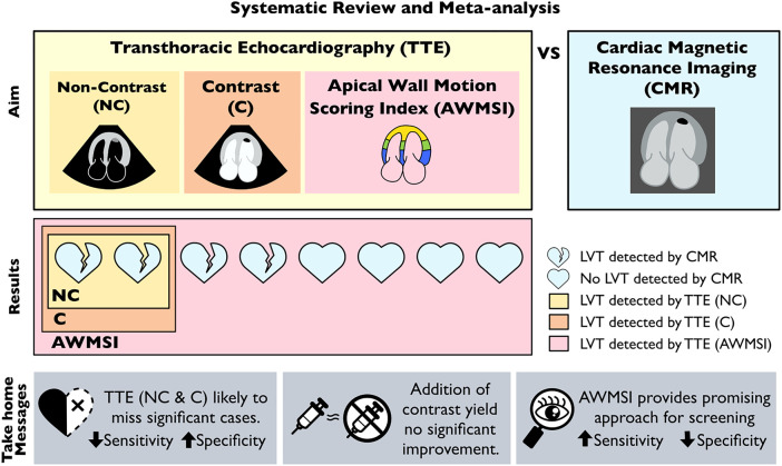
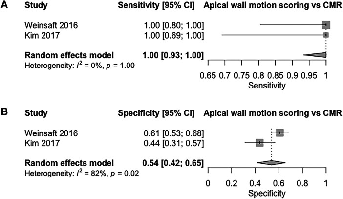
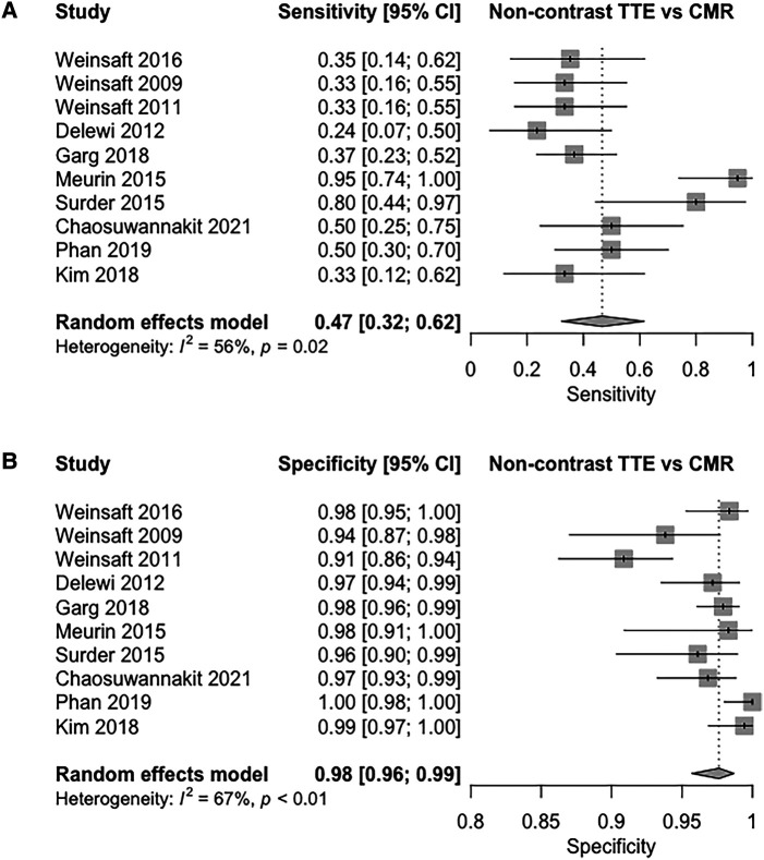
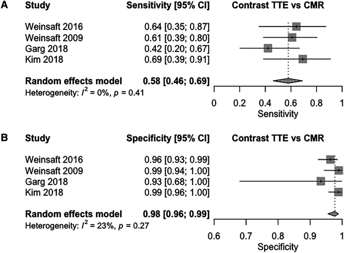
Transthoracic echocardiography (TTE) is the most commonly used imaging modality to diagnose left ventricular thrombus (LVT), however, cardiac magnetic resonance (CMR) remains the gold standard investigation. A comparison of the diagnostic performance between two modalities is needed to inform guidelines on a diagnostic approach towards LVT. We performed a systematic review and meta-analysis to investigate the diagnostic performance of three methods of TTE (non-contrast, contrast, and apical wall motion scoring) for the detection of LVT compared to CMR as a reference test. Studies comprising 2113 patients investigated for LVT using both TTE and CMR were included in the meta-analysis. For non-contrast TTE, pooled sensitivity and specificity were 47% [95% confidence interval (CI): 32-62%], and 98% (95% CI: 96-99%), respectively. In contrast, TTE pooled sensitivity and specificity values were 58% (95% CI: 46-69%), and 98% (95% CI: 96-99%), respectively. Apical wall motion scoring on non-contrast TTE yielded a sensitivity of 100% [95% CI: 93-100%] and a specificity of 54% (95% CI: 42-65%). The area under the curve (AUC) values from our summary receiver operating characteristic curve (SROC) for non-contrast and contrast TTE were 0.87 and 0.86 respectively, with apical wall motion studies having the highest AUC of 0.93. Despite high specificity, routine contrast and non-contrast TTE are likely to miss a significant number of LVT, making it a suboptimal screening tool. The addition of apical wall motion scoring provides a promising method to reliably identify patients requiring further investigations for LVT, whilst excluding others from unnecessary testing.

摘要

经胸超声心动图(TTE)是诊断左心室血栓(LVT)最常用的成像方式,然而,心脏磁共振成像(CMR)仍是金标准检查方法。需要比较这两种检查方式的诊断性能,以便为LVT的诊断方法制定指南提供依据。我们进行了一项系统评价和荟萃分析,以研究与作为参考检查的CMR相比,TTE的三种方法(非增强、增强和心尖壁运动评分)对LVT的诊断性能。荟萃分析纳入了2113例同时使用TTE和CMR检查LVT的患者。对于非增强TTE,汇总敏感性和特异性分别为47%[95%置信区间(CI):32 - 62%]和98%(95%CI:96 - 99%)。相比之下,增强TTE的汇总敏感性和特异性值分别为58%(95%CI:46 - 69%)和98%(95%CI:96 - 99%)。非增强TTE的心尖壁运动评分敏感性为100%[95%CI:93 - 100%],特异性为54%(95%CI:42 - 65%)。我们汇总的受试者工作特征曲线(SROC)中,非增强和增强TTE的曲线下面积(AUC)值分别为0.87和0.86,心尖壁运动研究的AUC最高,为0.93。尽管特异性较高,但常规的增强和非增强TTE可能会遗漏大量LVT,使其成为一种次优的筛查工具。增加心尖壁运动评分提供了一种有前景的方法,能够可靠地识别需要进一步检查LVT的患者,同时避免其他患者进行不必要的检查。

https://cdn.ncbi.nlm.nih.gov/pmc/blobs/dee6/11240154/9ec13d81f6a9/qyad041f3.jpg
https://cdn.ncbi.nlm.nih.gov/pmc/blobs/dee6/11240154/75516c5faeaa/qyad041_ga1.jpg
https://cdn.ncbi.nlm.nih.gov/pmc/blobs/dee6/11240154/314afb3f849c/qyad041f1.jpg
https://cdn.ncbi.nlm.nih.gov/pmc/blobs/dee6/11240154/d61b55d0d3d2/qyad041f2.jpg
https://cdn.ncbi.nlm.nih.gov/pmc/blobs/dee6/11240154/9ec13d81f6a9/qyad041f3.jpg

相似文献

1
A systematic review and meta-analysis of transthoracic echocardiogram vs. cardiac magnetic resonance imaging for the detection of left ventricular thrombus.经胸超声心动图与心脏磁共振成像检测左心室血栓的系统评价与Meta分析
Eur Heart J Imaging Methods Pract. 2023 Dec 7;1(2):qyad041. doi: 10.1093/ehjimp/qyad041. eCollection 2023 Sep.
2
Improved detection of echocardiographically occult left ventricular thrombi following ST-elevation myocardial infarction.经 ST 段抬高型心肌梗死治疗后,超声心动图隐匿性左心室血栓的检出率提高。
Eur Heart J Acute Cardiovasc Care. 2023 Oct 25;12(10):703-710. doi: 10.1093/ehjacc/zuad069.
3
Elusive left ventricular thrombus: Diagnostic role of cardiac magnetic resonance imaging-A case report and review of the literature.隐匿性左心室血栓:心脏磁共振成像的诊断作用——病例报告及文献综述
World J Clin Cases. 2018 Jun 16;6(6):127-131. doi: 10.12998/wjcc.v6.i6.127.
4
Determination of the prevalence and predictors of ventricular thrombus with assessment of the risk of systemic embolization to the CNS in patients after acute myocardial infarction using magnetic resonance imaging, echocardiography and cardiac markers - a prospective, unicentric, observational study.使用磁共振成像、超声心动图和心脏标志物,对急性心肌梗死后患者进行中枢神经系统全身性栓塞风险评估,以确定心室血栓的患病率和预测因素——一项前瞻性、单中心、观察性研究。
Biomed Pap Med Fac Univ Palacky Olomouc Czech Repub. 2023 Jan 8. doi: 10.5507/bp.2023.050.
5
LV thrombus detection by routine echocardiography: insights into performance characteristics using delayed enhancement CMR.常规超声心动图检测左心耳血栓:延迟增强磁共振成像对其性能特征的深入了解。
JACC Cardiovasc Imaging. 2011 Jul;4(7):702-12. doi: 10.1016/j.jcmg.2011.03.017.
6
Thrombus formation in the left ventricle after large myocardial infarction – assessment with cardiac magnetic resonance imaging.大面积心肌梗死后左心室血栓形成——心脏磁共振成像评估
Swiss Med Wkly. 2015 Jun 22;145:w14122. doi: 10.4414/smw.2015.14122. eCollection 2015.
7
Echocardiographic Algorithm for Post-Myocardial Infarction LV Thrombus: A Gatekeeper for Thrombus Evaluation by Delayed Enhancement CMR.心肌梗死后左心室血栓的超声心动图算法:延迟强化心脏磁共振成像评估血栓的守门人
JACC Cardiovasc Imaging. 2016 May;9(5):505-15. doi: 10.1016/j.jcmg.2015.06.017. Epub 2015 Oct 14.
8
Validity of echocardiography for detection of left ventricular thrombus with surgical validation in patients awaiting durable left ventricular assist device.超声心动图检测左心室血栓的有效性:在等待持久左心室辅助装置的患者中,与手术验证相吻合。
J Card Surg. 2021 Aug;36(8):2722-2728. doi: 10.1111/jocs.15662. Epub 2021 May 28.
9
Use of Transthoracic Echocardiogram to Detect Left Ventricular Thrombi.经胸超声心动图检测左心室血栓。
Ann Thorac Surg. 2021 Feb;111(2):556-560. doi: 10.1016/j.athoracsur.2020.05.106. Epub 2020 Jul 17.
10
Cardiac Magnetic Resonance Imaging and Transthoracic Echocardiography: Investigation of Concordance between the Two Methods for Measurement of the Cardiac Chamber.心脏磁共振成像和经胸超声心动图:两种方法测量心腔的一致性研究。
Medicina (Kaunas). 2019 Jun 9;55(6):260. doi: 10.3390/medicina55060260.

引用本文的文献

1
Left Ventricular Thrombosis in Ischemic and Non-Ischemic Cardiomyopathies: Focus on Evidence-Based Treatment.缺血性和非缺血性心肌病中的左心室血栓形成:聚焦循证治疗
J Clin Med. 2025 Feb 27;14(5):1615. doi: 10.3390/jcm14051615.
2
[Clinical, diagnostic and therapeutic profile of patients with left intraventricular thrombus in three high-complexity centers during the period 2000-2022].[2000年至2022年期间三个高复杂性中心左心室内血栓患者的临床、诊断和治疗概况]
Arch Peru Cardiol Cir Cardiovasc. 2024 Dec 11;5(4):207-214. doi: 10.47487/apcyccv.v5i4.430. eCollection 2024 Oct-Dec.
3
Direct oral anticoagulants or warfarin in patients with left ventricular thrombus after ST-elevation myocardial infarction: a pilot trial and a prespecified meta-analysis of randomised trials.

本文引用的文献

1
Management of Patients at Risk for and With Left Ventricular Thrombus: A Scientific Statement From the American Heart Association.左心室血栓风险患者及左心室血栓患者的管理:美国心脏协会科学声明
Circulation. 2022 Oct 11;146(15):e205-e223. doi: 10.1161/CIR.0000000000001092. Epub 2022 Sep 15.
2
Left Ventricular Thrombus Following Acute Myocardial Infarction: JACC State-of-the-Art Review.急性心肌梗死后左心室血栓:美国心脏病学会最新综述。
J Am Coll Cardiol. 2022 Mar 15;79(10):1010-1022. doi: 10.1016/j.jacc.2022.01.011.
3
Left Ventricular Thrombi: Insights from Cardiac Magnetic Resonance Imaging.
ST段抬高型心肌梗死后左心室血栓患者使用直接口服抗凝剂或华法林:一项试点试验及随机试验的预先设定的荟萃分析
EuroIntervention. 2025 Jan 6;21(1):82-92. doi: 10.4244/EIJ-D-24-00527.
4
Evolution of left ventricular thrombus on serial cardiovascular magnetic resonance imaging.连续心血管磁共振成像上左心室血栓的演变
Eur Heart J Cardiovasc Imaging. 2025 Jan 31;26(2):349-358. doi: 10.1093/ehjci/jeae271.
左心室血栓:心脏磁共振成像的新视角。
Tomography. 2021 May 12;7(2):180-188. doi: 10.3390/tomography7020016.
4
Preferred reporting items for journal and conference abstracts of systematic reviews and meta-analyses of diagnostic test accuracy studies (PRISMA-DTA for Abstracts): checklist, explanation, and elaboration.诊断试验准确性研究的系统评价和Meta分析的期刊及会议摘要的首选报告项目(PRISMA-DTA摘要版):清单、解释及详述
BMJ. 2021 Mar 15;372:n265. doi: 10.1136/bmj.n265.
5
Echocardiography versus computed tomography and cardiac magnetic resonance for the detection of left heart thrombosis: a systematic review and meta-analysis.超声心动图与计算机断层扫描和心脏磁共振成像在左心血栓检测中的比较:系统评价和荟萃分析。
Clin Res Cardiol. 2021 Nov;110(11):1697-1703. doi: 10.1007/s00392-020-01741-7. Epub 2020 Sep 13.
6
Long-Term Embolic Outcomes After Detection of Left Ventricular Thrombus by Late Gadolinium Enhancement Cardiovascular Magnetic Resonance Imaging: A Matched Cohort Study.心脏磁共振延迟钆增强成像检测左心室血栓后的长期栓塞结局:一项匹配队列研究。
Circ Cardiovasc Imaging. 2019 Nov;12(11):e009723. doi: 10.1161/CIRCIMAGING.119.009723. Epub 2019 Nov 11.
7
Incidence and predictors of left ventricular thrombus formation following acute ST-segment elevation myocardial infarction: A serial cardiac MRI study.急性ST段抬高型心肌梗死后左心室血栓形成的发生率及预测因素:一项心脏MRI系列研究
Int J Cardiol Heart Vasc. 2019 Jul 4;24:100395. doi: 10.1016/j.ijcha.2019.100395. eCollection 2019 Sep.
8
Predictors and prognosis of left ventricular thrombus in post-myocardial infarction patients with left ventricular dysfunction after percutaneous coronary intervention.经皮冠状动脉介入治疗后左心室功能不全的心肌梗死后患者左心室血栓形成的预测因素及预后
J Thorac Dis. 2018 Aug;10(8):4912-4922. doi: 10.21037/jtd.2018.07.69.
9
Assessment of LVEF using a new 16-segment wall motion score in echocardiography.在超声心动图中使用新的16节段壁运动评分评估左心室射血分数。
Echo Res Pract. 2018 Jun;5(2):63-69. doi: 10.1530/ERP-18-0006. Epub 2018 Mar 21.
10
2017 ESC Guidelines for the management of acute myocardial infarction in patients presenting with ST-segment elevation: The Task Force for the management of acute myocardial infarction in patients presenting with ST-segment elevation of the European Society of Cardiology (ESC).2017年欧洲心脏病学会(ESC)ST段抬高型急性心肌梗死患者管理指南:欧洲心脏病学会(ESC)ST段抬高型急性心肌梗死患者管理工作组
Eur Heart J. 2018 Jan 7;39(2):119-177. doi: 10.1093/eurheartj/ehx393.