• 文献检索
  • 文档翻译
  • 深度研究
  • 学术资讯
  • Suppr Zotero 插件Zotero 插件
  • 邀请有礼
  • 套餐&价格
  • 历史记录
应用&插件
Suppr Zotero 插件Zotero 插件浏览器插件Mac 客户端Windows 客户端微信小程序
定价
高级版会员购买积分包购买API积分包
服务
文献检索文档翻译深度研究API 文档MCP 服务
关于我们
关于 Suppr公司介绍联系我们用户协议隐私条款
关注我们

Suppr 超能文献

核心技术专利:CN118964589B侵权必究
粤ICP备2023148730 号-1Suppr @ 2026

文献检索

告别复杂PubMed语法,用中文像聊天一样搜索,搜遍4000万医学文献。AI智能推荐,让科研检索更轻松。

立即免费搜索

文件翻译

保留排版,准确专业,支持PDF/Word/PPT等文件格式,支持 12+语言互译。

免费翻译文档

深度研究

AI帮你快速写综述,25分钟生成高质量综述,智能提取关键信息,辅助科研写作。

立即免费体验

纵向 OCT 和 OCTA 监测显示视网膜变性 10(rd10)小鼠的玻璃体液血管加速消退。

Longitudinal OCT and OCTA monitoring reveals accelerated regression of hyaloid vessels in retinal degeneration 10 (rd10) mice.

机构信息

Department of Bioengineering, University of Illinois at Chicago, Chicago, IL, 60607, USA.

Department of Ophthalmology and Visual Sciences, University of Illinois at Chicago, Chicago, IL, 60612, USA.

出版信息

Sci Rep. 2019 Nov 13;9(1):16685. doi: 10.1038/s41598-019-53082-9.

DOI:10.1038/s41598-019-53082-9
PMID:31723168
原文链接:https://pmc.ncbi.nlm.nih.gov/articles/PMC6853881/
Abstract

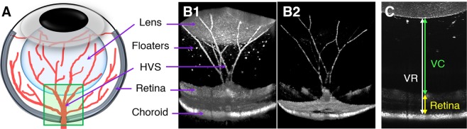
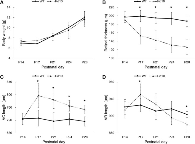
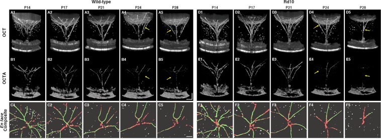
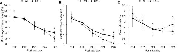
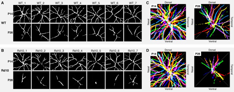
The hyaloid vascular system (HVS) is known to have an important role in eye development. However, physiological mechanisms of HVS regression and their correlation with developmental eye disorders remain unclear due to technical limitations of conventional ending point examination with fixed tissues. Here, we report comparative optical coherence tomography (OCT) and OCT angiography (OCTA) monitoring of HVS regression in wild-type and retinal degeneration 10 (rd10) mice. Longitudinal OCTA monitoring revealed accelerated regression of hyaloid vessels correlated with retinal degeneration in rd10. Quantitative OCT measurement disclosed significant distortions of both retinal thickness and the vitreous chamber in rd10 compared to WT mice. These OCT/OCTA observations confirmed the close relationship between HVS physiology and retinal neurovascular development. The distorted HVS regression might result from retinal hyperoxia or dopamine abnormality due to retinal remodeling in rd10 retina. By providing a noninvasive imaging platform for longitudinal monitoring of HVS regression, further OCT/OCTA study may lead to in-depth understanding of the physiological mechanisms of HVS regression in normal and diseased eyes, which is not only important for advanced study of the nature of the visual system but also may provide insights into the development of better treatment protocols of congenital eye disorders.

摘要

玻璃血管系统(HVS)在眼睛发育中具有重要作用。然而,由于传统的固定组织终点检查技术的限制,HVS 退化的生理机制及其与发育性眼病的相关性仍不清楚。在这里,我们报告了野生型和视网膜变性 10 型(rd10)小鼠玻璃血管系统(HVS)退化的比较光学相干断层扫描(OCT)和 OCT 血管造影(OCTA)监测。纵向 OCTA 监测显示,HVS 退化在 rd10 中与视网膜变性相关,呈加速趋势。与 WT 小鼠相比,定量 OCT 测量显示 rd10 中的视网膜厚度和玻璃体腔存在明显扭曲。这些 OCT/OCTA 观察结果证实了 HVS 生理学与视网膜神经血管发育之间的密切关系。rd10 视网膜中的视网膜过度氧化或多巴胺异常可能导致 HVS 退化的扭曲。通过提供 HVS 退化的纵向监测的非侵入性成像平台,进一步的 OCT/OCTA 研究可能深入了解正常和患病眼睛中 HVS 退化的生理机制,这不仅对视觉系统本质的深入研究很重要,还可能为先天性眼病的更好治疗方案的发展提供思路。

https://cdn.ncbi.nlm.nih.gov/pmc/blobs/22a8/6853881/047bd5890e3f/41598_2019_53082_Fig6_HTML.jpg
https://cdn.ncbi.nlm.nih.gov/pmc/blobs/22a8/6853881/a4d4bf146284/41598_2019_53082_Fig1_HTML.jpg
https://cdn.ncbi.nlm.nih.gov/pmc/blobs/22a8/6853881/cd57fff40b3c/41598_2019_53082_Fig2_HTML.jpg
https://cdn.ncbi.nlm.nih.gov/pmc/blobs/22a8/6853881/31145b8abc1b/41598_2019_53082_Fig3_HTML.jpg
https://cdn.ncbi.nlm.nih.gov/pmc/blobs/22a8/6853881/4c612bd6ea54/41598_2019_53082_Fig4_HTML.jpg
https://cdn.ncbi.nlm.nih.gov/pmc/blobs/22a8/6853881/8265cffc51e5/41598_2019_53082_Fig5_HTML.jpg
https://cdn.ncbi.nlm.nih.gov/pmc/blobs/22a8/6853881/047bd5890e3f/41598_2019_53082_Fig6_HTML.jpg

相似文献

1
Longitudinal OCT and OCTA monitoring reveals accelerated regression of hyaloid vessels in retinal degeneration 10 (rd10) mice.纵向 OCT 和 OCTA 监测显示视网膜变性 10(rd10)小鼠的玻璃体液血管加速消退。
Sci Rep. 2019 Nov 13;9(1):16685. doi: 10.1038/s41598-019-53082-9.
2
Long-term characterization of retinal degeneration in rd1 and rd10 mice using spectral domain optical coherence tomography.利用频域光学相干断层扫描对 rd1 和 rd10 小鼠的视网膜变性进行长期特征描述。
Invest Ophthalmol Vis Sci. 2012 Jul 10;53(8):4644-56. doi: 10.1167/iovs.12-9611.
3
Functional OCT angiography reveals early physiological dysfunction of hyaloid vasculature in developing mouse eye.功能光学相干断层扫描血管造影术揭示了发育中小鼠眼球中玻璃血管的早期生理功能障碍。
Exp Biol Med (Maywood). 2019 Jul;244(10):819-823. doi: 10.1177/1535370219850787. Epub 2019 May 24.
4
Comparative Optical Coherence Tomography Angiography of Wild-Type and rd10 Mouse Retinas.野生型和rd10小鼠视网膜的光学相干断层扫描血管造影对比
Transl Vis Sci Technol. 2018 Dec 28;7(6):42. doi: 10.1167/tvst.7.6.42. eCollection 2018 Nov.
5
Light-Dependent OCT Structure Changes in Photoreceptor Degenerative rd 10 Mouse Retina.光依赖性 OCT 结构变化在感光细胞退行性 rd10 鼠视网膜中。
Invest Ophthalmol Vis Sci. 2018 Feb 1;59(2):1084-1094. doi: 10.1167/iovs.17-23011.
6
Changes in morphology and visual function over time in mouse models of retinal degeneration: an SD-OCT, histology, and electroretinography study.视网膜变性小鼠模型中形态学和视觉功能随时间的变化:一项频域光学相干断层扫描、组织学和视网膜电图研究。
Jpn J Ophthalmol. 2016 Mar;60(2):111-25. doi: 10.1007/s10384-015-0422-0. Epub 2016 Jan 5.
7
Optical coherence tomography angiography: A comprehensive review of current methods and clinical applications.光学相干断层扫描血管造影:当前方法与临床应用的全面综述。
Prog Retin Eye Res. 2017 Sep;60:66-100. doi: 10.1016/j.preteyeres.2017.07.002. Epub 2017 Jul 29.
8
Different effects of valproic acid on photoreceptor loss in Rd1 and Rd10 retinal degeneration mice.丙戊酸对Rd1和Rd10视网膜变性小鼠光感受器丧失的不同影响。
Mol Vis. 2014 Nov 4;20:1527-44. eCollection 2014.
9
IMPROVED DETECTION AND DIAGNOSIS OF POLYPOIDAL CHOROIDAL VASCULOPATHY USING A COMBINATION OF OPTICAL COHERENCE TOMOGRAPHY AND OPTICAL COHERENCE TOMOGRAPHY ANGIOGRAPHY.采用光学相干断层扫描和光学相干断层扫描血管造影相结合的方法提高息肉状脉络膜血管病变的检测和诊断。
Retina. 2019 Sep;39(9):1655-1663. doi: 10.1097/IAE.0000000000002228.
10
Optical Coherence Tomography Angiography of Pigmented Paravenous Retinochoroidal Atrophy.色素性视网膜脉络膜静脉旁萎缩的光学相干断层扫描血管造影
Ophthalmic Surg Lasers Imaging Retina. 2018 May 1;49(5):381-383. doi: 10.3928/23258160-20180501-14.

引用本文的文献

1
Age-Related Retinal Layer Thickness Changes Measured by OCT in Mice: Implications for Alzheimer's Disease.OCT 测量小鼠年龄相关的视网膜层厚度变化:对阿尔茨海默病的影响。
Int J Mol Sci. 2024 Jul 27;25(15):8221. doi: 10.3390/ijms25158221.
2
OCT Imaging in Murine Models of Alzheimer's Disease in a Systematic Review: Findings, Methodology and Future Perspectives.阿尔茨海默病小鼠模型中的光学相干断层扫描成像系统评价:研究结果、方法与未来展望
Biomedicines. 2024 Feb 27;12(3):528. doi: 10.3390/biomedicines12030528.
3
Normative mice retinal thickness: 16-month longitudinal characterization of wild-type mice and changes in a model of Alzheimer's disease.

本文引用的文献

1
Functional OCT angiography reveals early physiological dysfunction of hyaloid vasculature in developing mouse eye.功能光学相干断层扫描血管造影术揭示了发育中小鼠眼球中玻璃血管的早期生理功能障碍。
Exp Biol Med (Maywood). 2019 Jul;244(10):819-823. doi: 10.1177/1535370219850787. Epub 2019 May 24.
2
An opsin 5-dopamine pathway mediates light-dependent vascular development in the eye.视蛋白 5-多巴胺途径介导眼睛中光依赖性血管发育。
Nat Cell Biol. 2019 Apr;21(4):420-429. doi: 10.1038/s41556-019-0301-x. Epub 2019 Apr 1.
3
Comparative Optical Coherence Tomography Angiography of Wild-Type and rd10 Mouse Retinas.
正常小鼠视网膜厚度:野生型小鼠16个月的纵向特征及阿尔茨海默病模型中的变化。
Front Aging Neurosci. 2023 Apr 6;15:1161847. doi: 10.3389/fnagi.2023.1161847. eCollection 2023.
4
Three-dimensional characterization of developing and adult ocular vasculature in mice using in toto clearing.使用整体透明法对小鼠发育中和成年期眼血管系统进行三维表征。
Commun Biol. 2022 Oct 27;5(1):1135. doi: 10.1038/s42003-022-04104-2.
5
An end-to-end network for segmenting the vasculature of three retinal capillary plexuses from OCT angiographic volumes.一种用于从光学相干断层扫描血管造影体积中分割三个视网膜毛细血管丛血管系统的端到端网络。
Biomed Opt Express. 2021 Jul 16;12(8):4889-4900. doi: 10.1364/BOE.431888. eCollection 2021 Aug 1.
6
In vivo imaging of the inner retinal layer structure in mice after eye-opening using visible-light optical coherence tomography.睁眼后利用可见光光学相干断层扫描对小鼠内视网膜层结构进行体内成像。
Exp Eye Res. 2021 Oct;211:108756. doi: 10.1016/j.exer.2021.108756. Epub 2021 Sep 4.
7
Concurrent OCT and OCT angiography of retinal neurovascular degeneration in the 5XFAD Alzheimer's disease mice.5XFAD阿尔茨海默病小鼠视网膜神经血管退变的光学相干断层扫描(OCT)与OCT血管造影同步检查
Neurophotonics. 2021 Jul;8(3):035002. doi: 10.1117/1.NPh.8.3.035002. Epub 2021 Jul 10.
8
Longitudinal normative OCT retinal thickness data for wild-type mice, and characterization of changes in the 3×Tg-AD mice model of Alzheimer's disease.正常野生型小鼠 OCT 视网膜厚度的纵向参考数据,以及阿尔茨海默病 3×Tg-AD 小鼠模型变化的特征描述。
Aging (Albany NY). 2021 Apr 2;13(7):9433-9454. doi: 10.18632/aging.202916.
9
Vascular morphology and blood flow signatures for differential artery-vein analysis in optical coherence tomography of the retina.视网膜光学相干断层扫描中用于动脉-静脉差异分析的血管形态和血流特征
Biomed Opt Express. 2020 Dec 15;12(1):367-379. doi: 10.1364/BOE.413149. eCollection 2021 Jan 1.
10
retinal imaging in translational regenerative research.转化性再生研究中的视网膜成像
Ann Transl Med. 2020 Sep;8(17):1096. doi: 10.21037/atm-20-4355.
野生型和rd10小鼠视网膜的光学相干断层扫描血管造影对比
Transl Vis Sci Technol. 2018 Dec 28;7(6):42. doi: 10.1167/tvst.7.6.42. eCollection 2018 Nov.
4
Gene expression in response to optical defocus of opposite signs reveals bidirectional mechanism of visually guided eye growth.光刺激的正负反馈对基因表达的影响揭示了视觉引导眼球生长的双向机制。
PLoS Biol. 2018 Oct 9;16(10):e2006021. doi: 10.1371/journal.pbio.2006021. eCollection 2018 Oct.
5
Histochemical characteristics of regressing vessels in the hyaloid vascular system of neonatal mice: Novel implication for vascular atrophy.新生鼠玻璃血管系统中退化血管的组织化学特征:血管萎缩的新意义。
Exp Eye Res. 2018 Jul;172:1-9. doi: 10.1016/j.exer.2018.03.024. Epub 2018 Mar 27.
6
A highly efficient murine model of experimental myopia.一种高效的实验性近视小鼠模型。
Sci Rep. 2018 Feb 1;8(1):2026. doi: 10.1038/s41598-018-20272-w.
7
Retinopathy of prematurity: the need for prevention.早产儿视网膜病变:预防的必要性。
Eye Brain. 2016 May 20;8:91-102. doi: 10.2147/EB.S99038. eCollection 2016.
8
Pan-retinal characterisation of Light Responses from Ganglion Cells in the Developing Mouse Retina.发育中小鼠视网膜神经节细胞的光反应全视网膜特征。
Sci Rep. 2017 Feb 10;7:42330. doi: 10.1038/srep42330.
9
Optical coherence tomography angiography of stimulus evoked hemodynamic responses in individual retinal layers.个体视网膜各层刺激诱发血流动力学反应的光学相干断层扫描血管造影术。
Biomed Opt Express. 2016 Jul 29;7(8):3151-62. doi: 10.1364/BOE.7.003151. eCollection 2016 Aug 1.
10
Developmental regression of hyaloid vasculature is triggered by neurons.神经元触发了玻璃体血管系统的发育性退化。
J Exp Med. 2016 Jun 27;213(7):1175-83. doi: 10.1084/jem.20151966. Epub 2016 Jun 20.