Center for Stem Cell and Regenerative Medicine, Tokyo Medical and Dental University, 1-5-45, Bunkyo-ku, Yushima, Tokyo, Japan.
Research & Development Center, Dai Nippon Printing Co., Ltd., Tokyo, Japan.
Sci Rep. 2019 Nov 14;9(1):16835. doi: 10.1038/s41598-019-53383-z.
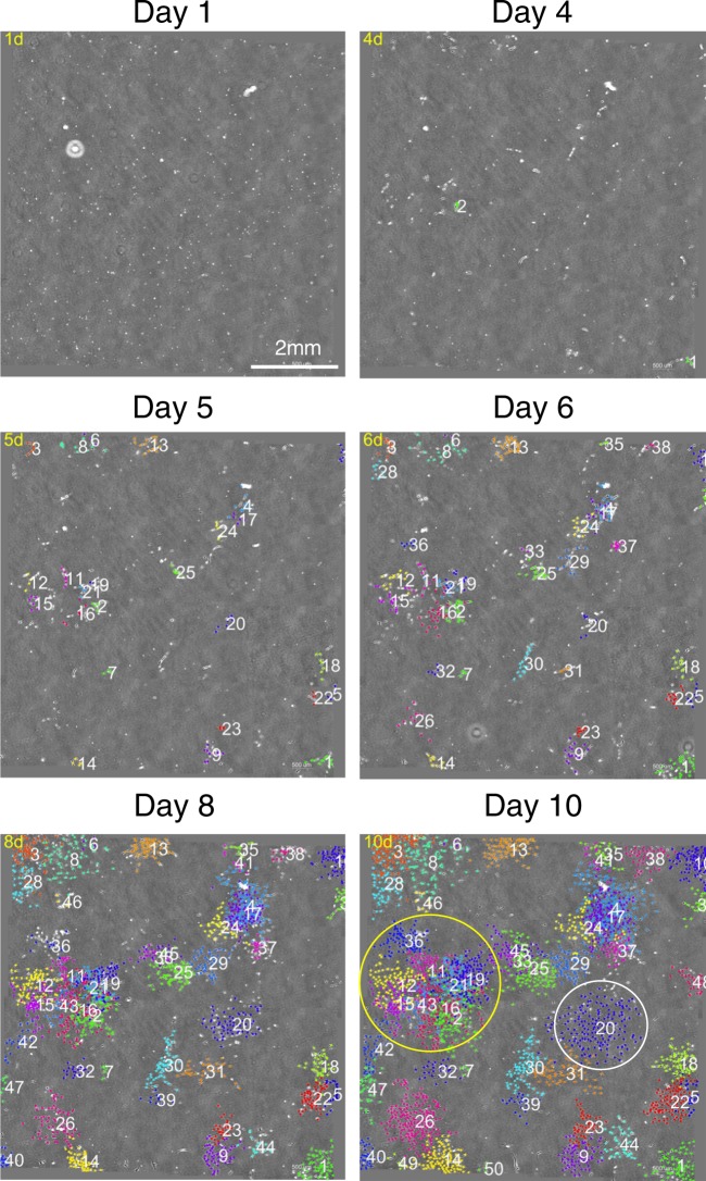
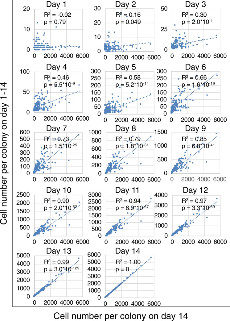
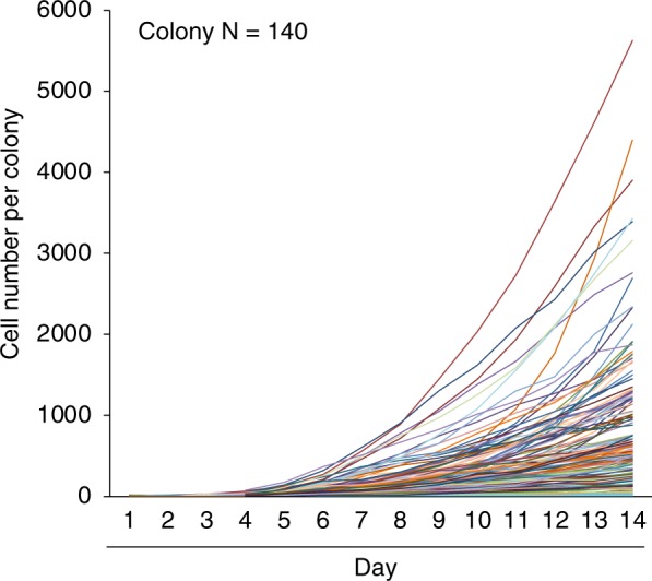
Mesenchymal stem cells from the synovium (synovial MSCs) are attractive for cartilage and meniscus regeneration therapy. We developed a software program that can distinguish individual colonies and automatically count the cell number per colony using time-lapse images. In this study, we investigated the usefulness of the software and analyzed colony formation in cultured synovial MSCs. Time-lapse image data were obtained for 14-day-expanded human synovial MSCs. The cell number per colony (for 145 colonies) was automatically counted from phase-contrast and nuclear-stained images. Colony growth curves from day 1 to day 14 (for 140 colonies) were classified using cluster analysis. Correlation analysis of the distribution of the cell number per colony at 14 days versus that number at 1-14 days revealed a correlation at 7 and 14 days. We obtained accurate cell number counts from phase-contrast images. Individual colony growth curves were classified into three main groups and subgroups. Our image analysis software has the potential to improve the evaluation of cell proliferation and to facilitate successful clinical applications using MSCs.
滑膜间充质干细胞(synovial MSCs)是软骨和半月板再生治疗的理想选择。我们开发了一种软件程序,可以使用延时图像区分单个菌落并自动计算每个菌落的细胞数量。在这项研究中,我们研究了该软件的实用性,并分析了培养的滑膜间充质干细胞的菌落形成情况。对 14 天培养的人滑膜间充质干细胞进行了延时图像数据采集。从相差和核染色图像自动计数每个菌落的细胞数量(145 个菌落)。使用聚类分析对第 1 天至第 14 天(140 个菌落)的菌落生长曲线进行分类。对第 14 天和第 1-14 天每个菌落细胞数量的分布进行相关性分析,发现第 7 天和第 14 天有相关性。我们从相差图像中获得了准确的细胞数量计数。单个菌落生长曲线分为三个主要组和亚组。我们的图像分析软件具有提高细胞增殖评估的潜力,并有助于使用间充质干细胞进行成功的临床应用。