• 文献检索
  • 文档翻译
  • 深度研究
  • 学术资讯
  • Suppr Zotero 插件Zotero 插件
  • 邀请有礼
  • 套餐&价格
  • 历史记录
应用&插件
Suppr Zotero 插件Zotero 插件浏览器插件Mac 客户端Windows 客户端微信小程序
定价
高级版会员购买积分包购买API积分包
服务
文献检索文档翻译深度研究API 文档MCP 服务
关于我们
关于 Suppr公司介绍联系我们用户协议隐私条款
关注我们

Suppr 超能文献

核心技术专利:CN118964589B侵权必究
粤ICP备2023148730 号-1Suppr @ 2026

文献检索

告别复杂PubMed语法,用中文像聊天一样搜索,搜遍4000万医学文献。AI智能推荐,让科研检索更轻松。

立即免费搜索

文件翻译

保留排版,准确专业,支持PDF/Word/PPT等文件格式,支持 12+语言互译。

免费翻译文档

深度研究

AI帮你快速写综述,25分钟生成高质量综述,智能提取关键信息,辅助科研写作。

立即免费体验

视网膜血流与青光眼视野进展的关系。

Association of Retinal Blood Flow with Progression of Visual Field in Glaucoma.

机构信息

Department of Ophthalmology, Seoul St. Mary's Hospital, College of Medicine, The Catholic University of Korea, Seoul, Republic of Korea.

出版信息

Sci Rep. 2019 Nov 14;9(1):16813. doi: 10.1038/s41598-019-53354-4.

DOI:10.1038/s41598-019-53354-4
PMID:31728047
原文链接:https://pmc.ncbi.nlm.nih.gov/articles/PMC6856104/
Abstract

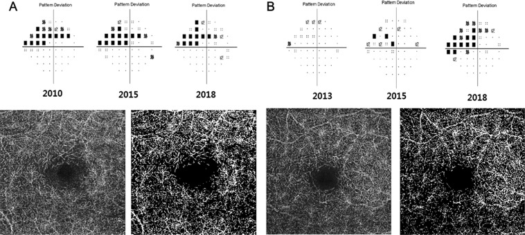
In the glaucoma clinic, patients with normal intraocular pressure (IOP) can sometimes show visual field (VF) progression. Therefore, clarification of relationship between vascular status and glaucomatous VF deterioration is a focus of interest. We used optical coherence tomography angiography (OCTA), with the aim of evaluating the relationship between vessel density (VD) and VF progression in glaucoma patients. We included 104 eyes with open angle glaucoma who were followed up for at least 5 years in this retrospective case-control study. Superficial and deep VD of macula were assessed by OCTA. Regression analysis and Cox proportional hazards model were used to identify factors significantly associated with VF progression. In logistic regression analysis determining VF progression from Guided Progression Analysis (GPA) program, initial IOP and deep macular VD were significantly associated with VF progression in multivariate analysis (P = 0.019 and 0.004). Cox proportional hazards model also identified deep macular VD as significantly related to VF progression (P = 0.035). In conclusion, initial IOP and deep VD were related to VF deterioration in glaucoma. Deep VD might be used as a surrogate of glaucomatous VF progression related with vascular incompetence.

摘要

在青光眼门诊中,眼压正常的患者有时会出现视野(VF)进展。因此,阐明血管状态与青光眼 VF 恶化之间的关系是研究的重点。我们使用光相干断层扫描血管造影(OCTA),旨在评估青光眼患者血管密度(VD)与 VF 进展之间的关系。在这项回顾性病例对照研究中,我们纳入了 104 只患有开角型青光眼且至少随访 5 年的眼。通过 OCTA 评估黄斑区浅层和深层 VD。回归分析和 Cox 比例风险模型用于确定与 VF 进展显著相关的因素。在确定 GPA 程序中 VF 进展的逻辑回归分析中,初始眼压和黄斑深层 VD 在多变量分析中与 VF 进展显著相关(P=0.019 和 0.004)。Cox 比例风险模型也确定黄斑深层 VD 与 VF 进展显著相关(P=0.035)。总之,初始眼压和黄斑深层 VD 与青光眼的 VF 恶化有关。深层 VD 可能可作为与血管功能不全相关的青光眼 VF 进展的替代指标。

https://cdn.ncbi.nlm.nih.gov/pmc/blobs/38f6/6856104/e1de35956d95/41598_2019_53354_Fig1_HTML.jpg

相似文献

1
Association of Retinal Blood Flow with Progression of Visual Field in Glaucoma.视网膜血流与青光眼视野进展的关系。
Sci Rep. 2019 Nov 14;9(1):16813. doi: 10.1038/s41598-019-53354-4.
2
OCTA vessel density changes in the macular zone in glaucomatous eyes.青光眼患者眼部黄斑区光学相干断层扫描血管密度的变化
Graefes Arch Clin Exp Ophthalmol. 2018 Aug;256(8):1499-1508. doi: 10.1007/s00417-018-3965-1. Epub 2018 Apr 10.
3
Projection-Resolved Optical Coherence Tomography Angiography of Macular Retinal Circulation in Glaucoma.青光眼黄斑视网膜循环的投影分辨光学相干断层扫描血管造影
Ophthalmology. 2017 Nov;124(11):1589-1599. doi: 10.1016/j.ophtha.2017.06.002. Epub 2017 Jul 1.
4
Association Between Parapapillary Choroidal Vessel Density Measured With Optical Coherence Tomography Angiography and Future Visual Field Progression in Patients With Glaucoma.光学相干断层扫描血管造影测量的脉络膜旁血管密度与青光眼患者未来视野进展的相关性。
JAMA Ophthalmol. 2019 Jun 1;137(6):681-688. doi: 10.1001/jamaophthalmol.2019.0422.
5
Optical Coherence Tomography Angiography Macular Vascular Density Measurements and the Central 10-2 Visual Field in Glaucoma.光学相干断层扫描血管造影黄斑血管密度测量与青光眼中心 10-2 视野。
J Glaucoma. 2018 Jun;27(6):481-489. doi: 10.1097/IJG.0000000000000964.
6
Decreased macular deep capillary plexus is associated with functional progression of normal tension glaucoma patients with unilateral visual field loss.黄斑深层毛细血管丛减少与单侧视野丧失的正常眼压青光眼患者的功能进展有关。
Br J Ophthalmol. 2024 Jan 29;108(2):188-194. doi: 10.1136/bjo-2022-322362.
7
Association of superficial macular vessel density with visual field progression in open-angle glaucoma with central visual field damage.在伴有中央视野损害的开角型青光眼患者中,浅层视网膜血管密度与视野进展的相关性。
Sci Rep. 2023 May 3;13(1):7190. doi: 10.1038/s41598-023-34000-6.
8
Multilayer Macula Vessel Density and Visual Field Progression in Glaucoma.青光眼的多层黄斑血管密度与视野进展。
Am J Ophthalmol. 2022 May;237:193-203. doi: 10.1016/j.ajo.2021.11.018. Epub 2021 Nov 19.
9
Optical Coherence Tomography Angiography of the Peripapillary Retina in Primary Angle-Closure Glaucoma.原发性闭角型青光眼视乳头周围视网膜的光学相干断层扫描血管造影
Am J Ophthalmol. 2017 Oct;182:194-200. doi: 10.1016/j.ajo.2017.07.024. Epub 2017 Aug 7.
10
Combining optical coherence tomography with visual field data to rapidly detect disease progression in glaucoma: a diagnostic accuracy study.结合光学相干断层扫描和视野数据快速检测青光眼疾病进展:一项诊断准确性研究。
Health Technol Assess. 2018 Jan;22(4):1-106. doi: 10.3310/hta22040.

引用本文的文献

1
Biomechanical and Vascular Metrics Between Eyes of Patients With Asymmetric Glaucoma and Symmetric Glaucoma.不对称青光眼和对称青光眼患者眼间的生物力学和血管计量学比较。
J Glaucoma. 2024 May 1;33(5):334-339. doi: 10.1097/IJG.0000000000002363. Epub 2024 Jan 9.
2
Foveal avascular zone vessel density is associated with visual field progression in early-stage glaucoma eyes with central visual field damage.中心视野损伤的早期青光眼眼中,中心凹无血管区血管密度与视野进展相关。
Sci Rep. 2023 Oct 25;13(1):18285. doi: 10.1038/s41598-023-45541-1.
3
Ocular blood flow biomarkers may predict long-term glaucoma progression.

本文引用的文献

1
Risk Factors for Fast Visual Field Progression in Glaucoma.青光眼视野快速进展的危险因素。
Am J Ophthalmol. 2019 Nov;207:268-278. doi: 10.1016/j.ajo.2019.06.019. Epub 2019 Jun 22.
2
Investigation of intraocular pressure fluctuation as a risk factor of glaucoma progression.眼压波动作为青光眼进展危险因素的研究。
Clin Ophthalmol. 2018 Dec 18;13:9-16. doi: 10.2147/OPTH.S186526. eCollection 2019.
3
Effect of Macular Vascular Density on Central Visual Function and Macular Structure in Glaucoma Patients.青光眼患者黄斑血管密度对中心视觉功能和黄斑结构的影响。
眼血流生物标志物可预测青光眼的长期进展。
Br J Ophthalmol. 2024 Jun 20;108(7):946-950. doi: 10.1136/bjo-2022-322644.
4
Factors Associated with Vascular Changes at the Level of Retinal Ganglion Cell Axon versus Soma/Dendrite in Glaucoma Patients.青光眼患者视网膜神经节细胞轴突与胞体/树突水平血管变化的相关因素
J Clin Med. 2023 Jun 22;12(13):4221. doi: 10.3390/jcm12134221.
5
Association of superficial macular vessel density with visual field progression in open-angle glaucoma with central visual field damage.在伴有中央视野损害的开角型青光眼患者中,浅层视网膜血管密度与视野进展的相关性。
Sci Rep. 2023 May 3;13(1):7190. doi: 10.1038/s41598-023-34000-6.
6
Association of macular vessel density and ganglion cell complex thickness with central visual field progression in glaucoma.青光眼中心视野进展与黄斑血管密度和神经节细胞复合体厚度的关系。
Br J Ophthalmol. 2023 Nov 22;107(12):1828-1833. doi: 10.1136/bjo-2022-321870.
7
Retinal Oxygen Extraction in Patients with Primary Open-Angle Glaucoma.原发性开角型青光眼患者的视网膜氧提取。
Int J Mol Sci. 2022 Sep 5;23(17):10152. doi: 10.3390/ijms231710152.
8
Attenuated Amplitude of Pattern Electroretinogram in Glaucoma Patients with Choroidal Parapapillary Microvasculature Dropout.脉络膜视乳头旁微血管缺失的青光眼患者图形视网膜电图振幅降低
J Clin Med. 2022 Apr 28;11(9):2478. doi: 10.3390/jcm11092478.
9
Association of choroidal blood flow with autonomic dysfunction in patients with normal tension glaucoma.正常眼压性青光眼患者脉络膜血流与自主神经功能障碍的相关性。
Sci Rep. 2022 Mar 24;12(1):5136. doi: 10.1038/s41598-022-09162-4.
10
Optic Disc Hemorrhage Is Not Associated with Global Choroidal Vessel Loss, but Is Associated with Localized Choroidal Vessel Loss in Glaucoma.视盘出血与全脉络膜血管丢失无关,但与青光眼患者的局部脉络膜血管丢失有关。
J Clin Med. 2022 Feb 18;11(4):1080. doi: 10.3390/jcm11041080.
Sci Rep. 2018 Oct 30;8(1):16009. doi: 10.1038/s41598-018-34417-4.
4
Macular and Optic Nerve Head Vessel Density and Progressive Retinal Nerve Fiber Layer Loss in Glaucoma.青光眼的黄斑和视神经头血管密度与进行性视网膜神经纤维层丢失。
Ophthalmology. 2018 Nov;125(11):1720-1728. doi: 10.1016/j.ophtha.2018.05.006. Epub 2018 Jun 12.
5
OCTA vessel density changes in the macular zone in glaucomatous eyes.青光眼患者眼部黄斑区光学相干断层扫描血管密度的变化
Graefes Arch Clin Exp Ophthalmol. 2018 Aug;256(8):1499-1508. doi: 10.1007/s00417-018-3965-1. Epub 2018 Apr 10.
6
Modern Diagnostic Techniques for the Assessment of Ocular Blood Flow in Myopia: Current State of Knowledge.评估近视患者眼血流的现代诊断技术:当前知识现状
J Ophthalmol. 2018 Jan 21;2018:4694789. doi: 10.1155/2018/4694789. eCollection 2018.
7
Optical coherence tomography angiography.光学相干断层扫描血管造影术。
Prog Retin Eye Res. 2018 May;64:1-55. doi: 10.1016/j.preteyeres.2017.11.003. Epub 2017 Dec 8.
8
Prediction of Visual Field Progression in Patients with Primary Open-Angle Glaucoma, Mainly Including Normal Tension Glaucoma.原发性开角型青光眼,主要包括正常眼压型青光眼患者视野进展的预测。
Sci Rep. 2017 Nov 8;7(1):15048. doi: 10.1038/s41598-017-15267-y.
9
Optical Coherence Tomography Angiography of the Superficial Microvasculature in the Macular and Peripapillary Areas in Glaucomatous and Healthy Eyes.青光眼患者和健康人眼睛黄斑区及视乳头周围区域浅表微血管的光学相干断层扫描血管造影
Invest Ophthalmol Vis Sci. 2017 Jul 1;58(9):3637-3645. doi: 10.1167/iovs.17-21846.
10
Optical coherence tomography angiography vessel density mapping at various retinal layers in healthy and normal tension glaucoma eyes.健康和正常眼压性青光眼患者眼睛不同视网膜层的光学相干断层扫描血管造影血管密度映射
Graefes Arch Clin Exp Ophthalmol. 2017 Jun;255(6):1193-1202. doi: 10.1007/s00417-017-3671-4. Epub 2017 Apr 20.