Zhang Shunhua, Wu Chan, Liu Liang, Jia Yali, Zhang Yao, Zhang Yang, Zhang Hua, Zhong Yong, Huang David
Ophthalmology Department, Peking Union Medical College Hospital, Peking Union Medical College, Chinese Academy of Medical Sciences, Beijing, China.
Casey Eye Institute, Oregon Health and Science University, Portland, Oregon.
Am J Ophthalmol. 2017 Oct;182:194-200. doi: 10.1016/j.ajo.2017.07.024. Epub 2017 Aug 7.
To measure the change of peripapillary retinal vessel density (VD) in eyes with a history of acute primary angle-closure glaucoma (PACG).
Case-control study.
Twenty-one consecutive Chinese patients with history of unilateral acute PACG were enrolled. Eyes with acute PACG constituted the case group, while the contralateral eyes without attack constituted the control. All patients underwent ophthalmic examinations including best-corrected visual acuity, intraocular pressure, and visual field (VF). Spectral-domain optical coherence tomography (SD-OCT) was used to obtain both structural OCT and OCT angiography (OCTA). Structural OCT scans provided thickness measurements of the peripapillary retinal nerve fiber layer (RNFL) and macular ganglion cell complex (GCC). OCTA was used to measure all-plexus peripapillary retinal VD.
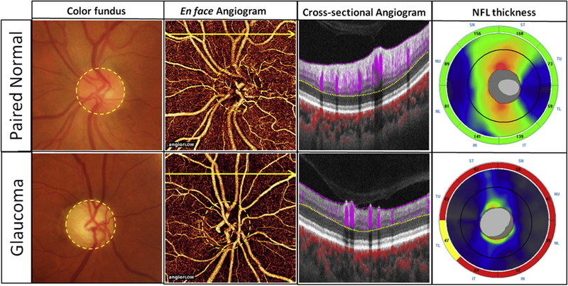
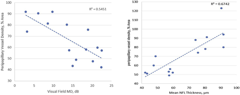
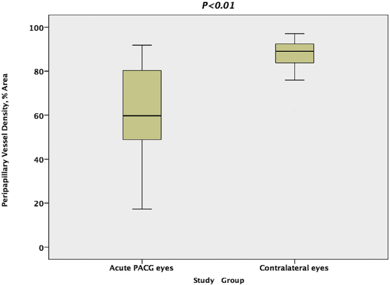
In unaffected eyes, a dense microvascular network surrounded the disc on all-plexus retinal OCTA. The vascular network was visibly attenuated and focal capillary dropout was evident in acute PACG eyes. The peripapillary VD in acute PACG eyes was 66.6% ± 17.3% (mean ± standard deviation), which was significantly (P < .01) reduced compared to 87.2% ± 8.6% in the unaffected eyes. In acute PACG eyes, peripapillary retinal VD was positively correlated with RNFL and GCC thicknesses (P < .001 each) and negatively correlated with VF mean deviation (P = .002) and cup-to-disc ratio (P = .0064). In unaffected eyes, there were no correlations between peripapillary retinal VD and glaucoma-related parameters.
In acute PACG eyes, peripapillary retinal VD decreased significantly compared with the contralateral unaffected eyes. Peripapillary retinal VD was significantly correlated with other glaucomatous changes.
测量有急性原发性闭角型青光眼(PACG)病史的眼睛中视乳头周围视网膜血管密度(VD)的变化。
病例对照研究。
纳入21例连续的有单侧急性PACG病史的中国患者。患有急性PACG的眼睛构成病例组,而未发作的对侧眼睛构成对照组。所有患者均接受眼科检查,包括最佳矫正视力、眼压和视野(VF)。使用光谱域光学相干断层扫描(SD-OCT)获取结构OCT和OCT血管造影(OCTA)。结构OCT扫描提供视乳头周围视网膜神经纤维层(RNFL)和黄斑神经节细胞复合体(GCC)的厚度测量值。OCTA用于测量视乳头周围全层视网膜VD。
在未受影响的眼睛中,全层视网膜OCTA上视盘周围有密集的微血管网络。在急性PACG眼中,血管网络明显变细,局部毛细血管缺失明显。急性PACG眼中视乳头周围VD为66.6%±17.3%(平均值±标准差),与未受影响眼睛中的87.2%±8.6%相比显著降低(P<.01)。在急性PACG眼中,视乳头周围视网膜VD与RNFL和GCC厚度呈正相关(均P<.001),与VF平均偏差呈负相关(P=.002),与杯盘比呈负相关(P=.0064)。在未受影响的眼睛中,视乳头周围视网膜VD与青光眼相关参数之间无相关性。
与对侧未受影响的眼睛相比,急性PACG眼中视乳头周围视网膜VD显著降低。视乳头周围视网膜VD与其他青光眼性改变显著相关。