• 文献检索
  • 文档翻译
  • 深度研究
  • 学术资讯
  • Suppr Zotero 插件Zotero 插件
  • 邀请有礼
  • 套餐&价格
  • 历史记录
应用&插件
Suppr Zotero 插件Zotero 插件浏览器插件Mac 客户端Windows 客户端微信小程序
定价
高级版会员购买积分包购买API积分包
服务
文献检索文档翻译深度研究API 文档MCP 服务
关于我们
关于 Suppr公司介绍联系我们用户协议隐私条款
关注我们

Suppr 超能文献

核心技术专利:CN118964589B侵权必究
粤ICP备2023148730 号-1Suppr @ 2026

文献检索

告别复杂PubMed语法,用中文像聊天一样搜索,搜遍4000万医学文献。AI智能推荐,让科研检索更轻松。

立即免费搜索

文件翻译

保留排版,准确专业,支持PDF/Word/PPT等文件格式,支持 12+语言互译。

免费翻译文档

深度研究

AI帮你快速写综述,25分钟生成高质量综述,智能提取关键信息,辅助科研写作。

立即免费体验

锥形束 CT 用于听力保护人工耳蜗植入术的围手术期成像——一项人体尸体研究。

Cone beam CT for perioperative imaging in hearing preservation Cochlear implantation - a human cadaveric study.

机构信息

Sunnybrook Research Institute, Sunnybrook Health Sciences Centre, University of Toronto, 2075 Bayview Ave., Room M1, Toronto, 102, Canada.

University of Western Australia, School of Surgery, Perth, Australia.

出版信息

J Otolaryngol Head Neck Surg. 2019 Nov 21;48(1):65. doi: 10.1186/s40463-019-0388-x.

DOI:10.1186/s40463-019-0388-x
PMID:31753027
原文链接:https://pmc.ncbi.nlm.nih.gov/articles/PMC6873551/
Abstract

BACKGROUND

Knowledge of the cochlear implant array's precise position is important because of the correlation between electrode position and speech understanding. Several groups have provided recent image processing evidence to determine scalar translocation, angular insertion depth, and cochlear duct length (CDL); all of which are being used for patient-specific programming. Cone beam computed tomography (CBCT) is increasingly used in otology due to its superior resolution and low radiation dose. Our objectives are as followed: 1.Validate CBCT by measuring cochlear metrics, including basal turn diameter (A-value) and lateral wall cochlear duct length at different angular intervals and comparing it against microcomputed CT (uCT).2.Explore the relationship between measured lateral wall cochlear duct length at different angular intervals and insertion depth among 3 different length electrodes using CBCT.

METHODS

The study was performed using fixed human cadaveric temporal bones in a tertiary academic centre. Ten temporal bones were subjected to the standard facial recess approach for cochlear implantation and imaged by CBCT followed by uCT. Measurements were performed on a three-dimensional reconstructed model of the cochlea. Sequential insertion of 3 electrodes (Med-El Flex24, 28 and Soft) was then performed in 5 bones and reimaged by CBCT. Statistical analysis was performed using Pearson's correlation.

RESULTS

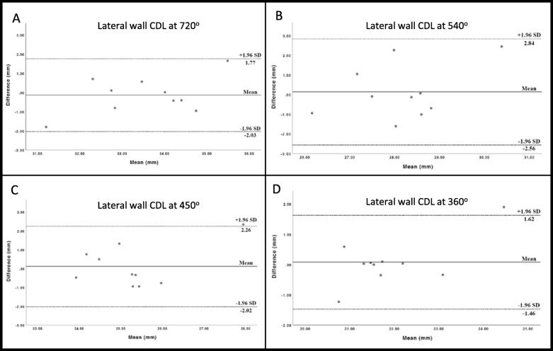
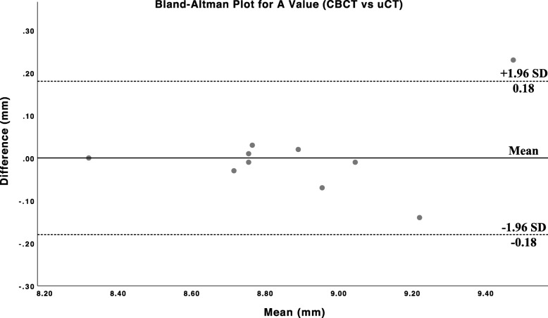
There was good agreement between CBCT and uCT for cochlear metrics, validating the precision of CBCT against the current gold standard uCT in imaging. The A-value recorded by both modalities showed a high degree of linear correlation and did not differ by more than 0.23 mm in absolute values. For the measurement of lateral wall CDL at various points along the cochlea, there was a good correlation between both modalities at 360 deg and 720 deg (r = 0.85, p < 0.01 and r = 0.79, p < 0.01). The Flex24 electrode displayed consistent insertion depth across different bones.

CONCLUSIONS

CBCT reliably performs cochlear metrics and measures electrode insertion depth. The low radiation dose, fast acquisition time, diminished metallic artifacts and portability of CBCT make it a valid option for imaging in cochlear implant surgery.

摘要

背景

由于电极位置与言语理解之间存在相关性,了解 Cochlear 植入物阵列的精确位置很重要。 有几个研究小组最近提供了图像处理证据,以确定标量移位、角插入深度和耳蜗管长度(CDL); 所有这些都用于患者特定的编程。 由于其优越的分辨率和低辐射剂量,锥形束计算机断层扫描(CBCT)在耳科学中越来越多地使用。 我们的目标如下:1. 通过测量耳蜗参数来验证 CBCT,包括在不同角度间隔处的基底转直径(A 值)和外侧壁耳蜗管长度,并将其与微计算机断层扫描(uCT)进行比较。2. 探索使用 CBCT 测量不同角度间隔处的外侧壁耳蜗管长度与 3 个不同长度电极的插入深度之间的关系。

方法

该研究在一家三级学术中心的固定人体尸体颞骨上进行。 10 个颞骨接受 Cochlear 植入的标准面神经隐窝入路,并通过 CBCT 成像,然后进行 uCT 成像。 在耳蜗的三维重建模型上进行测量。 然后在 5 个骨头中连续插入 3 个电极(Med-El Flex24、28 和 Soft),并通过 CBCT 重新成像。 使用 Pearson 相关进行统计分析。

结果

CBCT 和 uCT 对耳蜗参数的测量结果吻合良好,验证了 CBCT 在成像方面对当前金标准 uCT 的精确性。 两种模态记录的 A 值显示出高度的线性相关性,绝对值相差不超过 0.23 毫米。 对于耳蜗各部位外侧壁 CDL 的测量,两种模态在 360°和 720°之间相关性良好(r=0.85,p<0.01 和 r=0.79,p<0.01)。 Flex24 电极在不同骨头中具有一致的插入深度。

结论

CBCT 可靠地执行耳蜗参数测量和电极插入深度测量。 低辐射剂量、快速采集时间、减少的金属伪影和 CBCT 的便携性使其成为 Cochlear 植入手术成像的有效选择。

https://cdn.ncbi.nlm.nih.gov/pmc/blobs/0c1f/6873551/1a31bc53d261/40463_2019_388_Fig3_HTML.jpg
https://cdn.ncbi.nlm.nih.gov/pmc/blobs/0c1f/6873551/93003053efa8/40463_2019_388_Fig1_HTML.jpg
https://cdn.ncbi.nlm.nih.gov/pmc/blobs/0c1f/6873551/a84567219de7/40463_2019_388_Fig2_HTML.jpg
https://cdn.ncbi.nlm.nih.gov/pmc/blobs/0c1f/6873551/1a31bc53d261/40463_2019_388_Fig3_HTML.jpg
https://cdn.ncbi.nlm.nih.gov/pmc/blobs/0c1f/6873551/93003053efa8/40463_2019_388_Fig1_HTML.jpg
https://cdn.ncbi.nlm.nih.gov/pmc/blobs/0c1f/6873551/a84567219de7/40463_2019_388_Fig2_HTML.jpg
https://cdn.ncbi.nlm.nih.gov/pmc/blobs/0c1f/6873551/1a31bc53d261/40463_2019_388_Fig3_HTML.jpg

相似文献

1
Cone beam CT for perioperative imaging in hearing preservation Cochlear implantation - a human cadaveric study.锥形束 CT 用于听力保护人工耳蜗植入术的围手术期成像——一项人体尸体研究。
J Otolaryngol Head Neck Surg. 2019 Nov 21;48(1):65. doi: 10.1186/s40463-019-0388-x.
2
CT imaging-based approaches to cochlear duct length estimation-a human temporal bone study.基于 CT 成像的耳蜗管长度估计方法——一项人类颞骨研究。
Eur Radiol. 2022 Feb;32(2):1014-1023. doi: 10.1007/s00330-021-08189-x. Epub 2021 Aug 31.
3
Differences of radiological artefacts in cochlear implantation in temporal bone and complete head.颞骨和全头颅人工耳蜗植入中放射伪影的差异。
Cochlear Implants Int. 2014 Mar;15(2):112-7. doi: 10.1179/1754762813Y.0000000035. Epub 2013 Nov 25.
4
The use of cone-beam computed tomography to determine cochlear implant electrode position in human temporal bones.使用锥形束计算机断层扫描确定人工耳蜗电极在人类颞骨中的位置。
Otol Neurotol. 2014 Sep;35(8):1338-44. doi: 10.1097/MAO.0000000000000295.
5
Imaging of the human cochlea using micro-computed tomography before and after cochlear implantation: comparison with cone-beam computed tomography.使用微计算机断层扫描术在人工耳蜗植入前后对人耳蜗进行成像:与锥形束计算机断层扫描术的比较。
Eur Arch Otorhinolaryngol. 2023 Jul;280(7):3131-3140. doi: 10.1007/s00405-022-07811-y. Epub 2023 Jan 6.
6
Imaging cochlear implantation with round window insertion in human temporal bones and cochlear morphological variation using high-resolution cone beam CT.使用高分辨率锥形束CT对人颞骨中经圆窗插入的人工耳蜗植入进行成像及耳蜗形态变异研究。
Acta Otolaryngol. 2015 May;135(5):466-72. doi: 10.3109/00016489.2014.993090. Epub 2015 Feb 13.
7
Cone beam computed tomography and histological evaluations of a straight electrode array positioning in temporal bones.锥形束计算机断层扫描及颞骨中直电极阵列定位的组织学评估
Acta Otolaryngol. 2017 Mar;137(3):229-234. doi: 10.1080/00016489.2016.1227477. Epub 2016 Sep 22.
8
Cone-beam CT versus multi-slice CT systems for postoperative imaging of cochlear implantation--a phantom study on image quality and radiation exposure using human temporal bones.锥形束 CT 与多层 CT 系统在后发性人工耳蜗植入术后成像中的比较——基于人颞骨的图像质量和辐射剂量的体模研究。
Otol Neurotol. 2015 Apr;36(4):592-9. doi: 10.1097/MAO.0000000000000673.
9
Depth of electrode insertion and postoperative performance in humans with cochlear implants: a histopathologic study.人工耳蜗植入者电极插入深度与术后表现:一项组织病理学研究
Audiol Neurootol. 2010;15(5):323-31. doi: 10.1159/000289571. Epub 2010 Mar 4.
10
Comparison of Skull Radiograph and Computed Tomography Measurements of Cochlear Implant Insertion Angles.耳蜗植入体植入角度的颅骨 X 线片与计算机断层扫描测量值的比较。
Otol Neurotol. 2019 Mar;40(3):e298-e303. doi: 10.1097/MAO.0000000000002121.

引用本文的文献

1
The Role of Imaging Investigations in Evaluation of Cochlear Dimensions in Candidates for Cochlear Implantation-Our Experience.影像学检查在人工耳蜗植入候选者耳蜗尺寸评估中的作用-我们的经验。
Medicina (Kaunas). 2023 Nov 27;59(12):2086. doi: 10.3390/medicina59122086.
2
OTOPLAN, Cochlear Implant, and Far-Advanced Otosclerosis: Could the Use of Software Improve the Surgical Final Indication?耳托邦,人工耳蜗,以及极重度耳硬化症:软件的使用是否可以改善手术的最终适应证?
J Int Adv Otol. 2022 Jan;18(1):74-78. doi: 10.5152/iao.2022.21329.
3
Determination of Cochlear Duct Length With 3D Two-dimensional Methods: A Retrospective Clinical Study of Imaging by Computed Tomography and Cone Beam Computed Tomography.

本文引用的文献

1
Cochlear duct length along the outer wall vs organ of corti: Which one is relevant for the electrode array length selection and frequency mapping using Greenwood function?沿外壁的蜗管长度与柯蒂氏器:哪一个与使用格林伍德函数选择电极阵列长度和频率映射相关?
World J Otorhinolaryngol Head Neck Surg. 2018 Dec 29;5(2):117-121. doi: 10.1016/j.wjorl.2018.09.004. eCollection 2019 Jun.
2
A Novel Method for Clinical Cochlear Duct Length Estimation toward Patient-Specific Cochlear Implant Selection.一种用于临床耳蜗管长度估计以进行个性化人工耳蜗选择的新方法。
OTO Open. 2018 Oct 2;2(4):2473974X18800238. doi: 10.1177/2473974X18800238. eCollection 2018 Oct-Dec.
3
应用三维二维方法测量耳蜗管长度:基于计算机断层扫描和锥形束计算机断层扫描成像的回顾性临床研究。
In Vivo. 2021 Nov-Dec;35(6):3339-3344. doi: 10.21873/invivo.12631.
4
[The application progress of cone beam computed tomography in cochlear implantation].[锥形束计算机断层扫描在人工耳蜗植入中的应用进展]
Lin Chuang Er Bi Yan Hou Tou Jing Wai Ke Za Zhi. 2021 Sep;35(9):845-849. doi: 10.13201/j.issn.2096-7993.2021.09.018.
5
CT imaging-based approaches to cochlear duct length estimation-a human temporal bone study.基于 CT 成像的耳蜗管长度估计方法——一项人类颞骨研究。
Eur Radiol. 2022 Feb;32(2):1014-1023. doi: 10.1007/s00330-021-08189-x. Epub 2021 Aug 31.
6
[Progress of research on cone beam CT in cochlear implantation].[锥形束CT在人工耳蜗植入中的研究进展]
Lin Chuang Er Bi Yan Hou Tou Jing Wai Ke Za Zhi. 2021 Jun;35(6):567-572. doi: 10.13201/j.issn.2096-7993.2021.06.019.
7
A novel cochlear measurement that predicts inner-ear malformation.一种新型的耳蜗测量方法,可预测内耳畸形。
Sci Rep. 2021 Apr 1;11(1):7339. doi: 10.1038/s41598-021-86741-x.
An automated A-value measurement tool for accurate cochlear duct length estimation.
一种用于准确估计耳蜗管长度的自动 A 值测量工具。
J Otolaryngol Head Neck Surg. 2018 Jan 22;47(1):5. doi: 10.1186/s40463-018-0253-3.
4
Measuring Cochlear Duct Length - a historical analysis of methods and results.测量蜗管长度——方法与结果的历史分析
J Otolaryngol Head Neck Surg. 2017 Mar 7;46(1):19. doi: 10.1186/s40463-017-0194-2.
5
Automatic Cochlear Duct Length Estimation for Selection of Cochlear Implant Electrode Arrays.用于选择人工耳蜗电极阵列的耳蜗管长度自动估计
Otol Neurotol. 2017 Mar;38(3):339-346. doi: 10.1097/MAO.0000000000001329.
6
Cone Beam CT Versus Multislice CT: Radiologic Diagnostic Agreement in the Postoperative Assessment of Cochlear Implantation.锥形束CT与多层螺旋CT:人工耳蜗植入术后评估中的放射学诊断一致性
Otol Neurotol. 2016 Oct;37(9):1246-54. doi: 10.1097/MAO.0000000000001165.
7
Flat-Panel CT Imaging for Individualized Pitch Mapping in Cochlear Implant Users.用于人工耳蜗植入者个性化节距映射的平板CT成像
Otol Neurotol. 2016 Jul;37(6):672-9. doi: 10.1097/MAO.0000000000001060.
8
Imaging cochlear implantation with round window insertion in human temporal bones and cochlear morphological variation using high-resolution cone beam CT.使用高分辨率锥形束CT对人颞骨中经圆窗插入的人工耳蜗植入进行成像及耳蜗形态变异研究。
Acta Otolaryngol. 2015 May;135(5):466-72. doi: 10.3109/00016489.2014.993090. Epub 2015 Feb 13.
9
Radiologic and functional evaluation of electrode dislocation from the scala tympani to the scala vestibuli in patients with cochlear implants.人工耳蜗植入患者电极从鼓阶移位至前庭阶的放射学和功能评估
AJNR Am J Neuroradiol. 2015 Feb;36(2):372-7. doi: 10.3174/ajnr.A4189. Epub 2014 Nov 27.
10
Method to estimate the complete and two-turn cochlear duct length.估计完整和两圈蜗管长度的方法。
Otol Neurotol. 2015 Jun;36(5):904-7. doi: 10.1097/MAO.0000000000000620.